and finally acquisition of boundary specific properties (Irving et al., 1996). ..... Fujii, T., Pichel, J. G., Taira, M., Toyama, R., Dawid, I. B. and Westphal, H. (1994).
1215
Development 124, 1215-1226 (1997) Printed in Great Britain © The Company of Biologists Limited 1997 DEV2100
Segmental and neuronal architecture of the hindbrain of Krox-20 mouse mutants Sylvie Schneider-Maunoury1, Tania Seitanidou1, Patrick Charnay1* and Andrew Lumsden2 1Unité 368 de l’Institut National de la Santé et de la Recherche Médicale, Biologie Moléculaire du Développement, Ecole Normale Supérieure, 46, rue d’Ulm, 75230 Paris Cedex 05, France 2Division of Anatomy and Cell Biology, United Medical and Dental School, Guy’s Hospital, London SE1 9RT, UK
*Author for correspondence
SUMMARY The vertebrate hindbrain is transiently segmented during its early development with the formation of reiterated bulges, the rhombomeres (r). The Krox-20 gene, which encodes a zinc finger transcription factor, has been shown previously to be implicated in the maintenance of r3 and r5 (Schneider-Maunoury, S., Topilko, P., Seitanidou, T., Levi, G., Cohen-Tannoudji, M., Pournin, S., Babinet, C. and Charnay, P. (1993) Cell 75, 1199-1214; Swiatek, P. J. and Gridley, T. (1993) Genes Dev. 7, 2071-2084. However, it was not clear from these analyses how extensive the deletion of r3 and r5 was and whether the overall segmentation and internal architecture of the hindbrain was affected. We have now reinvestigated these issues by analysis of rhombomere boundaries, using both morphological and molecular markers, and of the fate of specific motor neuron populations, using retrograde and anterograde carbocyanine dye tracing. We conclude that r3 and r5 and their derivatives are completely eliminated in Krox-20−/− INTRODUCTION Hindbrain patterning involves a segmentation process that has been highly conserved during vertebrate evolution (Lumsden, 1990; Wilkinson, 1993; Keynes and Krumlauf, 1994). In this region of the neural tube, a series of 7-8 periodic swellings, called rhombomeres, appear transiently along the anteroposterior (AP) axis. Investigations of the properties and functions of rhombomeres during the last few years has indicated that they play an essential role in establishing the developmental pattern of both hindbrain and craniofacial (branchial) morphogenesis. Rhombomeres have been shown to constitute units of cell lineage restriction (Fraser et al., 1990; Birgbauer and Fraser, 1994; Wingate and Lumsden, 1996) as well as domains of specific gene expression (reviewed by Wilkinson, 1993). They also constitute metameric units of neurogenesis and neuronal organisation. The earliest neurons to differentiate in the hindbrain, the reticular neurons, first appear in the evennumbered rhombomeres r2, r4 and r6 and later in oddnumbered rhombomeres (Lumsden and Keynes, 1989). Their various phenotypes are reiterated with a one rhombomererepeat organisation (Clarke and Lumsden, 1993). A two rhom-
embryos while overall hindbrain segmentation is maintained. In addition, we show that the disappearance of these territories has important consequences for even-numbered rhombomeres as well, in particular on axonal navigation: (i) a population of r6 motoneurons, presumably normally fated to join the glossopharyngeal nerve, has its axons misrouted toward the facial exit point in r4; (ii) the trigeminal motor axons are also misrouted, presumably because of the proximity of the trigeminal and facial exit points. They fasciculate with facial axons outside the neural tube and enter the second branchial arch instead of the first arch. This navigational error could explain the disappearance, at around 17.5 dpc, of the trigeminal motor nucleus in Krox-20−/− embryos by inadequate supply of essential, possibly arch-specific survival factors. Key words: Krox-20, gene inactivation, hindbrain development, segmentation, cranial motor nerves
bomere-repeat organisation has been shown for hindbrain branchiomotor nuclei by retrograde axon tracing using lipophilic fluorescent dyes in chick and mouse embryos (Lumsden and Keynes, 1989; Marshall et al., 1992; Carpenter et al., 1993). The trigeminal (Vth), facial (VIIth) and glossopharyngeal (IXth) nerves exit the brain from an even-numbered rhombomere (r2, r4 and r6 respectively) and project into the corresponding branchial arch (BA1, BA2 and BA3 respectively); their nuclei lie within pairs of adjacent rhombomeres: r2 and r3 (trigeminal), r4 and r5 (facial) and r6 and r7 (glossopharyngeal). Finally, somatic motor nerves show a segmentspecific origin within the hindbrain, with some differences between species (Lumsden and Keynes, 1989; Gilland and Baker, 1993). The restriction of cell mixing between adjacent rhombomeres has been shown to be due primarily to intrinsic differences, presumably to do with adhesive properties, whose major determinant appears to vary with a two-rhombomere periodicity (Guthrie and Lumsden, 1991; Guthrie et al., 1993). Inter-rhombomere boundaries are thought to be generated by the apposition of cells with different surface properties. Later, rhombomere boundaries acquire specific properties, both struc-
1216 S. Schneider-Maunoury and others turally and functionally: boundary cells have a reduced rate of cell division and decreased interkinetic nuclear migration (Guthrie et al., 1991), enlarged intercellular space (Heyman et al., 1993), reduced gap junctions permeability (Martinez et al., 1992) and express specific markers (Lumsden and Keynes, 1989; Layer and Alber, 1990; Heyman et al., 1995; Mahmood et al., 1995). The boundaries also constitute preferential conduits for axonal growth (Heyman et al., 1993). A number of regulatory genes have been shown to have segment-specific expression patterns along the AP axis of the hindbrain. These include genes encoding transcription factors, such as Krox-20 (Wilkinson et al., 1989a) and several Hox genes (Murphy et al., 1989; Wilkinson et al., 1989b; Hunt et al., 1991, reviewed by Krumlauf et al., 1993), genes encoding proteins involved in intercellular communication such as growth factors (Wilkinson, 1990) and receptor-type tyrosine kinases (GilardiHebenstreit et al., 1992; Nieto et al., 1992; Becker et al., 1994; Ruiz and Robertson, 1994; Ellis et al., 1995) and genes involved in the cellular response to retinoic acid signalling (Ruberte et al., 1991, 1992). Krox-20 encodes a transcription factor with a DNA-binding domain consisting of three C2H2-type zinc fingers (Chavrier et al., 1988, 1990; Vesque and Charnay, 1992). In the hindbrain, it is activated before morphological segmentation in two non-adjacent stripes which prefigure, and then coincide with, rhombomeres 3 and 5 (Wilkinson et al., 1989a), an expression pattern that has been conserved during vertebrate evolution (Nieto et al., 1991; Oxtoby and Jowett, 1993). Krox20 has therefore been suggested to play a role in the process of segmentation (Gilardi et al., 1991). Indeed, it has been shown that Krox-20 directly activates the transcription of two Hox genes, Hoxb-2 (Sham et al., 1993) and Hoxa-2 (Nonchev et al., 1996), in rhombomeres 3 and 5. Furthermore, inactivation of the Krox-20 gene in the mouse leads to profound perturbation of hindbrain development and perinatal death (SchneiderMaunoury et al., 1993; Swiatek and Gridley, 1993). Although presumptive territories of rhombomeres 3 and 5 form normally at early stages in Krox-20−/− embryos, these two rhombomeres appeared not to be maintained. This interpretation was based on the conjunction of both molecular and anatomical data: the domains of Krox-20 expression, visualised by the activity of a Krox-20/β-galactosidase fusion protein, disappeared much earlier in homozygous mutant than in control embryos (Schneider-Maunoury et al., 1993); the length of the hindbrain was reduced by about 20%; the Vth and VIIth ganglia, derived partly from neural crest cells originating from r2 and r4 respectively, were brought together; the abducens (VIth) nerve, originating exclusively from r5 (in the mouse; Gilland and Baker, 1993), was absent; the expression patterns of several molecular markers in the hindbrain of Krox-20−/− embryos were also consistent with the absence or the strong reduction of r3 and r5. However, clear evidence for morphologic segmentation in the r2-r6 region were not obtained in these previous studies, which precluded the direct observation of rhombomere number and morphology. In particular, it could not be established whether r3 and r5 and their derivatives were totally eliminated in Krox-20−/−embryos. We have now pursued the analysis of Krox-20 mutant embryos to define more precisely the effect of the mutation on hindbrain anatomy and neuronal architecture. Our data are consistent with the complete disappearance of r3 and r5; additionally they reveal surprising behaviours and fates
of several neuronal populations, which shed some new light on their development. MATERIALS AND METHODS Embryo genotyping and X-gal staining Wild-type, heterozygous and homozygous embryos for the Krox-20 mutation were generated by crossing heterozygous mice carrying the Krox-20/lacZ fusion mutation described previously (SchneiderMaunoury et al., 1993). PCR genotyping of yolk sacs was performed as described (Schneider-Maunoury et al., 1993). During the course of these experiments, we never noticed any difference between wild-type and heterozygous mutant embryos, except for lacZ expression. Therefore we will refer to both types of embryos as controls. X-gal staining was performed on whole embryos as previously described (Schneider-Maunoury et al., 1993). For combined X-gal staining/immunocytochemistry, embryos were washed after X-gal staining in PBS containing 0.1% Tween 20 (PBT) and then processed for immunocytochemistry. For combined X-gal staining/in situ hybridisation, embryos were washed after X-gal staining in PBT, dehydrated gradually in methanol, and then processed for hybridisation. Immunocytochemistry and whole-mount in situ hybridisation Immunocytochemistry on whole-mount embryos was performed as described by Lumsden and Keynes (1989); Schneider-Maunoury et al. (1993). The 2H3, 4D5 and anti-chondroitin sulphate monoclonal antibodies were used at final dilutions of 1:5000, 1:200 and 1:50 respectively. For immunocytochemistry on cryostat sections, a polyclonal goat anti-ChAT antibody (Cappel) was used as described previously (Schneider-Maunoury et al., 1993). To reveal the binding of the monoclonal antibodies, we used a secondary anti-mouse horseradish peroxidase (HRP)-conjugated antibody (Sigma). HRP activity was detected with diaminobenzidine. Whole-mount in situ hybridisation was performed as described by Wilkinson (1992). RNA probes were derived from an 800 bp EcoRI fragment from the mouse Hoxb-1 cDNA (Wilkinson et al., 1989b), a 1.5 kb XhoI fragment from the mouse lim-1 gene (Fujii et al., 1994) and a 2.5 kb EcoRI fragment of the mouse PLZF gene (Cook et al., 1995). Retrograde and anterograde tracing of cranial motor nerves 10.5-11.5 dpc embryos were dissected in PBS and embryos were either injected immediately or else fixed and stored in 4% paraformaldehyde (PFA) at 4°C for up to 15 days before injection. Carbocyanine dyes DiI and DiO (Molecular Probes) were used at a final concentration of 5 mg/ml and 20 mg/ml, respectively, in dimethylformamide. Dyes were injected under a dissecting microscope using a micromanipulator and a pressure microinjector. Injected embryos were kept in the dark for 4 to 12 hours for unfixed embryos, and 24-36 hours for fixed embryos. For retrograde tracing, the cranial nerve roots were exposed from the ventral aspect by dissecting away the ventral endoderm and mesoderm. After injection, the hindbrain was mounted flat, pial side up, after removal of the pial membrane. For anterograde tracing, the hindbrain was opened along the dorsal midline to admit the micropipette to the IVth ventricle and a series of small focal injections were made into the basal (ventral) neuroepithelium, close to its border with the floor plate. At the end of the storage period, the embryos were mounted flat, as before, either with or without the attached subjacent branchial region.
RESULTS The Krox-20 mutation affects the hindbrain pattern of reticular neuron differentiation In 9.5 dpc mouse embryos (McKay et al., 1994), as in chick
Hindbrain architecture of Krox-20 mutants 1217 embryos at HH stages 13 to 14 (Lumsden and Keynes, 1989), rhombomeres 3 and 5 can be distinguished from evennumbered rhombomeres by their less advanced differentiation of reticular neurons, revealed by lower neurofilament immunoreactivity. In a first attempt to determine the extent of the deletion affecting r3 and r5 in homozygous Krox-20 mutants, we combined staining with X-gal (to reveal β-galactosidase activity) and the 2H3 antibody, directed against the 155×103 Mr component of neurofilaments. In control embryos, we found two regions of relatively lower 2H3 immunoreactivity within the hindbrain (Fig. 1A). The rostral region overlapped with the anterior domain of lacZ expression in heterozygous embryos, thereby corresponding precisely to r3 (Fig. 1A). In homozygous mutant embryos, no X-gal staining was observed in this region and the domain of high neuronal content was continuous between presumptive r2 and r4 and represented the AP extent of approximately two rhombomeres (Fig. 1B). In the caudal part of the hindbrain, the region of reduced neuronal content corresponded to both r5 (X-gal staining) and r6 in heterozygous embryos. The limited neuronal differentiation in r6 is contrary to previous reports and this may be due to differences in the mouse strains. In homozygous mutant embryos, the X-gal-stained region was reduced to remnants at 9.5 dpc (Fig. 1B, arrowhead), and correspondingly, the region of reduced neuronal content was limited to the AP length of about one rhombomere. These data are consistent with Krox-20 inactivation leading to the complete disappearance of r3 and a strong reduction of r5, but the maintenance of a part of r5 cannot be excluded by this criterion.
B
A
r2 r3 r4 r5 r6
Krox-20+/-
Krox-20-/-
Fig. 1. Combined X-gal and anti-neurofilament antibody staining of 9.5 dpc embryo hindbrains. In the heterozygous mutant embryo (A), there are two β-galactosidase positive regions, corresponding to r3 and r5. lacZ expression in r3 has already begun to fade at this stage. Two regions of reduced neuronal content are visible, one spanning r3 and the other spanning r5 and r6. In the homozygous mutant embryo (B), the β-galactosidase staining is restricted to a small region of the alar plate, which presumably corresponds to remnants of r5 (arrowhead). Only one region of lower neuronal content is detected, in the presumptive r6 region.
Examination of inter-rhombomere boundaries in Krox-20−/−− embryos suggests the complete disappearance of r3 and r5 Another way to address the possible disappearance of a rhombomere in the homozygous mutant is to analyse the number and identity of inter-rhombomere boundaries. The mouse lim1 gene has recently been reported to be expressed at a higher relative level in a series of transverse bands within the hindbrain (Fujii et al., 1994). However it was not determined whether these bands correspond to inter-rhombomere boundaries. We have performed in situ hybridisation studies on whole-mount embryos using the lim-1 probe. As previously described, we found that in the hindbrain of 10.5 dpc control embryos, lim-1 was in particular expressed in a longitudinal ventromedial domain with regular reinforcements in transverse bands (Fig. 2A). To determine the position of these bands relative to rhombomere boundaries, we combined lim-1 hybridisation with either detection of ß-galactosidase activity in heterozygous mutant embryos (Fig. 2E) or hybridisation with a Hoxb-1 probe, which specifically labels r4 (Fig. 2C). We found that three of the transverse lim-1 bands coincide precisely with the limits of expression of Krox-20 (in r5, since r3 expression is lost at that stage) or Hoxb-1, indicating that they marked the r3/r4, r4/r5 and r5/r6 boundaries. Besides these three lim-1 expression bands, three other transverse bands were observed, two more rostrally and the other more caudally, the latter delimiting a caudal domain of higher expression (Fig. 2A). In view of our results, it is very likely that these three additional bands correspond to the r1/r2, r2/r3 and r6/r7 boundaries, respectively. Two additional markers of rhombomere boundaries were used in our studies: PLZF mRNA (Cook et al., 1995) and chondroitin sulphate proteoglycan (CSPG) immunoreactivity (Heyman et al., 1995). They allowed labelling of the boundaries to a larger ventrolateral extent than lim-1 (Fig. 2F,H). Finally, we observed flat-mounted hindbrains of 10.5 dpc embryos under Nomarski optics which allowed visualisation of the morphological boundaries as darker transverse stripes in the neuroepithelium. Five stripes, namely the r1/r2 to r5/r6 boundaries, were clearly visible and the sixth, the r6/r7 boundary, was less prominent (Fig. 2J). These stripes delimited territories of the same size and shape as those revealed by the different in situ hybridisation experiments (Fig. 2A,C,E,F,H, note in particular the larger size of r2 and the triangular shape of r4). Together, these results indicate that the transverse bands revealed by the molecular markers and the morphological stripes correspond to the inter-rhombomere boundaries. In homozygous mutant embryos, only four bands of high relative lim-1 and PLZF expression and CSPG immunoreactivity were observed and were spaced at about one rhombomere apart (Fig. 2B,G,I). In the case of lim-1, the correlation of the positions of these bands with r4, as defined by the Hoxb-1 expression domain, was examined. The results of the double labelling experiment, together with the location of another lim1 longitudinal intense expression domain in the dorsolateral neuroepithelium (Fig. 2A-D) suggested that these bands mark the r1/r2, r2/r4, r4/r6 and r6/r7 boundaries. Direct observation of homozygous mutant hindbrains under Nomarski optics also revealed four transverse stripes regularly spaced along the AP axis (Fig. 2K). Two of them were prominent in all preparations: the rostral one was interpreted as the r1/r2 boundary and the
1218 S. Schneider-Maunoury and others caudal one, from its position just rostral to the otic vesicle, was interpreted as the r4/r6 boundary. The positions of the two other, less prominent stripes suggested that they corresponded to the r2/r4 and r6/r7 boundaries (Fig. 2K). These latter boundaries appeared also less labelled by the PLZF probe and the CSPG antibody (Fig. 2G,I). In the mutant the four morphological stripes appeared restricted to the basal plate, while in control embryos they spanned the entire width of the neuroep-
E
D
C
B
A
ithelium (Fig. 2J,K). This restriction may explain our previous difficulty in observing morphological bulges in coronal sections from Krox-20−/− embryos (Schneider-Maunoury et al., 1993). Using parasagittal sections, clear undulations on the ventricular/pial axis were observed in the homozygous mutant. However, consistent with the rhombomere boundary analysis, only five rhombomeres could be identified, instead of seven in the controls (Fig. 2L,M).
2 3
2
4
4
4
5
5
6
6
ctl
6
ctl
-/-
F
H
G
I
2
2
2
3
+/-
-/-
2
3
4
4
5
6
4
4
6
5 6
6
ctl
J
-/-
K
-/-
ctl
M
L
2 2
3 4
2
3
2
4
4 5
7
5
6
6
4
6
6 7
ctl
-/-
ctl
-/-
Fig. 2. Visualisation of inter-rhombomere boundaries in 10.5 dpc embryos, in flat mounts (A-K) or in parasagittal sections (L,M). (A,B) In situ hybridisation with the lim-1 probe. In control embryos (A), lim-1 is expressed in two longitudinal domains, ventromedial and dorsolateral, and in a posterior very dorsal domain. In the ventromedial region, the six transverse bands corresponding with inter-rhombomere boundaries are more intensely stained. In Krox-20−/− embryos (B), only four rhombomere boundaries are observed. (C,D) Double in situ hybridisation with the lim-1 and Hoxb-1 probes. In control embryos (C), the r4 (Hoxb-1-positive) domain is flanked by two transverse lim-1 bands, which correspond to the r3/r4 and r4/r5 boundaries. In homozygous mutant embryos (D), the position of the four lim-1 transverse bands relative to the Hoxb-1 positive domain suggest that they correspond to r1/r2, r2/r4, r4/r6 and r6/r7 boundaries. (E) lim-1 in situ hybridisation combined with X-gal staining on a Krox-20+/− embryo. The r5 (β-galactosidase-positive) domain is precisely flanked by two transverse lim-1 bands, which thus correspond with the r4/r5 and r5/r6 boundaries. The blue spots rostral and caudal to the r5 domain are remnants of the facial and glossopharyngeal roots, which contain Krox-20-expressing cells at this stage (Schneider-Maunoury et al., 1993). (F,G) In situ hybridisation with the mouse PLZF probe. Six boundaries are labelled in control embryos (F) and only four in homozygous mutant embryos (G). (H,I) Immunohistochemistry with a monoclonal antibody directed against chondroitin sulphate. Six rhombomere boundaries are stained in the control embryo (H), and only four in the homozygous mutant embryo (I). The labelling of the r2/r3 and r6/r7 boundaries in the control embryo and of the r2/r4 and r6/r7 boundaries in the Krox-20−/− embryo is fainter. (J,K) Observation under Nomarski optics of inter-rhombomere boundaries in flat-mounted hindbrains from wild-type (J) and Krox-20−/− (K) embryos. Note the reduction of the number of the boundaries from six to four in the homozygous mutant embryo. The r6/r7 boundary in the control and the r2/r4 and r6/r7 boundaries in the mutant are fainter. (L,M) Parasagittal paraffin sections of wild-type (L) and Krox-20−/− (M) embryos. The number of rhombomeres separated by bulges of the neuroepithelium into the neurocoele is decreased from seven in the wild-type embryo to five in the homozygous mutant. Rhombomere numbers are indicated.
Hindbrain architecture of Krox-20 mutants 1219
Fig. 3. Anti-155×103 Mr neurofilament (2H3) antibody staining of peripheral nerves in the hindbrain region of 10.5 dpc embryos. (A) Side view of a wild-type embryo, at a deep level of focus, showing the Vth and VIIth motor roots. (B) Homozygous mutant embryo, same view and focus as in A, showing that the Vth and VIIth motor roots exit the hindbrain at separate but proximal positions and then fasciculate. Vep, VIIep: exit points of the trigeminal and facial motor nerves, respectively; Vm, VIIm: trigeminal and facial motor roots, respectively; ov, otic vesicle.
and VIIth motor roots, which lie ventromedial to the sensory roots and enter the first and second branchial arch, respectively (Fig. 3A). In Krox-20−/− embryos, the Vth and VIIth motor roots exit the brain at separate points but, surprisingly, they fasciculate and enter the second branchial arch as a single nerve (Fig. 3B). The organisation of the branchiomotor nuclei was analysed following retrograde labelling with lipophilic fluorescent dyes. DiI was injected into the Vth motor nerve root and DiO into the VIIth nerve root. Consistent with previous studies of mouse (Marshall et al., 1992; Carpenter et al., 1993), DiI injection in control embryos labelled trigeminal neurons located mainly in r2 and r3, with a few in r1 (Fig. 4A). DiO injections labelled facial neurons, which include three subpopulations: r4 neurons whose axons migrate laterally to the r4 exit point, r5 neurons whose axons migrate first anteriorly along the lateral border of the floor plate and then laterally (AL population), and r5 neurons whose axons extend first laterally in r5 and then anteriorly (LA population) (Fig. 4A). Together, the three populations form a characteristic fan-shaped nucleus, as previously described (Carpenter et al., 1993). As expected in homozygous mutant embryos, since the Vth and VIIth nerve roots fasciculate, injection of DiI into the Vth nerve root and of DiO into the VIIth nerve root labelled the same group of neurons (Fig. 4B,C), which could be divided into two populations. The caudal one formed a nucleus whose shape and size was characteristic of the facial nucleus. The rostral population contained neurons with axonal trajectories characteristic of trigeminal neurons. These rostral neurons
In conclusion, the analysis of Krox-20−/− embryos indicates that the number of inter-rhombomere boundaries is reduced to four as compared to six in control embryos. This is consistent with the complete disappearance of two rhombomeres. Together with the study of the domains of reduced neuronal content and our previous analyses, these data suggest that the territories corresponding to r3 and r5 disappear completely in Krox-20 homozygous mutants. Morphology of the Vth and VIIth motor nuclei in Krox-20−/−− embryos Complete elimination of r3 and r5 should have profound consequences on the development of the trigeminal and facial motor nuclei and nerves. We have therefore investigated this issue in detail. We had previously shown that in homozygous mutant embryos, the Vth and VIIth sensory roots and ganglia are brought close together (Schneider-Maunoury et al., 1993). Stereomicroscopic observation of control embryos stained with the 2H3 antibody revealed the Vth
Fig. 4. Carbocyanine dye injections into the Vth and VIIth roots in 10.5 dpc embryos. DiI was injected in the Vth motor root and DiO into the VIIth motor root. DiI fluoresces yellow under blue excitation and red under green excitation. DiO is visible only under blue excitation, as a green label. Rostral is to the top and the floor plate is to the right. (A) Control embryo under blue excitation. Cell bodies of the trigeminal nerve are located mainly in r2 and r3 and a few in r1. Cell bodies of the facial nerve correspond to three neuronal populations: r4 neurons and the LA and AL populations of r5 neurons. (B) Homozygous mutant embryo, viewed under blue excitation. Labelled neurons can be divided into two subpopulations, whose axons exit the brain at two different but very close exit points. The rostral population contains neurons with intraepithelial axonal trajectories of trigeminal neurons. The caudal population contains the three characteristic types of facial neurons. (C) Same embryo as in B, viewed under green excitation, showing that all neurons are labelled with DiI and DiO. Vep, VIIep: exit points of the trigeminal and facial motor nerves, respectively; V/VIIep: juxtaposed exit points of the trigeminal and facial nerves; the LA and AL facial neuron populations are indicated.
1220 S. Schneider-Maunoury and others were less numerous than the normal population of trigeminal neurons, which was consistent with the disappearance of r3. Even though their axonal trajectory outside the neural tube does not correspond with that of normal trigeminal neurons, we will refer to these neurons as trigeminal because they lie in r2 territory. To try to separate the trigeminal and facial neuronal components in Krox-20−/− embryos, we then injected the dyes into the distal part of the nerves, within the branchial arches. When DiI was injected into the first branchial arch and DiO into the second branchial arch, no DiI labelling was observed in motor neurons in the neural tube in about half of the embryos. In the other half, a very small population of variably located neurons was labelled (data not shown). By contrast, a large population comprising both trigeminal and facial neurons was labelled with DiO (data not shown). These results confirm that most trigeminal neurons extend axons to the second branchial arch and that there is little or no motor innervation in the first arch. Presence of facial motoneurons in r6 The presence of an apparently normal facial nucleus in 10.5 dpc Krox-20−/− embryos was surprising in view of the elimination of r5, as suggested by the analysis of inter-rhombomere boundaries. We therefore studied the rhombomere location of the two facial motor neuron populations (AL and LA) that are normally specific to r5. Combined observation of DiI-labelled neurons and of the morphological stripes marking the rhombomere boundaries indicated that in Krox20−/− embryos both populations were located in r6 (Fig. 5D,E), instead of r5 in the wild-type animal (Fig. 5A,B). Rhombomere 6 normally contains branchiomotor neurons of the glossopharyngeal nerve. To investigate whether the facial and glossopharyngeal neuronal populations might overlap within this rhombomere in the homozygous mutant, we performed double labelling experiments at 11.5 dpc, injecting DiI and DiO into the distal parts of the 2nd arch (VIIth nerve) and 3rd arch (IXth nerve), respectively. In control embryos, the two labelled populations are segregated (Fig. 5C), whereas in homozygous mutant embryos they overlapped (Fig. 5F). In conclusion, our data indicate that, in addition to the apparently normal population of glossopharyngeal neurons, r6 of homozygous mutants contains two other neuronal populations whose axons exit the brain via the facial root in r4, thus mimicking the behaviour of the r5-specific LA and AL populations of control embryos.
Krox-20−/−− embryos lack visceromotor innervation originating in r5 To further study hindbrain neuronal architecture, we performed anterograde labelling experiments by DiI injection into the basal plate of 11.5 dpc embryos. In control embryos, tracer injection into the r2/r5 region labelled motor axons in the (mixed sensory/motor) mandibular nerve (Vth) which enters the mandibular process of BA1, the abducens nerve (VIth), the facial nerve (VIIth), the vestibulo-acoustic nerve (VIIIth; data not shown) and the greater petrosal and chorda tympani nerves (Fig. 6B and data not shown). The two latter nerves are branches of the facial nerve which carry preganglionic vis-
Fig. 5. Combined observation of dye labelling and morphological boundaries. Rostral is to the top. (A) 10.5 dpc heterozygous embryo, in which DiI was injected into the VIIth nerve root and DiO into the IXth nerve root. Pictures taken under Nomarski, fluorescein and rhodamine optics were superimposed on top of each other, in order to see the relationship of rhombomere boundaries with motor nuclei. The AL and LA populations of facial neurons are in r5. (B) Same view as in A, where only the fluorescent views were superimposed. (C) 11.5 dpc heterozygous embryo, where DiO was injected into the 2nd arch (VIIth nerve) and DiI into the 3rd arch (IXth nerve). Note that the two neuronal populations are segregated. (D) 10.5 dpc homozygous mutant embryo, in which DiI was injected into the Vth/VIIth nerve roots. Pictures taken under Nomarski and rhodamine optics were superimposed. (E) Same embryo and view as in D, where only the rhodamine view is shown. Note that facial motor neurons span r4 and r6 and that the LA and AL populations are now located in r6. (F) 11.5 dpc homozygous mutant embryo, in which DiO was injected into the 2nd arch nerve and DiI into the 3rd arch nerve. The two neuronal populations overlap. VIIep, IXep: exit points of the facial and glossopharyngeal motor nerves, respectively; V/VIIep: juxtaposed exit points of the trigeminal and facial nerves; the LA and AL facial neuron populations are indicated.
Hindbrain architecture of Krox-20 mutants 1221 eventually form the superior salivatory nucleus. The abducens cell bodies are also located exclusively in r5 in the mouse (Gilland and Baker, 1993) and are located in a medial position between the LA neurons and the floor plate. In Krox-20−/− embryos, DiI injection into the basal plate revealed that there are no labelled axons in the mandibular nerve in most of the embryos (Fig. 6C). In some embryos, a very faint labelling of the mandibular nerve was observed (Fig. 6D). This confirmed the absence of innervation of the first arch. The facial nerve was labelled by basal plate tracer injections, but no labelling corresponding to the greater petrosal, chorda tympani or abducens nerves was observed (Fig. 6C), except for a few neurons in some embryos (Fig. 6D). Therefore all the motor nerves receiving exclusive contributions from r5 neurons are eliminated in Krox-20−/− embryos. A possible exception is the AL subpopulation, but available evidence (see below) suggests that these cells originate in r4 before migrating caudally into r5.
Fig. 6. Anterograde labelling of cranial motor nerves by DiI injection into the basal plate of 11.5 dpc embryos. Rostral is to the top. (A) Schematic representation of the wild-type motor axonal pathways. The facial nucleus contains both branchiomotor and visceromotor components. The branchiomotor components are located in r4 and r5; their axons exit the brain through the motor root, join the facial nerve and innervate the second branchial arch (BA2). The preganglionic visceromotor components project in the chorda tympani (chorda tymp. n.) to the submandibular ganglion and in the greater superficial petrosal nerve (greater petr. n.) to the pterygopalatine ganglion. The cell bodies of these visceromotor subpopulations are located exclusively in r5 and their axons exit the brain through the sensory root of the facial nerve, also called intermediate nerve (interm. n.). (B) Control embryo: the mandibular (Vm) and facial (VII) branchiomotor nerves and the chorda tympani (VIIct), greater petrosal (VIIgp) and abducens (VI) nerves are labelled. (C,D) Homozygous mutant embryos: the mandibular nerve is not labelled in about half of the embryos whereas in the other half only a few neurons are labelled (arrowhead in D). The facial nerve is labelled, but neither the VIIth visceromotor neurons nor the abducens nerve can be detected, except for a few chorda tympani axons in some embryos (arrow in D). pter. g., pterygopalatine ganglion; submn. g., submaxillary ganglion; facial n., facial nerve; VII motor n., facial motor root; mandibular n., mandibular nerve; BA1, first branchial arch; gV, trigeminal ganglion; gVII, facial (geniculate) ganglion.
ceromotor efferents whose cell bodies are located exclusively in r5 (Fritzsch and Nichols, 1993) and correspond to the LA population described above (Fig. 6A). These neurons will
Fate of the trigeminal nucleus in Krox-20−/−− embryos Our present data demonstrate that in 10.5-11.5 dpc homozygous mutant embryos, the trigeminal nucleus is present although slightly reduced in size, and projects into BA2 instead of BA1. However, previous work had established that at 18.5 dpc the trigeminal nucleus was either severely reduced or absent in mutant embryos (Schneider-Maunoury et al., 1993). This led us to study the fate of the trigeminal nucleus during development to determine when it disappears in Krox-20−/− embryos. Hindbrain motor neurons are first distributed as a single column located ventrally along the lateral border of the floor plate. Later, trigeminal neurons migrate laterally and condense to form the trigeminal nucleus (Heaton and Moody, 1980; Simon et al., 1994) close to the trigeminal exit point in r2. To follow the fate of trigeminal neurons, we first performed an immunostaining analysis with the 4D5 antibody directed against the LIM homeobox protein Islet 1. Islet 1 is an early marker of developing spinal motor neurons (Ericson et al., 1992; Tsuchida et al., 1994) and has been shown to be expressed in cranial motor neurons (Jungbluth and Lumsden, unpublished data). In control embryos stained with the 4D5 antibody, lateral migration of trigeminal neurons was observed from 10.5 dpc (Fig. 7A) and was completed at 11.5 dpc (Fig. 7C and data not shown). In homozygous mutant embryos, the migration and condensation of trigeminal neurons occurred normally (Fig. 7B,D,F), except for a small population of cells (arrowhead in Fig. 7D) which separated from the main part of the Vth nucleus. The distance between the facial and trigeminal nuclei was reduced as compared to the control at all stages, indicating that the deletion created by the elimination of r3 was not compensated at later stages of development (Fig. 7C-F). These data demonstrate that, despite the fasciculation of trigeminal and facial axons, the trigeminal neurons migrate and condense normally and that the integrity of the trigeminal nucleus is maintained at least until 13.5 dpc. The trigeminal nuclei were then analysed between 15.5 and 18.5 dpc by cresyl-thionine staining and anti-choline acetyl transferase (ChAT) immunocytochemistry. In homozygous embryos they appeared normal or slightly reduced in size at 15.5 dpc and at 16.5 dpc the reduction became more dramatic (Fig. 7G-I and data not shown). At 17.5 dpc the presence of the trigeminal nucleus was inconstant in Krox-20−/− embryos. In some cases, it
1222 S. Schneider-Maunoury and others was present on one side and absent or strongly reduced on the other, as shown by the analysis of serial transverse sections (Fig. 7J-L). Finally at 18.5 dpc it was almost completely eliminated on both sides in homozygous mutant embryos (Schneider-Maunoury et al., 1993). In conclusion, these data indicate that the Vth nucleus disappears at around 17.5 dpc in Krox20−/− embryos, a late phenomenon in development as compared with the disappearance of r3.
tion of the hindbrain of Krox-20−/− embryos in great detail and found clear evidence of morphological segmentation (Fig. 8). (i) Undulations corresponding to r1, r2, r4, r6 and r7 are observed on the ventricular/pial surfaces of the neural tube in
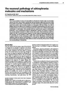
DISCUSSION Several regulatory genes have been directly implicated in the control of the morphogenesis of the hindbrain (Carpenter et al., 1993; Dolle et al., 1993; Mark et al., 1993; SchneiderMaunoury et al., 1993; Swiatek and Gridley, 1993; Cordes and Barsh, 1994; Zhang et al., 1994; Xu et al., 1995). In the cases when a loss-offunction mutation resulted in an elimination of part of the hindbrain, neither the precise extent of this deletion nor the cellular mechanisms responsible for it could be defined. These are, however, important issues when considering the function of candidate genes and the strategies employed by the hindbrain in the delimitation of territories and specification of their positional identity in the process of segmentation. Among these genes Krox-20 has been shown to be required for the normal development of r3 and r5 and to control the expression of a number of other regulatory genes in these rhombomeres (Sham et al., 1993; Nonchev et al., 1996; Vesque et al., 1996; Seitanidou, T., SchneiderMaunoury, S., Desmarquet, C., Wilkinson, D. G. and Charnay, P., unpublished data). We have shown that Krox-20 inactivation leads to complete disappearance of r3 and r5, while segmentation in the rest of the hindbrain appears to be unaffected. Nevertheless, the mutation does affect the organisation of evennumbered rhombomeres in which the gene is not expressed. Our conclusions are schematically represented in Fig. 8 and are detailed below. Segmental organisation of the hindbrain of Krox-20−/−− embryos We have investigated the organisa-
Fig. 7. Disappearance of the trigeminal nucleus around 17.5 dpc in Krox-20−/− embryos. Rostral is to the top. A-F) At early stages of hindbrain development, the motor neurons were revealed with the 4D5 antibody directed against Islet1. (A) Wild-type (A) and (B) Krox-20−/− 10.5 dpc embryos. Vth neurons start migrating laterally. (C) Wild-type (C) and (D) Krox-20−/− 12.5 dpc embryos. Trigeminal neuron migration is completed and the nucleus has condensed normally in the homozygous mutant, except for a small population of neurons (arrow) which lies caudal to the main body of the nucleus. (E) Wild-type and (F) Krox-20−/− 13.5 dpc embryos: facial neurons have now completed their migration. Note that the distance between the Vth and VIIth nuclei is smaller than in control embryos, due to the absence of r3. (G-I) Transverse cryostat sections through the brains of a 16.5 dpc wild-type embryo (G) and of 15.5 dpc (H) and 16.5 (I) dpc homozygous mutant embryos. The sections were stained with cresyl-thionine. The trigeminal nuclei are indicated by arrows. In the homozygous mutant embryos, they appear normal at 15.5 dpc (H), but are reduced in size at 16.5 dpc (I). In this latter case, the plane of section is not perfectly transverse and only one Vth nucleus is visible, but the other one was also observed on different sections. (JL) Serial transverse cryostat sections of a 17.5 dpc homozygous mutant embryo, stained with cresyl-thionine (J) or with an anti-ChAT antibody (K,L). Note that the trigeminal nucleus is reduced to remnants (arrowheads) on one side while still maintained on the other (arrow). V: trigeminal motor nucleus. VII: facial motor nucleus.
Hindbrain architecture of Krox-20 mutants 1223 parasagittal sections. (ii) Morphological structures correSwiatek and Gridley, 1993). We now provide evidence sugsponding to four rhombomere boundaries, namely r1/r2, r2/r4, gesting that this modification involves the complete disappearr4/r6 and r6/r7, are observed in the ventral half of the ance of these rhombomeres from about 10 dpc (Fig. 8). (i) As hindbrain. (iii) These limits are labelled by the rhombomere indicated above, morphological segmentation is maintained in boundary markers lim-1, PLZF and CSPG. the ventral half of Krox-20−/− embryos and the number and Transplantation experiments in chick have shown that the positions of the persisting boundaries indicate the absence of confrontation of rhombomeres of the same parity does not r3 and r5 in this region. (ii) Combined neurofilament and Xusually lead to the reformation of rhombomere boundaries and gal staining suggests the absence of r3 and is consistent with that cells of the same parity can mix relatively freely (Guthrie the loss of r5. (iii) Retrograde tracing of the trigeminal motor and Lumsden, 1991; Guthrie et al., 1993; Wizenmann and nucleus at 10.5 dpc shows that it is reduced in size. The trigemLumsden, unpublished data). The inactivation of Krox-20 inal motor population, unlike the facial and glossopharyngeal might therefore be expected to lead to profound defects in ones, is composed solely of branchiomotor neurons. The establishing boundaries; the disappearance of r3 and r5 would reduction of the nucleus is therefore consistent with the loss of be expected to allow the cells of even-numbered rhombomeres the r3 contingent of trigeminal branchiomotor neurons. (iv) to approximate and mix. Although our data do not address the Our data indicate that all motoneuron populations originating possibility of increased cell mixing, they do indicate that morfrom r5 are affected by the mutation: the somatomotor phological boundaries form normally at least in the ventral abducens nucleus is eliminated (this work, and Schneiderregion. A possible explanation for this apparent disparity is that Maunoury et al., 1993); the visceromotor population is also establishment of boundaries might be a multi-step process, affected as shown by anterograde tracing experiments which reveal the absence of the chorda tympani and greater petrosal involving first the definition of fuzzy territories of specific gene branches of the facial nerve. As indicated below, we think that expression, then progressive restriction to segmental domains, this effect actually reflects the disappearance of this latter popand finally acquisition of boundary specific properties (Irving ulation. et al., 1996). The transient confrontation of prospective evenThe elimination of all r5-derived motoneurons in Kroxand odd-numbered territories before the disappearance of r3 20−/− embryos deserves further discussion since retrograde and r5 in Krox-20−/− embryos might suffice to engage this process of boundary formation irreversibly. Upon disappeartracing analysis of the facial nerve at 10.5 dpc revealed a ance of the r3 and r5 territories, the even/odd boundaries might then fuse to generate the even/even boundaries which Control Krox-20 -/are observed at 10.5 dpc. Our observations of the presence of rhombomere boundaries and of sharp limits of expression for rhombomerespecific genes in Krox-20−/− embryos is also surprising in view of the recent r1 r1 implication of the Sek-1 receptor tyrosine gV r2 r2 kinase in the maintenance of rhomgV bomere-restricted gene expression in r4 r3 gVII Xenopus and zebrafish embryos (Xu et BA1 BA1 gVII r6 ov r4 al., 1995), since we have shown that Sekr7 1 expression in mouse r3 and r5 requires BA2 r5 BA2 ov the presence of the Krox-20 protein (Seir6 tanidou et al., unpublished data). BA3 BA3 However, it may be that in absence of r7 Krox-20 the deprivation of Sek-1 might BA4 BA4 not have the same consequences in term of restriction of cell movements or of ability to switch segmental identity. Another possibility is that the functional target of the dominant negative construct Fig. 8. Schematic representation of the segmental and neuronal organisation of the hindbrain of 10.5 dpc control and Krox-20−/− embryos. In Krox-20−/− embryos, rhombomeres 3 and 5 used by Xu and colleagues is not only are completely deleted while segmentation is otherwise maintained, the Vth motor neuron Sek-1 but also another member of this pool (red) is reduced due to the loss of r3 contribution, and trigeminal motor axons family of receptor tyrosine kinases. Complete disappearance of r3 and r5 in homozygous mutant embryos Previous work has indicated that the inactivation of the Krox-20 gene results in a profound alteration of r3 and r5 (Schneider-Maunoury et al., 1993;
fasciculate with facial motor axons (dark blue) to enter BA2 instead of BA1. The r5-derived population of facial neurons (light blue), which will constitute the superior salivatory nucleus and send axons to the chorda tympani and greater petrosal nerves in control embryos, is absent in homozygous mutants. However, a population of facial neurons of presumed r6 origin (purple) are observed in these embryos, resulting in a normal-looking fan-shaped nucleus. The abducens neurons (salmon-pink), derived exclusively from r5, are also absent in the homozygous mutant. Finally, in Krox-20−/− embryos, the IXth (green) and Xth (pink) motor nerves are partially fused, but their motor neurons appear normal and the glossopharyngeal and facial components overlap within r6.
1224 S. Schneider-Maunoury and others nucleus of normal size and morphology (Figs 4 and 5). In particular, the putative AL and LA neuronal populations, which are normally specific to r5 were present. However, our analysis of boundaries indicated that these cells were located in r6 in the homozygous mutant. This observation suggests an interpretation compatible with the elimination of r5. Firstly, the AL population, although normally located in r5, may be generated in r4: thus, the AL neurons express the r4 marker Hoxb-1 (Marshall et al., 1992) and are observed in retrograde tracing studies to occupy successively more caudal positions at successive stages of development. At 9.5 dpc they lie in medial r4, thereafter appearing to migrate posteriorly along the floor plate border into r5 and eventually into caudal r5 and rostral r6, where they turn laterally (McKay and Lumsden, unpublished data). The path taken by the posteriorly migrating cell bodies thus forms a loop around the abducens nucleus, the facial nerve genu. We propose that in the Krox-20−/− mutant, in the absence of an r5 territory, this migration now proceeds completely into r6. Secondly, the LA population is supposed to be generated in r5. We propose that this population does not form in the homozygous mutant. Instead, due to the abnormal proximity of the facial nerve exit point in r4, some axons belonging to r6 branchiomotor neurons and normally fated to join the glossopharyngeal nerve are misrouted and reach the facial exit point, thereby mimicking LA axons. Several observations support this interpretation: (1) glossopharyngeal and ‘facial’ neurons overlap in r6 in the Krox-20−/− mutant; (2) the LA-like neurons are unable to send axons into the greater petrosal and chorda tympani branches of the facial nerve suggesting that they do not have a correct preganglionic visceromotor identity; (3) experiments involving explant culture, ablation of the r4 exit point, or rhombomere transplantation or reversal have suggested that intrinsic mechanisms direct the initial lateral orientation of hindbrain motor axons, but that chemotropic cues from nerve exit points might be responsible for their later rostral or caudal orientation (Guthrie and Lumsden, 1991; Chang et al., 1992; Guthrie and Lumsden, 1992; Guthrie and Pini, 1995). Similar mechanisms might therefore govern the extension of the axons of r6-derived motoneurons. In addition, it is possible that r5 constitutes a barrier for growth cone migration of r6 neurons during normal development. Such a barrier would be eliminated in Krox-20−/− embryos. Another possible explanation for the presence of LA-like neurons in r6 can nevertheless be envisaged. These neurons could have become specified in the presumptive r5 territory before its disappearance and then migrated into r6 where they would develop their axons. This new environment or abnormal specification resulting from the lack of exposure to Krox-20 might modify their properties, resulting in the inability of their axons to join the chorda tympani and the greater petrosal. In this respect it is interesting to note that several members of the EPH family of tyrosine kinase receptors, which may be involved in axonal guidance, are specifically expressed in r5 (Becker et al., 1994). The Krox-20 mutation affects even-numbered rhombomeres and their derivatives Whatever the explanation for the presence of facial-like neurons in r6, our data indicates that this even-numbered rhombomere is affected by the mutation, a phenomenon that is not restricted to r6. We have noticed that r4, as defined by Hoxb-
1 expression, is significantly narrower in Krox-20−/− embryos as compared to controls (Fig. 2C,D). In the case of r2, it is the trajectory of trigeminal motor axons that is modified in Krox20−/− embryos. Although they exit the brain at their normal exit point in r2, they then fasciculate with facial axons and enter the second branchial arch instead of the first arch. In addition, as discussed below, the trigeminal nucleus completely disappears in Krox-20−/− embryos at later stages of embryogenesis. An interesting question concerning these phenotypes of rhombomeres that do not express Krox-20, is whether they are directly caused by the lack of expression of Krox-20 or rather are mere ‘mechanical’ consequences of the disappearance of r3 and r5. Although there is now direct evidence for the existence of communication between adjacent rhombomeres (Graham et al., 1993, 1994; Graham and Lumsden, 1996), we favour the second hypothesis for explaining modified axonal trajectories. The misrouting of both trigeminal and glossopharyngeal axons could be related simply to the abnormal proximity of the r4 exit point, due to the absence of r3 and r5 respectively. In any case, our data further support the view of an integrated organisation of the hindbrain, where rhombomeres do not develop completely autonomously. Possible branchial arch specificity of neurotrophic factors The misrouting of trigeminal axons in Krox-20−/− embryos suggests that there is no strong specificity for a particular branchial arch in the guidance of branchiomotor axons, since r2 neurons can send axons to BA2. Similarly, it has been shown using r4 to r2 transplantations in chick embryos, that a subpopulation of r4 neurons (the vestibulo-acoustic nerve efferents) can send axons into BA1 (Simon et al., 1995). The fate of the misrouted trigeminal motor neurons was investigated between 10.5 and 18.5 dpc. They were shown to migrate and condense normally and, apart from an initial size reduction, did not show major abnormality until 15.5 dpc. By 17.5 dpc, however, they appeared to be dramatically affected and were always absent or reduced to remnants by 18.5 dpc. This suggests that most trigeminal motoneurons die around 17.5 dpc in Krox-20−/− embryos. This massive death therefore appears to occur precisely within the normal period of motor neuron cell death in the mouse (Lance Jones, 1982); in the facial nucleus, selective neuronal death occurs between 17 and 20 dpc (Ashwell and Watson, 1983). Interestingly, a similar late death of misrouted neurons occurs in transplantation experiments which involve anteroposterior reversal of rhombomere 3 in chick embryos and result in r3-derived motor neurons exiting the brain via the facial root in r4 and migrating into BA2 instead of BA1 (J. Warrilow and S. Guthrie, personal communication). These observations suggest that the late death of the misrouted trigeminal motor neurons may be due to inadequate survival factors during this critical period. This could be due either to their incapacity to establish functional synapses with muscle cells from the second arch or to the fact that the second arch targets would provide them with trophic factors different from those of the first arch. In any case, the data suggest the existence of specificity in the interaction between a branchiomotor nerve and its corresponding branchial arch. We thank Dr T. Jessel for the gift of the 4D5 antibody, Dr H.
Hindbrain architecture of Krox-20 mutants 1225 Westphal for the mouse lim-1 probe, Dr R. Krumlauf for the Hoxb-1 probe, Dr A. Zelent for the PLZF probe and Drs I. McKay and M. Wassef for critically reading the manuscript. The 2H3 monoclonal antibody was obtained from the Developmental Studies Hybridoma Bank. Work in the Charnay lab was supported by grants from INSERM, MESR, EEC, ARC, LNFCC, AFM and ARSEP. T. S. was supported by a fellowship from Recherche et Partage. Work in the Lumsden lab was supported by grants from the MRC, the Wellcome Trust and the Howard Hughes Medical Institute, of which A. L. is an International Research Scholar.
REFERENCES Ashwell, K. W. and Watson, C. R. (1983). The development of facial motoneurones in the mouse: neuronal death and the innervation of the facial muscles. J. Embryol. Exp. Morphol. 77, 117-41. Becker, N., Seitanidou, T., Murphy, P., Mattei, M. G., Topilko, P., Nieto, M. A., Wilkinson, D. G., Charnay, P. and Gilardi Hebenstreit, P. (1994). Several receptor tyrosine kinase genes of the Eph family are segmentally expressed in the developing hindbrain. Mech. Dev. 47, 3-17. Birgbauer, E. and Fraser, S. E. (1994). Violation of cell lineage restriction compartments in the chick hindbrain. Development 120, 1347-1356. Carpenter, E. M., Goddard, J. M., Chisaka, O., Manley, N. R. and Capecchi, M. R. (1993). Loss of Hox-A1 (Hox-1.6) function results in the reorganization of the murine hindbrain. Development 118, 1063-1075. Chang, S., Fan, J. and Nayak, J. (1992). Pathfinding by cranial nerve VII (facial) motorneurons in the chick hindbrain. Development 114, 815-823. Chavrier, P., Zerial, M., Lemaire, P., Almendral, J., Bravo, R. and Charnay, P. (1988). A gene encoding a protein with zinc fingers is activated during G0/G1 transition in cultured cells. EMBO J. 7, 29-35. Chavrier, P., Vesque, C., Galliot, B., Vigneron, M., Dolle, P., Duboule, D. and Charnay, P. (1990). The segment-specific gene Krox-20 encodes a transcription factor with binding sites in the promoter region of the Hox-1.4 gene. EMBO J. 9, 1209-1218. Clarke, J. D. W. and Lumsden, A. (1993). Segmental repetition of neuronal phenotype sets in the chick embryo hindbrain. Development 118, 151-162. Cook, M., Gould, A., Brand, N., Davies, J., Strutt, P., Shaknovich, R., Licht, J., Waxman, S., Chen, Z., Gluecksohn-Waelsch, S., Krumlauf, R. and Zelent, A. (1995). Expression of the zinc-finger gene PLZF at rhombomere boundaries in the vertebrate hindbrain. Proc. Natl. Acad. Sci. USA 92, 22492253. Cordes, S. P. and Barsh, G. S. (1994). The mouse segmentation gene Kr encodes a novel basic domain-leucine zipper transcription factor. Cell 79, 1025-1034. Dolle, P., Lufkin, T., Krumlauf, R., Mark, M., Duboule, D. and Chambon, P. (1993). Local alterations of Krox-20 and Hox gene expression in the hindbrain suggest lack of rhombomeres 4 and 5 in homozygote null Hoxa-1 (Hox-1.6) mutant embryos. Proc. Natl. Acad. Sci. USA 90, 7666-7670. Ellis, J., Liu, Q., Breitman, M., Jenkins, N. A., Gilbert, D. J., Copeland, N. G., Tempest, H. V., Warren, S., Muir, E., Schilling, H. and et al. (1995). Embryo brain kinase: a novel gene of the eph/elk receptor tyrosine kinase family. Mech. Dev. 52, 319-341. Ericson, J., Thor, S., Edlund, T., Jessell, T. M. and Yamada, T. (1992). Early stages of motor neuron differentiation revealed by expression of homeobox gene Islet-1. Science 256, 1555-1560. Fraser, S., Keynes, R. and Lumsden, A. (1990). Segmentation in the chick embryo hindbrain is defined by cell lineage restrictions. Nature 344, 431435. Fritzsch, B. and Nichols, D. H. (1993). DiI reveals a prenatal arrival of efferents at the differentiating otocyst of mice. Hear Res. 65, 51-60. Fujii, T., Pichel, J. G., Taira, M., Toyama, R., Dawid, I. B. and Westphal, H. (1994). Expression patterns of the murine LIM class homeobox gene lim1 in the developing brain and excretory system. Dev. Dyn. 199, 73-83. Gilardi, P., Schneider Maunoury, S. and Charnay, P. (1991). Krox-20: a candidate gene for the regulation of pattern formation in the hindbrain. Biochimie 73, 85-91. Gilardi-Hebenstreit, P., Nieto, M. A., Frain, M., Mattei, M. G., Chestier, A., Wilkinson, D. G. and Charnay, P. (1992). An Eph-related receptor protein tyrosine kinase gene segmentally expressed in the developing mouse hindbrain. Oncogene 7, 2499-2506. Gilland, E. and Baker, R. (1993). Conservation of neuroepithelial and
mesodermal segments in the embryonic vertebrate head. Acta Anat. Basel 148, 110-123. Graham, A., Heyman, I. and Lumsden, A. (1993). Even-numbered rhombomeres control the apoptotic elimination of neural crest cells from oddnumbered rhombomeres in the chick hindbrain. Development 119, 233-245. Graham, A., Francis West, P., Brickell, P. and Lumsden, A. (1994). The signalling molecule BMP4 mediates apoptosis in the rhombencephalic neural crest. Nature 372, 684-686. Graham, A. and Lumsden, A. (1996). Interactions between rhombomeres modulate Krox-20 and follistatin expression in the chick embryo hindbrain. Development 122, 473-480. Guthrie, S., Butcher, M. and Lumsden, A. (1991). Patterns of cell division and interkinetic nuclear migration in the chick embryo hindbrain. J. Neurobiol. 22, 742-754. Guthrie, S. and Lumsden, A. (1991). Formation and regeneration of rhombomere boundaries in the developing chick hindbrain. Development 112, 221-229. Guthrie, S. and Lumsden, A. (1992). Motor neuron pathfinding following rhombomere reversals in the chick embryo hindbrain. Development 114, 663-673. Guthrie, S., Prince, V. and Lumsden, A. (1993). Selective dispersal of avian rhombomere cells in orthotopic and heterotopic grafts. Development 118, 527-538. Guthrie, S. and Pini, A. (1995). Chemorepulsion of developing motor axons by the floor plate. Neuron 14, 1117-1130. Heaton, M. B. and Moody, S. A. (1980). Early development and migration of the trigeminal motor nucleus in the chick embryo. J. Comp. Neurol. 189, 6199. Heyman, I., Kent, A. and Lumsden, A. (1993). Cellular morphology and extracellular space at rhombomere boundaries in the chick embryo hindbrain. Dev. Dyn. 198, 241-253. Heyman, I., Faissner, A. and Lumsden, A. (1995). Cell and matrix specialisations of rhombomere boundaries. Dev. Dyn. 204, 301-315. Hunt, P., Gulisano, M., Cook, M., Sham, M. H., Faiella, A., Wilkinson, D., Boncinelli, E. and Krumlauf, R. (1991). A distinct Hox code for the branchial region of the vertebrate head. Nature 353, 861-864. Irving, C., Nieto, M. A., DasGupta, R., Charnay, P. and Wilkinson, D. G. (1996). Progressive spatial restriction of Sek-1 and Krox-20 gene expression during hindbrain segmentation. Dev. Biol. 173, 26-38. Keynes, R. and Krumlauf, R. (1994). Hox genes and regionalization of the nervous system. Annu. Rev. Neurosci. 17, 109-132. Krumlauf, R., Marshall, H., Studer, M., Nonchev, S., Sham, M. H. and Lumsden, A. (1993). Hox homeobox genes and regionalisation of the nervous system. J. Neurobiol. 24, 1328-1340. Lance Jones, C. (1982). Motoneuron cell death in the developing lumbar spinal cord of the mouse. Brain Res. 256, 473-479. Layer, P. G. and Alber, R. (1990). Patterning of chick brain vesicles as revealed by peanut agglutinin and cholinesterases. Development 109, 613624. Lumsden, A. (1990). The cellular basis of segmentation in the developing hindbrain. Trends Neurosci. 13, 329-335. Lumsden, A. and Keynes, R. (1989). Segmental patterns of neuronal development in the chick hindbrain. Nature 337, 424-428. Mahmood, R., Kiefer, P., Guthrie, S., Dickson, C. and Mason, I.J. (1995). Multiple roles for FGF-3 during cranial neural development of the chicken. Development 121, 1399-1410. Mark, M., Lufkin, T., Vonesch, J. L., Ruberte, E., Olivo, J. C., Dolle, P., Gorry, P., Lumsden, A. and Chambon, P. (1993). Two rhombomeres are altered in Hoxa-1 mutant mice. Development 119, 319-338. Marshall, H., Nonchev, S., Sham, M. H., Muchamore, I., Lumsden, A. and Krumlauf, R. (1992). Retinoic acid alters hindbrain Hox code and induces transformation of rhombomeres 2/3 into a 4/5 identity. Nature 360, 737-741. Martinez, S., Geijo, E., Sanchez Vives, M. V., Puelles, L. and Gallego, R. (1992). Reduced junctional permeability at interrhombomeric boundaries. Development 116, 1069-1076. McKay, I. J., Muchamore, I., Krumlauf, R., Maden, M., Lumsden, A. and Lewis, J. (1994). The kreisler mouse: a hindbrain segmentation mutant that lacks two rhombomeres. Development 120, 2199-2211. Murphy, P., Davidson, D. R. and Hill, R. E. (1989). Segment-specific expression of a homoeobox-containing gene in the mouse hindbrain. Nature 341, 156-159. Nieto, M. A., Bradley, L. C. and Wilkinson, D. G. (1991). Conserved segmental expression of Krox-20 in the vertebrate hindbrain and its relationship to lineage restriction. Development 2, 59-62.
1226 S. Schneider-Maunoury and others Nieto, M. A., Gilardi Hebenstreit, P., Charnay, P. and Wilkinson, D. G. (1992). A receptor protein tyrosine kinase implicated in the segmental patterning of the hindbrain and mesoderm. Development 116, 1137-1150. Nonchev, S., Vesque, C., Maconochie, M., Seitanidou, T., ArizaMcNaughton, L., Frain, M., Marshall, H., Sham, M. H., Krumlauf, R. and Charnay, P. (1996). Segmental expression of Hoxa-2 in the hindbrain is directly regulated by Krox-20. Development 122, 543-554. Oxtoby, E. and Jowett, T. (1993). Cloning of the zebrafish krox-20 gene (krx20) and its expression during hindbrain development. Nucl. Acids Res. 21, 1087-1095. Ruberte, E., Dolle, P., Chambon, P. and Morriss Kay, G. (1991). Retinoic acid receptors and cellular retinoid binding proteins. II. Their differential pattern of transcription during early morphogenesis in mouse embryos. Development 111, 45-60. Ruberte, E., Friederich, V., Morriss Kay, G. and Chambon, P. (1992). Differential distribution patterns of CRABP I and CRABP II transcripts during mouse embryogenesis. Development 115, 973-987. Ruiz, J. C. and Robertson, E. J. (1994). The expression of the receptor-protein tyrosine kinase gene, eck, is highly restricted during early mouse development. Mech. Dev. 46, 87-100. Schneider-Maunoury, S., Topilko, P., Seitanidou, T., Levi, G., Cohen Tannoudji, M., Pournin, S., Babinet, C. and Charnay, P. (1993). Disruption of Krox-20 results in alteration of rhombomeres 3 and 5 in the developing hindbrain. Cell 75, 1199-1214. Sham, M. H., Vesque, C., Nonchev, S., Marshall, H., Frain, M., Gupta, R. D., Whiting, J., Wilkinson, D., Charnay, P. and Krumlauf, R. (1993). The zinc finger gene Krox20 regulates HoxB2 (Hox2.8) during hindbrain segmentation. Cell 72, 183-196. Simon, H., Guthrie, S. and Lumsden, A. (1994). Regulation of SC1/DMGRASP during the migration of motor neurons in the chick embryo brain stem. J. Neurobiol. 25, 1129-1143. Simon, H., Hornbruch, A. and Lumsden, A. (1995). Independent assignment of antero-posterior and dorso-ventral positional values in the developing chick hindbrain. Curr. Biol. 5, 205-214. Swiatek, P. J. and Gridley, T. (1993). Perinatal lethality and defects in hindbrain development in mice homozygous for a targeted mutation of the zinc finger gene Krox20. Genes Dev. 7, 2071-2084.
Tsuchida, T., Ensini, M., Morton, S. B., Baldassare, M., Edlund, T., Jessell, T. M. and Pfaff, S. L. (1994). Topographic organization of embryonic motor neurons defined by expression of LIM homeobox genes. Cell 79, 957-970. Vesque, C. and Charnay, P. (1992). Mapping functional regions of the segment-specific transcription factor Krox-20. Nucl. Acids Res. 20, 24852492. Vesque, C., Maconochie, M., Nonchev, S., Ariza-McNaughton, L., Kuroiwa, A., Charnay, P. and Krumlauf, R. (1996). Hoxb-2 transcriptional activation in rhombomeres 3 and 5 requires an evolutionarily conserved cisacting element in addition to the Krox-20 binding site. EMBO J. 19, 53835396. Wilkinson, D. G. (1990). Segmental gene expression in the developing mouse hindbrain. Sem. Dev. Biol. 1, 127-134. Wilkinson, D. G. (1992). Whole-mount in situ hybridisation of vertebrate embryos. In In Situ Hybridisation: a practical approach (ed. D. G. Wilkinson), pp. 75-83. Oxford: IRL Press. Wilkinson, D. G. (1993). Molecular mechanisms of segmental patterning in the vertebrate hindbrain. Perspect Dev. Neurobiol. 1, 117-125. Wilkinson, D. G., Bhatt, S., Chavrier, P., Bravo, R. and Charnay, P. (1989a). Segment-specific expression of a zinc-finger gene in the developing nervous system of the mouse. Nature 337, 461-464. Wilkinson, D. G., Bhatt, S., Cook, M., Boncinelli, E. and Krumlauf, R. (1989b). Segmental expression of Hox-2 homoeobox-containing genes in the developing mouse hindbrain. Nature 341, 405-409. Wingate, R. and Lumsden, A. (1996). Persistence of rhombomeric organisation in the postsegmental avian hindbrain. Development 122, 21432152. Xu, Q., Alldus, G., Holder, N. and Wilkinson, D. G. (1995). Expression of truncated Sek-1 receptor tyrosine kinase disrupts the segmental restriction of gene expression in the Xenopus and zebrafish hindbrain. Development 121, 4005-4016. Zhang, M., Kim, H. J., Marshall, H., Gendron Maguire, M., Lucas, D. A., Baron, A., Gudas, L. J., Gridley, T., Krumlauf, R. and Grippo, J. F. (1994). Ectopic Hoxa-1 induces rhombomere transformation in mouse hindbrain. Development 120, 2431-2442. (Accepted 5 January 1997)