rd
The 3 European Medical and Biological Engineering Conference EMBEC'05
November 20 – 25, 2005 Prague, Czech Republic
SEGMENTATION OF THE PIG HEAD M. Hannula*, T. Arola*, J. Hyttinen*, P. Kauppinen*, M. Silfverhuth**, V. Jäntti***, J. Malmivuo* * Ragnar Granit Institute, Tampere University of Technology, Tampere, Finland ** Diagnostic Radiology / Clinical Neurophysiology, Oulu University Hospital, Oulu, Finland *** Clinical Neurophysiology, Tampere University Hospital, Tampere, Finland
[email protected] Abstract: A detailed porcine finite difference head model was constructed for bioelectric and high frequency electromagnetic field Specific Absorption Rate (SAR) calculations. Magnetic Resonance (MR) data set was obtained of the pig’s head. These data was segmented using various techniques including thresholding, region growing and active contours. The segmentation algorithms and user interface were implemented in Matlab. The result is a detailed model of the pig head consisting of 11 labeled tissues and 2309164 voxels or computational elements. The model is now used in SAR calculations and furthermore it will be used in forward and inverse bioelectric solutions. Introduction Nowadays people are interested in the radiation affects of various devices such as mobile phones. Radiation dose can be calculated using SAR techniques [4, 11, 12]. Models are used as a basis for these calculations. More detailed models are needed in order to get more accurate results. Furthermore in various brain studies accurate models are needed to study the bioelectric phenomena of the brain and the neurons. Porcine model has been used in testing in various biomedical applications in vivo [1, 3, 10]. In addition, pig model is employed in a study for obtaining the effects of high energy mobile phone fields on brain functions (http://www.uku.fi/hermo/). In order to calculate the SAR in brain areas and to assess the bioelectric fields a volume conductor model of the pig head was required. A segmentation of a pig head was produced for high frequency electromagnetic field SAR calculation and bioelectric problems. In addition, the pig model was considered to be important because pig is used as a test animal for various medical and biomedical experiments. Creating a model of a pig enables us to do simulations and calculations in a complete pig head and compare the results with experimental data. An MR image set of a pig head was obtained for segmentation. The goal was accurate segmentation of the pig head with as many different tissues as possible. This paper discusses the steps and difficulties encountered during the segmentation process.
IFMBE Proc. 2005 11(1)
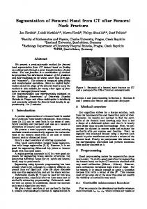
Material and Methods Material for this study was obtained from a project where the pig model was used to study the development of an epileptic focus in a pig brain under anesthesia. The pig was anesthetized with isoflurane to EEG burst suppression level. Penicillin was then injected intracortically to produce focal epileptic discharge. EEG and functional MR imaging (fMRI) were recorded simultaneously. The MR images were used for the basis of the developed model [1]. MR imaging was performed with a GE (Milwaukee, WI, USA) Sigma EchoSpeed 1.5 T scanner. The 3D MR data set consisted of 124 transversal slices with 1.5mm slice thickness in 256x256 resolution (Figure 1).
Figure 1: MR image of the pig’s head While being a lot different from human head in anatomy, the pig’s head proved to be quite a challenge in segmentation. Pig’s brain is a lot smaller than the human brain. On the other hand the muscles are much larger in pig’s head. Also the bone structure is much more complicated in pig’s head. Segmentation procedure started with tissues that are relatively straightforward to segment such as the eyes and skin. Volumes defined by those segmentations were
ISSN: 1727-1983 © 2005 IFMBE
rd
The 3 European Medical and Biological Engineering Conference EMBEC'05
subtracted from the original data in order to make the remaining tasks less complicated. Starting point was to extract the whole head volume from the background. Thresholding was the simplest and most efficient technique for this duty. Morphological methods such as erosion and dilation were used with the skin extraction [2]. The brain was extracted with the help of active contours i.e. snakes [5]. Snake methods applied included Balloon method [7], Gradient Vector Flow (GVF) [8] method and Generalized Gradient Vector Flow (GGVF) [9] method. White matter and gray matter were separated by using thresholding. The complexity of bare bone structure was the first obstacle. Bone has quite a poor appearance in MR images compared to Computed Tomography (CT) images. To make the task easier, we obtained a pig’s skull shown in Figure 2 and had a CT data set taken from it (Figure 3). The skull was from a different specimen as the MR data thus the CT data was only used as a guideline for the most difficult parts of the bone area segmentation. 3D region growing [6] was used to segment the eyes and muscles since the method is suitable for extracting areas that are smoothly continuous and relatively homogeneous. Some manual segmentation had to be done for both tissues e.g. to fill holes. Difficulties were encountered in the segmentation of the muscles as those exist in a large area and some edges are quite weak. Thus the region growing often leaked to undesired areas. In our 2D segmentation methods like active contour the fact that consecutive slices look much alike helped a lot. Result of the current slice can be used as an initial value for the next slice. This approach was used for extracting e.g. the skull and the brain.
November 20 – 25, 2005 Prague, Czech Republic
Figure 3: CT images of the pig’s skull helped in identifying the skull also from the MR image data set. Bone tissue is clearly visible in CT images. All the previous algorithms were implemented in Matlab in our institute. Software used in segmentation was mainly Matlab based, but also other software was used e.g. a 3D visualization engine developed in our institute [13]. 2D segmentation included a careful segmentation in three orthogonal planes keeping in mind the segmentation results in the other two planes. Thus step-shaped results and holes in solid surfaces were avoided effectively. Throughout the segmentation procedure the result was carefully examined with the 3D visualization engine to validate the extracted surfaces and volumes. Morphological methods were used often in order to finalize the result. In addition to the semiautomatic methods, manual segmentation had to be done too, especially with the skull, because of poor appearance in MR images. Segmentation procedure and used methods are shown in Figure 4.
Figure 2: Pig’s skull was obtained for guidance to the segmentation of the bone from the MR images. Figure 4: Segmentation procedure. Boxes in the middle present different segmentation tasks and those are in performance order from left to right. Oval shaped objects illustrate used segmentation methods marked with different colors. Boxes have a color that corresponding method is mainly used in segmentation.
IFMBE Proc. 2005 11(1)
ISSN: 1727-1983 © 2005 IFMBE
rd
The 3 European Medical and Biological Engineering Conference EMBEC'05
November 20 – 25, 2005 Prague, Czech Republic
Results The final product, the segmented pig’s head consists of 11 different tissues. Segmented tissues were gray matter, white matter, marrow bone, cortical bone, vitreous humor, sclera and lenses in the eyes, muscle, skin, fat and a remaining mixture of blood, fat, and muscle (Figure 5). The model obtained from the segmentation was converted to Finite Difference Time Domain (FDTD) – format in order to allow SAR calculations as well as for our own FDM format for bioelectric applications.
Figure 6: Segmented pig head in 3D visualization. The figure shows bone structure as bone marrow and cortical bone, the brain and the eyes. Skin is shown highly transparent to enhance the perspective of the image.
Figure 5: Segmented pig’s head in 2D illustration. The head consists of 11 tissues showing in different colors. Segmentation also enables volumetric calculations of the tissues. Tissues and corresponding volumes are presented in Table 1. 3D visualization of the complete segmentations can be seen in Figure 6 and Figure 7. Table 1: Different tissues listed with some volumes and percentage values calculated. Tissue brain, gray matter brain, white matter bone marrow bone cortical eye, vitreous humor eye, sclera eye, lens muscle skin fat mixture: blood, fat, muscle
IFMBE Proc. 2005 11(1)
Volume/ml 61,3 18,8 42,6 279,8 5,9 0,3 0,4 145,6 153,5 206,6
% 2,91 0,89 2,02 13,28 0,28 0,01 0,02 6,91 7,28 9,80
1192,5
56,59
Figure 7: Volume rendering of pig’s head showing cortical bone, fat and the eyes. Discussion Segmentation of the whole pig head was quite a laborious task. Applied segmentation techniques included thresholding, region growing, active contour and morphological methods. During the segmentation valuable first hand information on the segmentation techniques was achieved. According to this information, our present segmentation methods can be improved and new ones need to be developed. Most importantly it was observed that 2D segmentation methods really are not adequate for obtaining high quality 3D model data and thus 3D segmentation methods and tools are needed. To obtain solid surfaces and continuous volumes 3D visualization must be utilized with 2D segmentation. Without this the
ISSN: 1727-1983 © 2005 IFMBE
rd
The 3 European Medical and Biological Engineering Conference EMBEC'05
2D segmentation will produce artifacts such as holes in thin areas such as the skull. This can result in large errors in calculated bioelectric fields. It was also noticed very clearly that there is not a segmentation method that would work in every case. Different segmentation methods have to work together for desired result. At least, methods often have to be adjusted for different purposes and user has to set some limitations in order to get acceptable results. For example working with region growing it is sometimes a matter of finding the balance between too small or leaking results. 3D methods such as region growing work better with a data set where the gap between two consecutive slices is as small as possible. If the desired volume changes much in horizontal plane methods may not continue to the right area and the result is incorrect. These kinds of situations need observation during the segmentation. Hence faulty processes can be stopped in the beginning and time consuming iterations can be avoided. Active contour methods worked well with the brain. Two difficulties sometimes arise with active contour: how to obtain the initial curve and how to stop the curve at the right edge. Using achieved result from the previous slice an initial curve for the current slice was obtained easily. There were no major changes between two consecutive slices in brain border. Thus the right border was also found nicely. In general, pig anatomy is so different from the human anatomy that a person with special knowledge on pig anatomy was found necessary in order to obtain correct segmentation. Now, similar kinds of assignments are easier to complete with a priori knowledge of anatomy and suitable methods for segmentation.
November 20 – 25, 2005 Prague, Czech Republic
[2]
[3]
[4]
[5]
[6]
[7]
[8]
[9]
[10]
Conclusions We have segmented a full pig’s head with 11 tissue types with 124 slices in 256x256 resolution. Our goal has been a detailed, valid porcine head model. This has been accomplished by using various segmentation methods combined with 3D visualization. The segmentation produced can be used for several purposes. The original need was a model for high frequency electromagnetic field specific absorption rate calculation. With the completed model various bioelectric applications will be studied. For example, with the FDM solver implemented in our institute, the full resolution of 2309164 voxels can be applied to simulate EEG and source localization in penicillin induced epilepsy in pig’s brain during anesthesia.
References [1]
MÄKIRANTA M. (2004), 'EEG and BOLDcontrast fMRI in brain Cerebrovascular reactivity, suppression of neuronal activity, global and local brain injury', Department of Diagnostic Radiology, Department of Anaesthesiology, University of Oulu, Oulu
IFMBE Proc. 2005 11(1)
[11]
[12]
[13]
GONZALEZ R. C. and WOODS R. E. (2002): 'Digital image processing', Prentice Hall, Upper Saddle River, N.J. RIMPILÄINEN J. (2001), 'Biochemical and reperfusion targeting strategies to improve brain protection during prolonged hypothermic circulatory arrest', Department of Surgery, University of Oulu, Oulu GANDHI O. (1990): 'Biological Effects and Medical Applications of Electromagnetic Energy', Prentice Hall, Englewood Cliffs KASS M., WITKIN A. P. and TERZOPOULOS D. (1987): 'SNAKES: Active Contour Models', International Journal of Computer Vision, pp.321-331 ADAMS R. and BISCHOF L. (1994): 'Seeded Region Growing', IEEE Transactions on Pattern Analysis and Machine Intelligence, vol.16, pp.641-647 COHEN L. D. (1991): 'On Active Contour Models and Balloons', CVGIP: Image Understanding, vol.53, pp.211-218 XU C. and PRINCE J. L. (1998): 'Snakes, Shapes, and Gradient Vector Flow', IEEE Transactions on Image Processing, vol.7, pp.359-369 XU C. and PRINCE J. L. (1998): 'Generalized Gradient Vector Flow External Forces for Active Contours', Signal Processing, vol.71, pp.131-139 AOKI M., NOMURA F., STROMSKI M. E., TSUJI M. K., FACKLER J. C., HICKEY P. R., HOLTZMAN D. and JONAS R. A. (1994): 'Effects of MK-801 and NBQX on acute recovery of piglet cerebral metabolism after hypothermic circulatory arrest', Journal of Cerebral Blood Flow & Metabolism, vol.14, pp.156-65 GERHARDT D. (2003): 'Definition of a parameter for a typical specific absorption rate under real boundary conditions of cellular phones in a GSM network', Advances in Radio Science, pp.335-338 KIVEKAS O., OLLIKAINEN J., LEHTINIEMI T., VAINIKAINEN P. (2004): 'Bandwidth, SAR, and efficiency of internal mobile phone antennas', IEEE Transactions on Electromagnetic Compatibility, vol.46, pp.71– 86 AROLA T., HANNULA M., HYTTINEN J., DASTIDAR P., SOIMAKALLIO S., MALMIVUO J. (2005): 'A Java-based Multiplatform High Performance 2D and 3D Visualization Engine for Medical Image Data Sets: Abdominal Aortic Aneurysm as an Example', Submitted to 3rd European Medical Conference on Biomedical Engineering, Prague, Czech Republic, November 20-25, 2005
ISSN: 1727-1983 © 2005 IFMBE


![[PDF] The Story of Peppa Pig (Peppa Pig) - Google Sites](https://m.moam.info/img/260x300/pdf-the-story-of-peppa-pig-peppa-pig-google-sites_64779475097c47a9708bdebe.jpg)


