Selection for a CEACAM Receptor-Specific Binding Phenotype during Neisseria gonorrhoeae Infection of the Human Genital Tract Anna Sintsova,a Henry Wong,a* Kelly S. MacDonald,b Rupert Kaul,b Mumtaz Virji,c
Scott D. Gray-Owena
a
Department of Molecular Genetics, University of Toronto, Toronto, Ontario, Canada ; Department of Medicine, University of Toronto, Toronto, Ontario, Canadab; Department of Cellular and Molecular Medicine, University of Bristol, Bristol, United Kingdomc
N
eisseria gonorrhoeae has persisted in the human population despite all attempts to limit the spread of infection (1). The alarming rise in antibiotic-resistant strains and increase in the global incidence of infection have put N. gonorrhoeae at the forefront of national and international public health agendas (2). N. gonorrhoeae is a sexually transmitted pathogen that most commonly colonizes the urogenital mucosa, although it may also be found on nasopharyngeal, rectal, and ocular surfaces. Disease manifestations vary greatly between men and women. Infections in men are commonly characterized by acute urethritis with profuse purulent discharge. This pus largely consists of polymorphonuclear leukocytes (PMNs), potently phagocytic cells responsible for bacterial elimination but with the potential to damage the surrounding tissues in the process (3). In women, N. gonorrhoeae colonizes the endocervix, where it also has the potential to cause painful inflammation and cervical discharge; however, most infections in women are asymptomatic (2). If left untreated, the gonococci may ascend into the female upper genital tract to promote a pathogenic inflammatory response that can precipitate severe health issues, including pelvic inflammatory disease (PID), ectopic pregnancies, and infertility (3). N. gonorrhoeae is a human-restricted pathogen that has evolved sophisticated mechanisms to facilitate colonization and persistence within its host. Essential for these processes are specialized adhesins that allow N. gonorrhoeae attachment to receptors expressed exclusively on human mucosal tissues. The type IV pilus mediates the initial bacterial attachment to the host cell. By virtue of its ability to retract, the pilus overcomes mucosal flow and brings the bacterium into close proximity to the epithelial cell (4) to facilitate a more intimate association and/or cellular invasion (5). Studies using male human volunteers indicate that the pilus is not required for initial infection, although it may contribute to disease manifestations (6).
1372
iai.asm.org
The colony opacity-associated (Opa) proteins were recognized by their effects on interbacterial aggregation and leukocyte association (7, 8) and were later shown to mediate a tight secondary association between N. gonorrhoeae and the epithelia (9). Each gonococcal isolate possesses ⬃11 different opa genes, each encoding antigenically and phenotypically distinct variants that reversibly turn expression “off” and “on” at a rate estimated to be 10⫺3 to 10⫺4/cell/generation (10, 11). N. gonorrhoeae isolates from both naturally infected men and women are predominantly Opa⫹, as are isolates obtained from men experimentally infected with transparent (Opa phase-varied off) colonies (12–14). Most gonococcal Opa variants bind to one or more members of the human CEACAM family of receptors (15–20). CEACAM receptors are members of the immunoglobulin (Ig) superfamily, containing an Ig variable-region-like N-terminal domain followed by a varying number of Ig constant-region-like domains exposed at the cell surface (21, 22). CEACAM1, CEACAM3, CEACAM5 (carcino-
Received 22 December 2014 Returned for modification 5 January 2015 Accepted 13 January 2015 Accepted manuscript posted online 20 January 2015 Citation Sintsova A, Wong H, MacDonald KS, Kaul R, Virji M, Gray-Owen SD. 2015. Selection for a CEACAM receptor-specific binding phenotype during Neisseria gonorrhoeae infection of the human genital tract. Infect Immun 83:1372–1383. doi:10.1128/IAI.03123-14. Editor: R. P. Morrison Address correspondence to Scott D. Gray-Owen,
[email protected]. * Present address: Henry Wong, Kingston General Hospital, Kingston, Ontario, Canada. A.S. and H.W. contributed equally to this work. Copyright © 2015, American Society for Microbiology. All Rights Reserved. doi:10.1128/IAI.03123-14
Infection and Immunity
April 2015 Volume 83 Number 4
Downloaded from http://iai.asm.org/ on October 21, 2015 by guest
Infections by Neisseria gonorrhoeae are increasingly common, are often caused by antibiotic-resistant strains, and can result in serious and lasting sequelae, prompting the reemergence of gonococcal disease as a leading global health concern. N. gonorrhoeae is a human-restricted pathogen that primarily colonizes urogenital mucosal surfaces. Disease progression varies greatly between the sexes: men usually present with symptomatic infection characterized by a painful purulent urethral discharge, while in women, the infection is often asymptomatic, with the most severe pathology occurring when the bacteria ascend from the lower genital tract into the uterus and fallopian tubes. Classical clinical studies demonstrated that clinically infectious strains uniformly express Opa adhesins; however, their specificities were unknown at the time. While in vitro studies have since identified CEACAM proteins as the primary target of Opa proteins, the gonococcal specificity for this human family of receptors has not been addressed in the context of natural infection. In this study, we characterize a collection of low-passage-number clinicalspecimen-derived N. gonorrhoeae isolates for Opa expression and assess their CEACAM-binding profiles. We report marked in vivo selection for expression of phase-variable Opa proteins that bind CEACAM1 and CEACAM5 but selection against expression of Opa variants that bind to the neutrophil-restricted decoy receptor CEACAM3. This is the first study showing phenotypic selection for distinct CEACAM-binding phenotypes in vivo, and it supports the opposing functions of CEACAMs that facilitate infection versus driving inflammation within the genital tract.
CEACAM-Binding Specificity of Neisseria gonorrhoeae
MATERIALS AND METHODS Reagents. Lipofectamine and hygromycin B were obtained from Invitrogen (Burlington, Ontario, Canada). Heparin sodium salt, -mercaptoethanol (BME), paraformaldhyde (PFA), gelatin, and p-nitrophenyl phos-
April 2015 Volume 83 Number 4
phate (pNPP) were obtained from Sigma (Oakville, Ontario, Canada). Fraction V bovine serum albumin (BSA), 5-bromo-4-chloro-3-indolyl phosphate disodium salt (BCIP), nitrotetrazolium blue chloride (NBT), and G418 sulfate were obtained from Bioshop (Burlington, Ontario, Canada). Monoclonal antibody 4B12/C11, which recognizes all the gonococcal Opa variants described, was generously provided by M. Achtman (Max-Planck-Institut für Infektionsbiologie) (49). The monoclonal antibody against the gonococcal pilus was a kind gift from M. So (Oregon Health Sciences University) (50). The anti-gonococcal polyclonal antibody UTR01 was generated by three subcutaneous immunizations with killed N. gonorrhoeae N302 (Opa⫺) as previously described (41). Alkaline phosphatase (AP)-conjugated goat anti-rabbit IgG(H⫹L), horseradish peroxidase (HRP)-conjugated goat anti-mouse IgG(H⫹L) and AP-conjugated goat anti-human IgG(H⫹L) antibodies were purchased from Jackson ImmunoResearch Laboratories (Mississauga, Ontario, Canada). The soluble CEACAM1-Fc fusion protein was prepared as described previously (23). The CEACAM-specific phycoerythrin (PE) conjugate (CD66-PE; clone B1.1) was obtained from Becton Dickinson. MicorTest 96 tissue culture plates were obtained from Becton Dickinson Labware (New Jersey, USA). Bacterial strains and growth conditions. LPCIs of N. gonorrhoeae were cultured from urethral and endocervical swabs in a sexually transmitted disease clinic or as part of a longitudinal study of 302 commercial sex workers in the lower-socioeconomic-class district of Pumwani in Nairobi, Kenya, from July 1991 to December 1995 (48). All male patients were symptomatic at the time of culture isolation. The swabs were streaked onto Thayer-Martin medium and incubated at 37°C and 5% CO2 for 48 h before further characterization. N. gonorrhoeae was identified by colony morphology, oxidase test, and Gram stain reaction. Specimens were subcultured once after multiple colonies were picked, resuspended in skim milk containing 10% glycerol, and frozen at ⫺70°C. Colony phenotypes were routinely monitored using a binocular microscope. Each colony phenotype representing more than 5% of the colonies from these primary stocks was selected for analysis. The isogenic N. gonorrhoeae MS11-derived strains N302 (Opa⫺), N303 (Opa50, which binds heparan sulfate proteoglycan [HSPG]-specific receptors), N309 (Opa52, which binds CEACAM1, -3, -5, and -6), N311 (Opa54, which binds CEACAM1 and -5), and N313 (Opa57, which binds CEACAM1, -3, -5, and -6), and N496 (Opa⫺ pilus⫹) were described previously (9, 20) and were generously provided by T. F. Meyer (Max-Planck-Institut für Infektionsbiologie, Berlin, Germany). Apart from strain N496, these derivatives of N. gonorrhoeae strain MS11 are pilus deficient and contain a chromosomal deletion of the opaC30 locus encoding the only Opa protein variant that mediates HSPG-dependent host cellular invasion by the strain (9). Due to the phase-variable expression of pilus and Opa proteins, the gonococcal strains were cultured from frozen stock onto GC agar 2 days prior to experimentation, and colonies with the desired Opa and pilus phenotypes were visually identified with a binocular microscope and then subcultured to provide bacteria for experimentation on the subsequent day. Opa expression of gonococcal isolates used for infection experiments was confirmed by immunoblot analyses of total bacterial lysates using monoclonal antibody 4B12/C11, which recognizes all the Opa protein variants described (49), and pilus expression was monitored using 10H5.1.1 antibody, which recognizes the conserved SM1 epitope (50). Cell lines. All cell lines were grown in RPMI 1640 containing L-glutamine and 5% fetal bovine serum (FBS) (Invitrogen) in a 37°C humidified incubator with 5% CO2. Lec11 cells, a CHO cell derivative (51), were used for expression of CEACAM receptors. CEACAM1 (52), CEACAM3 (53), CEACAM5, CEACAM6, CEACAM8 (54), and the expression vector alone were transfected into Lec11 cells with Lipofectamine according to the manufacturer’s protocol. The transfected cell lines were maintained in medium containing 400 g/ml G418-sulfate (CEACAM1, -5, -6, and -8 and vector alone) or 200 g/ml hygromycin B (CEACAM3). Ten million CEACAM-transfected cells were stained with the carcinoembryonic antigen (CEA) antibody (Dako, Denmark), which binds CEACAM1,
Infection and Immunity
iai.asm.org
1373
Downloaded from http://iai.asm.org/ on October 21, 2015 by guest
embryonic antigen [CEA]), and CEACAM6 (but apparently not other CEACAMs) are each capable of mediating neisserial adherence to and engulfment by the various tissues on which they are differentially expressed (16–20, 23, 24). Epithelial CEACAMs (CEACAM1, CEACAM5, and CEACAM6) are all presumed to facilitate bacterial colonization (25, 26). In the female genital tract, squamous epithelia express CEACAM5, whereas CEACAM1 is expressed on columnar epithelia of the endocervix and uterus (27), allowing each to be accessible for direct docking by the gonococci. Moreover, CEACAM1 is widely expressed on lymphocytes, and CEACAM1-induced signaling can influence immune cell activation (28–35), potentially providing a mechanism for immune evasion by N. gonorrhoeae. While no study to date has looked at CEACAM expression within the male urethra, a transgenic mouse line expressing human CEACAMs in a manner that closely reflects the spatiotemporal expression pattern in humans expresses CEACAM5 on the urethral mucosal surface (36). Despite an abundance of evidence for the importance of OpaCEACAM interaction for N. gonorrhoeae infection in vitro, the contribution of this association is challenging to assess in vivo. Most characterized gonococcal isolates bind to human CEACAM1 (15), but their ability to bind other CEACAMs remains uncertain. Due to the host specificity of Neisseria, mouse models remain limited. However, expression of human CEACAM1 in a mouse allowed persistent colonization by N. meningitidis (37), and a mouse model expressing human CEACAM5 showed increased gonococcal recovery from the lower genital tract (38). In contrast, Opa binding to neutrophil CEACAM3 drives inflammation and gonococcal clearance (39–47). The specific contribution of Opa binding to individual CEACAMs for N. gonorrhoeae colonization and pathogenesis in humans and how differences in CEACAM distribution between the sexes might affect the outcome of infection have not been addressed. In this study, we sought to characterize gonococcal CEACAMbinding phenotypes expressed within the human urogenital tract. To this end, we obtained a collection of primary low-passagenumber clinical isolates (LPCIs) of N. gonorrhoeae from male urethral swabs or endocervical specimens taken from a sexually transmitted disease (STD) clinic (48). Since binding specificity cannot be predicted by the antigenically hypervariable Opa protein sequences, we developed a high-throughput binding assay with transfected cells expressing individual human CEACAMs to define the specificity of each colony phenotype apparent within each low-passage-number clinical specimen. We observed clear selection for expression of Opa variants that bind to epithelial and leukocyte-expressed CEACAMs with concomitant selection against a CEACAM3-binding phenotype. Consistent with the detrimental effects of CEACAM3-dependent binding by neutrophils, a phase variant that binds to CEACAMs was actively phagocytosed, activated production of reactive oxygen species, promoted degranulation, and activated the release of proinflammatory cytokines from neutrophils, while its Opa-deficient variant did not. Together, these findings support a model in which Opa protein phase variation allows in vivo selection for neisserial phenotypes that facilitate persistence within the mucosal tissues.
Sintsova et al.
1374
iai.asm.org
N302 N303 N309 N313 N496
Recombinant
4034
4058
OI A B C D OI A B C
Opa Pilin
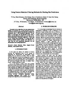
FIG 1 Opa protein expression by LPCIs. Shown is a representative immunoblot with Opa-specific (4B12/C11) and pilin-specific (10H5.1.1) antibodies showing Opa and pilus expression in control MS11 strains (Opa⫺, Opa50, Opa52, Opa57, and Opa⫺ pilus⫹) and low-passage-number strains 4034 and 4058. 4034A to -D and 4058A to -C variants were derived from original isolates (OI) based on distinct colony morphology. The immunoblot results for all the isolates are summarized in Table 1.
Statistical analysis. The bacterial strains were tested in groups with MS11 strains as controls. In order to present and compare all the data, binding data from the various plates were normalized to the MS11 controls. Data from two MS11 strains that fall within the dynamic range of the substrate reaction were chosen for the transformation of binding data from individual experiments. Briefly, the averages for the two MS11 control strains were first normalized to their respective cumulative means. A normalization factor for each experiment was calculated by taking the average of the two normalized MS11 averages. The data from each experiment are presented as the average among the triplicate wells, subtracting the averages from the no-GC control wells, and transformed according to the normalization factor. The standard deviation was calculated from the triplicate wells and adjusted with the same normalization factor. Primary neutrophil isolation. Human neutrophils were isolated from citrated whole blood taken from healthy volunteers by venipuncture using Ficoll-Paque Plus (Amersham Biosciences, Buckinghamshire, England). Contaminating erythrocytes were removed by dextran sedimentation and hypotonic shock, as described previously (41). Bacterial infections for immunofluorescence microscopy. PMNs (5 ⫻ 105) were centrifuged onto coverslips at 514 ⫻ g for 10 min. Cells were infected at an MOI of 10 (for binding and internalization studies) in a volume of 500 l, recentrifuged for 5 min at 57 ⫻ g to facilitate bacterial association with the cells, and then incubated at 37°C for the indicated durations. Postinfection, samples were washed with Hanks balanced salt solution (HBSS) and fixed using 3.7% paraformaldehyde. The cells were stained for CEACAM and bacteria and observed as described previously (18). Intracellular bacteria were differentiated from extracellular bacteria via exclusion of N. gonorrhoeae-specific antibody, as described previously (41). Oxidative-burst and degranulation assays. For the chemiluminescence-based oxidative-burst assay, 5 ⫻ 105 cells were incubated with 25 g/ml 5-amino-2,3-dihydro-1,4-phthalazinedione (luminol) (Sigma) in a volume of 100 l and then treated with agonists in a total volume of 200 l, with each sample done in triplicate. Infections proceeded for 60 min at 37°C, after which luminescence was read using a Tecan plate reader with i-control software. For the flow cytometry-based degranulation assay (CEACAM surface expression), 106 PMNs were treated with agonists in 500 l of HBSS for 30 min at 37°C. The infections were stopped by centrifugation at 2,400 ⫻ g for 3 min at RT. The cell pellets were fixed in 1% PFA and stained with 1.25 g of PE-conjugated rat anti-mouse CEACAM in a total volume of 200 l. Elastase and lactoferrin release assays were performed essentially as described previously (55). Briefly, 106 PMNs were exposed to agonists in a total volume of 500 l and then incubated for 1 h at 37°C. The cells were then pelleted, and the supernatants were collected. For the elastase assay, 50 l of supernatant was diluted 2-fold in PBS, incubated with 100 l DQ elastin substrate conjugated to Bodipy FL (from the EnzCheck Elastase kit; Molecular Probes), and then incubated for 2 h at RT before reading the fluorescence with excitation at 488 nm and emission at 515 nm. For the elastase assays, the percent release is shown,
Infection and Immunity
April 2015 Volume 83 Number 4
Downloaded from http://iai.asm.org/ on October 21, 2015 by guest
CEACAM3, CEACAM5, and CEACAM6 (20), followed by Bodipy FLconjugated goat anti-rabbit IgG(H⫹L) (Molecular Probes) for fluorescence-activated cell sorting using a FACSCalibur flow cytometer (BD Biosciences, Mississauga, Ontario, Canada) in order to remove the cells that did not express CEACAMs. CEACAM receptor expression. Flow cytometric analyses were carried out in order to confirm the surface localization of CEACAM receptors in Lec11 cells. Briefly, approximately 1 million trypsinized cells were incubated with 0.2 ml of phosphate-buffered saline (PBS) containing 1 mM MgCl2, 0.5 mM CaCl2, 0.22-m filtered (PBS-Mg-Ca), with 3% FBS (PBS-Mg-Ca-FBS) and 10 g/ml anti-CEA (Dako) for 1 h on ice. Unbound antibody was removed by washing the cell pellet with 3 changes of PBS-Mg-Ca-FBS. The cell pellets were then incubated with 0.2 ml of PBSMg-Ca-FBS containing 4 g/ml Bodipy FL-conjugated goat-anti-rabbit IgG(H⫹L), and unbound antibody was removed as described above. The stained cells were fixed with PBS containing 1% PFA, and fluorescence was measured with a FACSCalibur. In 96-well binding assays, Lec11 monolayers were mock infected with serum-free RPMI medium containing 200 g/ml heparin and 0.2% BSA (RPMI-B-H) alone and stained with anti-CEA (Dako), followed by AP-conjugated goat anti-rabbit IgG. Substrate development was carried out as described for bacterial binding. Ninety-six-well binding assay. The gonococcal isolates were divided into groups for experimentation in order to ease processing. For each binding experiment, the MS11 gonococcal strains, with a defined Opa phenotype, were included as experimental controls. Each gonococcal strain was tested in triplicate wells, and the experiment was repeated to ensure reproducibility. Ninety-six-well plates were coated with PBS-Mg-Ca containing 0.2% gelatin (0.22-m filtered) for 30 min at 37°C prior to being seeded with Lec11 cells at 80% confluence. The next morning, the serum present in the growth medium was removed by washing with PBS-Mg-Ca 3 times. The cells were then incubated with RPMI-B-H at 37°C and 5% CO2 until infection with bacteria, with the soluble heparin maintained throughout infection to prevent HSPG-dependent bacterial binding to the mammalian cells (20). The gonococci were washed with PBS-Mg-Ca to remove outer membrane blebs and then resuspended in RPMI-B-H for density measurements. The bacterial suspensions were applied to each cell line at a multiplicity of infection (MOI) of 50. To synchronize infection, bacteria were centrifuged onto the monolayers at 67 ⫻ g using a tabletop centrifuge at room temperature for 5 min. Infection was carried out at 37°C and 5% CO2 for 2 h, at which point nonadherent bacteria were removed by washing the wells 3 times with PBS-Mg-Ca. The monolayers were then fixed with PBS-Mg-Ca containing 1% PFA. To detect the presence of bound gonococci, PFA was first removed by 3 washes with PBS, and the monolayers were permeabilized with PBS containing 0.4% Triton X-100 for 10 min, followed by 3 washes with PBS. PBS containing 0.2% BSA was used to block the wells for 15 min prior to the addition of antibody UTR01 at 1:800 dilution for 1 h. Unbound antibodies were removed by 3 washes with PBS, followed by the addition of AP-conjugated goat anti-rabbit IgG at 1:10,000 for 1 h and a final 3 washes with PBS. pNPP was used to reveal bound bacteria. Absorbance at 405 nm was recorded 3 and 20 h postpNPP addition using a Titertek Multiskan Plus plate reader. All staining steps were carried out at room temperature (RT). Whole-cell dot blot. Gonococcal cells were washed once with PBSMg-Ca. Half a million cells in a 3-l volume were applied to a nitrocellulose membrane and air dried. The membrane was incubated in PBS containing 0.05% Tween 20 (PBS-T) and 5% skim milk powder overnight at 4°C. The next morning, the membrane was incubated with 0.5 g/ml of CEACAM1-Fc in blocker for 2 h at room temperature. The membrane was extensively washed prior to incubation with AP-conjugated goat antihuman IgG antibody at a 1:5,000 dilution. Excess antibody was removed by repeated washing in PBS-T. The alkaline phosphatase reaction was carried out by preequilibrating the membrane in 0.1 M Tris, pH 9.5, and subsequently incubating it in a solution containing 0.1 M Tris, pH 9.5, 7 mM MgCl2, 135 M BCIP, and 122 M NBT.
CEACAM-Binding Specificity of Neisseria gonorrhoeae
TABLE 1 Adhesin expression by a collection of low-passage-number clinical N. gonorrhoeae isolatesa No. of distinct bandsc
Original strainb
Male urethra
2171 2171A 2171B 2171C 2172 2173 2173A 2173B 2173C 2174 2174A 2174B 2174C 2175 2175A 2175B 2176 2176A 2176B 2177 2177A 2177B 2178 2178A 2178B 2179 2179A 2179B 2180 2180A 2180B 2180C
2057 2085 2086 2087 2058 2059 2088 2089 2090 2060 2091 2092 2093 2061 2094 2095 2062 2096 2097 2063 2098 2099 2064 2100 2101 2065 2102 2103 2066 2104 2105 2106
3 3 3 3 1 1 0 0 0 1 1 1 1 0 0 0 2 2 2 1 5 1 2 3 2 1 0 0 2 3 0 2
4001 4001A 4002 4002A 4002B 4004 4004A 4004B 4004C 4005 4005A 4005B 4005C 4007 4007A 4007B 4007C 4014 4014A 4014B 4014C 4015 4015A 4015B 4026 4026A
2067 2107 2068 2108 2109 2069 2110 2111 2112 2070 2113 2114 2115 2071 2116 2117 2118 2072 2119 2120 2121 2073 2112 2123 2074 2124
1 2 2 1 2 1 1 1 2 1 1 1 1 1 1 1 1 1 1 1 1 1 1 2 2 2
Cervix
April 2015 Volume 83 Number 4
Opa
Culture site
Pilus
1 1 1
1
⬎4 ⬎4 ⬎4 1
Original strainb
SGO strain no.d
4029 4029A 4029B 4033 4033A 4034 4034A 4034B 4034C 4034D 4039 4039A 4039B 4039C 4058 4058A 4058B 4058C 4077 4077A 4077B 4102 4102A 4102B 4102C 4121 4121A 4174 4174A 4174B 4174C 4180 4180A 4180B
2075 2125 2126 2076 2127 2077 2128 2129 2130 2131 2078 2132 2133 2134 2079 2135 2136 2137 2080 2138⫹/⫺ 21382081 2139 2140 2141 2082 2142 2083 2143 2144 2145 2084 2146 2147
No. of distinct bandsc Opa
Pilus
2 2 0 1 1 1 1 1 0 0 1 0 2 0 2 1 2 2 2 2 0 2 1 1 1 4 4 3 3 3 0 2 4 1
1 1
1
1
2 2
1 2 1 1 1 1
a The data represent a summary of immunoblot analyses for Opa protein and pilin expression in low-passage-number clinical N. gonorrhoeae isolates. b Colonies with distinct colony morphologies present within each original specimen were assigned alphabetic designations, e.g., colony phenotype variants derived from the original strain 4034 were labeled 4034A, 4034B, 4034C, and 4034D and analyzed alongside the original strain. c Number of distinct bands identified by Western blotting. d SGO strain no. indicates the strain designation assigned in the Scott Gray-Owen lab.
1
1 1
calculated as the amount of the protein in the supernatant divided by the amount released after phorbol myristate acetate (PMA) treatment. Lactoferrin release from PMN granules was assayed by enzyme-linked immunosorbent assay (ELISA), as described previously (56). For CEACAM release, 106 PMNs were infected for 1 h and fixed in 1% PFA. The cells were stained with CEACAM-PE, and fluorescence was measured with a FACSCalibur. Cytokine measurements. Cells (5 ⫻ 105) were infected with N. gonorrhoeae at an MOI of 10 and incubated at 37°C for 3 h. The infections were then stopped by centrifugation at 2,400 ⫻ g for 5 min at 4°C, and the supernatants were collected. Quantitative measurements of cytokines (interleukin 8 [IL-8] and tumor necrosis factor [TNF]) were performed using ELISA kits from BD Biosciences.
RESULTS 1
Most low-passage-number clinical isolates display Opa protein expression. Since the receptor specificity of Opa variants cannot
Infection and Immunity
iai.asm.org
1375
Downloaded from http://iai.asm.org/ on October 21, 2015 by guest
Culture site
SGO strain no.d
TABLE 1 (Continued)
Sintsova et al.
1376
iai.asm.org
FIG 2 Assay establishing 96-well CEACAM binding. (A) CEACAM expression by Lec11 cells. Lec11 cells transfected with the empty expression vector (V) or encoding individual CEACAM receptors were grown in gelatin-treated 96-well plates that also included control wells without cells but with gelatin treatment (Gelatin) or medium (Medium) alone. The plates were stained for CEACAM expression using anti-CEA (Dako) and AP-conjugated goat antirabbit secondary antibody and then detected using a colorimetric reaction. The means of results from triplicate wells and their standard deviations are presented. (B) Flow cytometric analysis of CEACAM expression on the Lec11 cell surface. Lec11 cell lines were trypsinized and stained with Dako CEA (black bars) or Dako isotype control (white bars) antibodies, followed by Bodipy FL-conjugated goat anti-rabbit antibody. The Lec11 cells were gated based on forward and side scatter profiles, and the mean FL1 (Bodipy FL) fluorescence is presented as histograms. (C) MS11 strains (MOI, 50) were used to infect Lec11 cells in a 96-well binding experiment. At 2 h postinfection, nonadherent bacteria were removed by washing, the monolayers were fixed and permeabilized, and bound gonococci were detected using UTR01 antibody, followed by the goat anti-rabbit AP-conjugated secondary antibody. Averages of the absorbances at 405 nm from triplicate wells after developing with colorimetric reagent, with the standard deviations, are presented.
Lec11 cells maintain their Opa binding specificity and to validate our multiplexed approach, we tested the binding of well-defined Opa variants expressed by recombinant N. gonorrhoeae MS11 strains to these cell lines in a 96-well format binding assay. Since gonococci can express adhesins that bind HSPG receptors, including certain individual Opa variants (18, 58, 59), soluble heparin was maintained within the culture medium to block HSPG (but not CEACAM) binding (18). The CEACAM-binding pattern observed for each strain tended to correlate with what was previously reported (20) (Fig. 2C). Wells that were mock infected (no GC) and gonococci that do not express Opa (Opa⫺) allowed us to establish background signals for N. gonorrhoeae and/or antibody binding to the Lec11 cell lines. Opa52-expressing gonococci bound CEACAM1-, CEACAM3-, CEACAM5-, and CEACAM6-expressing cells. While Opa57 is generally considered to also bind all four
Infection and Immunity
April 2015 Volume 83 Number 4
Downloaded from http://iai.asm.org/ on October 21, 2015 by guest
be predicted from their protein sequences, we set out to assess the CEACAM-binding phenotype of isolates minimally passaged from clinical specimens. We obtained stocks passaged twice (once from the specimen and once for freezing) from clinical samples taken from the male urethra or female endocervix. The 28 isolates selected represent the predominant gonococcal serovars circulating in Nairobi during the study period (48). Western blot analysis using 4B12/C11 antibody, which cross-reacts with all known N. gonorrhoeae Opa variants, revealed that 96% of the isolates (27 out of 28 strains) expressed at least one Opa protein (Fig. 1 and Table 1). This is consistent with previous reports indicating that gonococcal strains isolated from natural infections express Opa proteins (12–14). We detected pilus in only 8 of 28 strains (29%) (Fig. 1 and Table 1). Since piliation is generally considered to be present in fresh clinical isolates, it is possible that strain piliation may have been lost by pilE recombination and/or pilC phase variation during in vitro subculture (57). Microscopic examination of the LPCIs grown on GC agar revealed that each contained multiple distinct colony morphologies. Since gonococcal colony phenotypes often reveal changes in Opa and/or pilus expression, we considered each colony type separately. Sixty-four morphologically distinct gonococcal isolates were derived from the original 28 specimens, and each was then tested for Opa and pilus expression. Since phase variation is an ongoing process, subculturing was performed using a bottom-lit binocular dissecting microscope to confirm selection of colonies reflecting the originally observed phenotype and to exclude any visually apparent phase variants arising during passage. Western blot analyses showed that 64% (14 out of 22) of male-derived isolates expressed detectable Opa variants, while 83% (35 out of 42) of female-derived strains displayed one or more Opa variants (Table 1). Thus, while 96% of people in the cohort had Opaexpressing gonococci and the majority of colonies obtained from most specimens were Opa⫹ (Table 1), the infections often (6 of 18 female infections and 4 of 10 in males) also contained Opa⫺ N. gonorrhoeae. While most isolates displayed a single Opa and/or pilus variant band on the immunoblots, it is pertinent to note that some of the isolates expressed more than one discernible Opa protein and/or pilin variant (Table 1). Indeed, since variants with similar electrophoretic mobilities would not be discriminated, it is possible that this occurs more frequently than is apparent. Establishing a high-throughput CEACAM-binding assay. We hypothesized that the high prevalence of Opa expression in our LPCIs suggests that Opa proteins facilitate infection. However, since each Opa variant may have a different spectrum of CEACAM binding, since the spectrum of binding has the potential to drastically affect the host cellular response to infection (28, 30, 32, 38, 43, 44, 47), and since the pattern of CEACAM expression varies between sexes and cell types, we sought to characterize the binding specificity of each N. gonorrhoeae isolate. For this purpose, we transfected the Chinese hamster ovaryderived Lec11 cell line with individual CEACAM receptors (CEACAM1, CEACAM3, CEACAM5, CEACAM6, and CEACAM8). Since N. gonorrhoeae adhesins, such as pilus, have strict specificity for human receptors, this rodent cell line was chosen to eliminate any binding with endogenously expressed cellular receptors. Expression and maintenance of human CEACAM receptors on the cell surface was confirmed by in situ staining (Fig. 2A) and flow cytometry (Fig. 2B). In order to verify that the CEACAM receptors expressed on
CEACAM-Binding Specificity of Neisseria gonorrhoeae
Male isolates - + + ++ 2.0 1.5 1.0
Female isolates
+ + + +++ + + + - + + + +++ + + ++ + + +++ + + + + + + + - + + ++ + - + + ++ + + + ++ + + +++ + +
* ** ** * * * * ** * *
0.5
* ** *
* * * *
** * *** * * * *
*
CEACAM Binding (89%) - - - + + + ++ + + + - - + + + + + + ++ ++ + + + - + + ++ + + - + + +
CEACAM1 (80%)
* * * * *** ***** ****** *
*** * ****
**
*
* ** ** *** * * * ** * **
0
*
AP Signal (A405nm)
1.0 0.5
*
2.0 1.5
* *
*
0
* * **
* *
*
* * * ** **** * *
* * * * ** * * * * ** *
**
**
***
* * * ** *** *
***
1.0 0.5
! ! " #
* * *
*
** **
*** *
**** CEACAM5 (74%)
** * * * * **
*
*
**
0
* 2.0 1.5
*
** **
* *
*
CEACAM6 (51%)
1.0 0.5
* *** *
0 0.5 OpaOpa50 Opa52 Opa54 Opa57 Opa-Pil+
* * ***
***
*
*
***
* CEACAM8 (7%)
2171 A B C 2172 2173 A B C 2174 A B C 2175 A B 2176 A B 2177 A B 2178 A B 2179 A B 2180 A B C 4001 A 4002 A B 4004 A B C 4005 A B C 4007 A B C 4014 A B C 4015 A B 4026 A 4029 A B 4033 A 4034 A B C D 4039 A B C 4058 A B C 4077 A B 4102 A B C 4121 A 4174 A B C 4180 A B
*
0
*
** ** * * * * *** * * * * *****
FIG 3 Low-passage-number clinical gonococcal isolates show distinct CEACAM-binding profiles. Shown are binding data from 96-well binding experiments performed with Lec11 cells expressing the indicated human CEACAMs. The gray bars represent the cumulative means of MS11 controls, and the black bars are the original low-passage-number clinical specimens, with their respective phenotypically selected colony variants represented by the adjacent white bars. The threshold for binding was set at 1 standard deviation above the MS11 control strain and is represented by the horizontal dotted line. The asterisks indicate strains that were found to display significant levels of binding in two independent experiments. The percentages indicate the proportions of the total (original isolates plus individual colony variants) shown to bind a specific receptor. A plus symbol denotes binding to one or more CEACAMs, while a minus symbol denotes no observed binding to CEACAMs.
of these CEACAMs, its binding to CEACAM3 was modest in this assay system. As expected, Opa54-expressing gonococci bound only CEACAM1- and CEACAM5-expressing cells. Gonococci that express pili but not Opa (Opa⫺ pilus⫹) did not bind Lec11 cells but instead showed evidence of binding to wells without Lec11 cells (“gelatin” and “medium” wells). Opa50-expressing gonococci showed preference for Lec11 CEACAM1 cells and none of the other cell lines. Although Opa50 interacts specifically with HSPG receptors (18, 60), it does display a low level of CEACAM1 binding (20). Since Opa50 binds the carbohydrate component of HSPGs, the difference in binding could plausibly be attributed to distinct CEACAM1 glycosylation patterns between the Lec11 cell lines and those previously used; however, detailed analysis of
April 2015 Volume 83 Number 4
CEACAM glycosylation in these cells versus other cells has not been performed. LPCIs show distinct CEACAM-binding patterns. Next, we used the high-throughput binding assay to determine the CEACAM-binding phenotypes of the gonococcal strains recovered from low-passage-number clinical samples (Fig. 3). Eightytwo of 92 isolates (89%) bind to one or more CEACAMs. While most of them bind to both CEACAM1 and CEACAM5, 10 strains are specific for CEACAM1 (2173B, 2174A, 2177A, 2177B, 2179A, 4002B, 4015, 4015A, 4121, and 4174) and 2 are specific for CEACAM5 (2176 and 2174B). Despite the high level of sequence identity between CEACAM1 and CEACAM3 (88% identical within the N-terminal domain that binds Opa), far fewer strains
Infection and Immunity
iai.asm.org
1377
Downloaded from http://iai.asm.org/ on October 21, 2015 by guest
CEACAM3 (27%)
1.5
Sintsova et al.
profiles. (A) Whole-cell dot blot of the original (mixed colony phenotype) low-passage-number clinical specimens. Gonococci were immobilized on nitrocellulose filters and allowed to bind soluble CEACAM1-Fc, followed by AP-conjugated goat anti-human IgG and substrate development. Control strains expressing no Opa (Opa⫺) and Opa57 are shown at the top left. (B) Frequencies of binding to the indicated CEACAMs based on original lowpassage-number clinical strains isolated from males or females, as determined by CEACAM1-Fc whole-cell dot blotting and a 96-well binding assay. The binding frequencies for combined isolates are shown above the bars.
(27%) bound CEACAM3, and the level of binding to CEACAM3expressing cells tended to be considerably less than that to the other CEACAMs (Fig. 3), suggesting that there was selection against Opa variants that bind this neutrophil-restricted receptor. Among the original 28 LPCIs, CEACAM6 binding was more common in strains isolated from female (67%) than male (50%) infections (Fig. 4B). More strikingly, CEACAM5 binding was evident for all (100%) male isolates but for only 67% of femalederived specimens (Fig. 4B). This suggests a stronger selection for CEACAM5-specific Opa variants in men versus women, though the relatively small sample size (10 male versus 18 female samples) makes it inappropriate to draw a definitive conclusion from this relationship. Finally, it is pertinent to note that, while none of the recombinant Opa variants tested to date are able to bind to CEACAM8 (16, 18, 19), several isolates showed very low-level binding to
1378
iai.asm.org
Infection and Immunity
April 2015 Volume 83 Number 4
Downloaded from http://iai.asm.org/ on October 21, 2015 by guest
FIG 4 Male and female isolates show no difference in CEACAM-binding
CEACAM8-expressing cells (Fig. 3), whereas none bound to the untransfected Lec11 cell line (data not shown). Given that CEACAMs are heavily glycosylated, it is plausible that this interaction is distinct from the protein-protein-mediated binding between Opa and the other receptors (53). However, since each of these isolates also bound to other human CEACAMs, it also seems feasible that certain variants have very low affinity for this closely related receptor. To validate the results of the Lec11-based infection assay, we used an established whole-cell blotting assay using soluble CEACAM1-Fc fusion protein (23) to reassess the CEACAM1binding specificity of the commonly used MS11 strains versus the LPCIs. As expected, recombinant MS11 strains expressing Opa57 N. gonorrhoeae showed binding to CEACAM1-Fc, while Opa⫺ N. gonorrhoeae did not (Fig. 4A). The vast majority of the original (mixed-phenotype) LPCIs (91% of the isolates) also bound to CEACAM1-Fc, corroborating the results from the 96-well assay (Fig. 4B), although certain variants displayed clearer binding to either the soluble (2174 and 4001) or cell-expressed (4005) CEACAM1. Overall, these results suggest that there is a selective advantage for expressing Opa variants that bind CEACAM1 and CEACAM5 in vivo, presumably because they are expressed by mucosal epithelia. In contrast, the low level of binding to CEACAM3, which is a neutrophil-expressed phagocytic receptor, indicates that it may be detrimental to the bacteria. CEACAM binding affects human neutrophil responses to LPCIs. Characterization of individual CEACAM function in neutrophilic cell lines showed that Opa-mediated neutrophil activation is mediated by CEACAM3 signaling, while CEACAM1 and CEACAM6 allowed bacterial internalization without triggering production of reactive oxygen species (ROS) or release of toxic granule proteins (44). However, all of the studies focusing on neutrophil responses to Opa-CEACAM binding have been done exclusively with the commonly studied N. gonorrhoeae strains MS11 (44, 45, 47, 61, 62) and FA1090 (63, 64). The presence of naturally occurring gonococcal phase variants that either do or do not express Opa proteins, obtained from a single low-passage-number clinical specimen, allowed us to establish whether the CEACAM-binding specificity of primary LPCIs elicited cellular responses reminiscent of those seen with the recombinant strains. Isolate 4034B expresses Opa (here referred to as the Opa⫹ isolate) (Fig. 1) and shows binding to CEACAM1, CEACAM3, CEACAM5, and CEACAM6 (Fig. 3), while 4034C lacks Opa expression (here referred to as the Opa⫺ isolate) (Fig. 1) and does not bind CEACAM receptors (Fig. 3). When we assessed association of these two bacterial variants with human PMNs, the Opa⫹ isolate was bound and internalized to a greater extent than the Opa⫺ isolate (Fig. 5A and B). Moreover, the Opa⫹ isolate was killed faster than the Opa⫺ strain (Fig. 5C). Next, we assessed the activation of neutrophil antimicrobial responses after infection with the two phenotypic variants. The Opa⫹ variant activated production of higher levels of ROS than did the variant that lacks CEACAM-binding capacity (Fig. 6A and B). The Opa⫹ variant also drives a robust degranulation of both primary and secondary granules, evident from the release of primary (elastase) and secondary (lactoferrin) granule contents, while infection with the Opa⫺ variant did not lead to increased release of any granule proteins over uninfected controls (Fig. 6C). While CEACAMs are normally expressed on the surfaces of neu-
CEACAM-Binding Specificity of Neisseria gonorrhoeae
A
Actin
Ngo
Overlay
B # of bacteria/cell
8
Opa-
Opa
Total Intracellular
**
6
*
4 2
+
C
100
pa
60 40 20 0 0
20
40
60
80
Time, min
FIG 5 CEACAM-binding LPCI is readily phagocytosed by human neutrophils. (A) Human neutrophils were infected with primary specimen-derived phase variants 4034B (Opa⫹) and 4034C (Opa⫺). The cells were fixed 30 min postinfection and visualized by staining for actin with Texas Red-phalloidin, and bacteria (UTR101) are shown in green. Note that bacteria within the image depicting the Opa⫺ strain are rarely associated with PMNs, whereas the Opa⫹ bacteria accumulate within the cells. Shown is a representative of 3 independent experiments. (B) Intracellular and total PMN-associated bacteria were differentially stained and quantified via immunofluorescence microscopy. n ⫽ 3; the error bars represent standard errors of the mean (SEM). One-way analysis of variance (ANOVA) (with Tukey’s posttest) was performed for relevant samples. *, P ⬍ 0.05; **, P ⬍ 0.01. (C) Human PMNs kill Opa⫺ and Opa⫹ isolates with different kinetics. Adherent PMNs were infected with either Opa⫺ or Opa⫹ N. gonorrhoeae at an MOI of 1. Bacterial survival over time was evaluated as the number of CFU present in PMN lysates at each time point relative to the number of bacterial CFU present at time zero. n ⫽ 3; the error bars represent SEM.
trophils, they also comprise a major component of neutrophil secondary granules. Consequently, the Opa-dependent neutrophil degranulation also increased the expression of CEACAMs on the neutrophil surface (Fig. 6D), whereas infection with the Opa⫺ strain did not. We recently revealed that CEACAM3 engagement activates a proinflammatory transcriptional program in neutrophils, which results in production of proinflammatory cytokines via a signaling pathway that is independent of bacterial phagocytosis (47). Consistent with this, infection with the Opa⫹ phase variant led to significantly higher levels of both TNF and IL-8 (Fig. 6E). Considered together, these results indicate that the CEACAM-binding phenotype of primary LPCIs confers cell association and cellular response outcomes reflecting those seen with commonly used recombinant strains and consistent with the expression of Opa variants that bind to CEACAM3 on neutrophils being detrimental to the gonococci. DISCUSSION
N. gonorrhoeae is highly adapted to life in humans. Its narrow host range is largely defined by its adhesin proteins and immune evasion mechanisms, which are specific for human cellular receptors and serum proteins (59). Pilus facilitates initial bacterial attachment to the urogenital mucosa (4), where other adhesins can then confer a tighter secondary binding to epithelium-expressed receptors (3, 18, 58, 59). Of these, the Opa proteins mediate tight adherence and transcytosis to the subepithelial space (25, 26). The phase variability of the genes encoding Opa proteins and pilus
April 2015 Volume 83 Number 4
allows N. gonorrhoeae to change the patterns of expression of these adhesins randomly. Consequently, during a natural infection there may exist a mixed population of bacteria varying in piliated phenotypes and/or the numbers and types of Opa variants expressed. This ongoing diversification of phenotypes is balanced by ongoing phenotypic selection of variants expressing adhesins or other factors that facilitate infection within an individual and/or within a particular tissue-specific niche. In vitro cell culture and primary cell-based systems have clearly established that most Opa variants can bind one or more human CEACAMs, and transgenic mouse-based studies corroborate the importance of this binding for the establishment of infection (37, 38). Experimental human urethral model studies also suggest the importance of Opa proteins and pilus (6, 12–14, 65–67), yet the binding specificity of Opa variants expressed during human infection had not been addressed before this study. The frequent phase-variable switching on and off of pilus and Opa expression necessitated that isolates be minimally passaged before phenotypic studies. To achieve this, we obtained primary LPCIs collected as part of a longitudinal study of commercial sex workers and STD clinic patients in Nairobi, Kenya. The analysis of pilus and Opa expression showed that the majority of the isolates expressed one or more Opa variants. All 11 chromosomally located opa alleles are constitutively transcribed regardless of whether their respective translated proteins are phase varied on or off (68). Moreover, due to the hypervariable nature of the surfaceexposed loops of each Opa variant, their binding specificities can-
Infection and Immunity
iai.asm.org
1379
Downloaded from http://iai.asm.org/ on October 21, 2015 by guest
Opa+ Opa-
80
% Survival
O
O pa +
0
Sintsova et al.
10000
6000 5000 4000
RLU
6000
*
7000
Uninfected Opa+ Opa-
8000
RLU
**
B
A
3000
4000
2000 2000
1000 pa
U
ni
nf
Time, min
O
100
pa
80
O
60
d
40
te
20
ec
0
+
0
0
C
CEACAM (MFI)
60 40
600 400 200
20 pa O
pa O
te ec nf
e
ni U
yp ot Is
d
G Ig
rin er of ct La
El as ta se
+
0
0
E 1000
80
*** TNF , pg/ml
600 400
**
60 40 20
200 0
+ O
pa
pa
pa O
O
pa
-
+
0 O
IL-8, pg/ml
800
FIG 6 Neutrophil bactericidal and inflammatory responses to phenotypic variants of N. gonorrhoeae. Human PMNs were infected with 4034B (Opa⫹) and
4034C (Opa⫺) at an MOI of 10. The neutrophil oxidative burst, degranulation, and cytokine release were measured as described in Materials and Methods. (A and B) Oxidative-burst response to N. gonorrhoeae infection, illustrating the kinetics of response from one representative donor (A) and the means with SEM calculated based upon independent experiments with 3 different donors (B). One-way ANOVA (with Tukey’s posttest) was performed for relevant samples. *, P ⬍ 0.05; **, P ⬍ 0.01. RLU, relative light units. (C) Elastase and lactoferrin release in response to 4034B and 4034C is shown as percent release, calculated as the amount of the protein in the supernatant divided by the amount released after PMA treatment. n ⫽ 3; the error bars represent SEM. (D) Flow cytometric analysis of CEACAM expression on the human PMN cell surface. After infection with the Opa⫹ or Opa⫺ variant, the PMNs were fixed and stained with CEACAM-PE. The mean fluorescence intensity (MFI) from one representative donor is shown. (E) Human neutrophils infected with Opa⫹ N. gonorrhoeae show increased levels of IL-8 and TNF relative to PMNs infected with Opa⫺ bacteria. An unpaired Student t test was performed for relevant samples. **, P ⬍ 0.01; ***, P ⬍ 0.001.
not be inferred from the protein sequences. Since a bioinformatics-based approach was not feasible, and since the presence or absence of binding function is more important than the specific Opa alleles that confer binding, we sought to characterize the CEACAM-binding phenotype of each isolate. To this end, we developed a high-throughput binding assay that allows quantitative assessment of CEACAM-binding specificity. This revealed that most isolates bound both CEACAM1 and CEACAM5, perhaps reflecting the need for transmission between niches expressing these two receptors. However, when considered more closely, it is noteworthy that all the isolates from men bound CEACAM5 whereas only two-thirds of the isolates from women did so. Interestingly, in this regard, squamous cells of the lower genital tract tend to express CEACAM5 whereas columnar epithelial cells of
1380
iai.asm.org
the female endocervical and upper genital tract instead express CEACAM1 (27). While CEACAM family expression within the male urethra has not been mapped, the difference in receptor specificities of the recovered isolates makes it enticing to consider whether there is selection for CEACAM5 binding at this site. In contrast to the selection for CEACAM1 and CEACAM5 binding, we observed an apparent selection against binding to the neutrophil CEACAM3 receptor, with only 27% of all isolates showing measurable binding to CEACAM3. Moreover, for those isolates that did bind to CEACAM3, the detected signal strength was generally weaker than to the other CEACAMs. This difference in binding cannot be explained by lower levels of CEACAM3 expression by the transfected cell line, as all CEACAMs were expressed on the surface at similar levels. Instead, the selection
Infection and Immunity
April 2015 Volume 83 Number 4
Downloaded from http://iai.asm.org/ on October 21, 2015 by guest
80
%release
D 800
Unstimulated Opa+ Opa-
100
CEACAM-Binding Specificity of Neisseria gonorrhoeae
10. 11.
12. 13. 14.
15.
16.
17.
18.
ACKNOWLEDGMENTS Funding for this project was provided by CIHR grant MOP-15499. We thank Frank Plummer (University of Manitoba) for provision of clinical N. gonorrhoeae isolates, and we are grateful to Shannon McCaw and Helen Sarantis for helpful discussions during the course of this study. We report no conflict of interest.
19.
REFERENCES
21.
1. World Health Organization. 2012. Global action plan to control the spread and impact of antimicrobial resistance in Neisseria gonorrhoeae. http://www.who.int/entity/reproductivehealth/publications/rtis/978924 1503501/en/. 2. World Health Organization. 2012. Global incidence and prevalence of selected curable sexually transmitted infections—2008. http://www.who .int/iris/bitstream/10665/75181/1/9789241503839_eng.pdf. 3. Edwards J, Apicella M. 2004. The molecular mechanisms used by Neisseria gonorrhoeae to initiate infection differ between men and women. Clin Microbiol Rev 17:965. http://dx.doi.org/10.1128/CMR.17.4.965-981 .2004. 4. Biais N, Ladoux B, Higashi D, So M, Sheetz M. 2008. Cooperative retraction of bundled type IV pili enables nanonewton force generation. PLoS Biol 6:e87. http://dx.doi.org/10.1371/journal.pbio.0060087. 5. Faulstich M, Bottcher JP, Meyer TF, Fraunholz M, Rudel T. 2013. Pilus phase variation switches gonococcal adherence to invasion by caveolin-1dependent host cell signaling. PLoS Pathog 9:e1003373. http://dx.doi.org /10.1371/journal.ppat.1003373. 6. Hobbs MM, Sparling PF, Cohen MS, Shafer WM, Deal CD, Jerse AE. 2011. Experimental gonococcal infection in male volunteers: cumulative experience with Neisseria gonorrhoeae strains FA1090 and MS11mkC. Front Microbiol 2:123. http://dx.doi.org/10.3389/fmicb.2011.00123. 7. Swanson J. 1978. Studies on gonococcus infection. XIV. Cell wall protein differences among color/opacity colony variants of Neisseria gonorrhoeae. Infect Immun 21:292–302. 8. King GJ, Swanson J. 1978. Studies on gonococcus infection. XV. Identification of surface proteins of Neisseria gonorrhoeae correlated with leukocyte association. Infect Immun 21:575–583. 9. Kupsch EM, Knepper B, Kuroki T, Heuer I, Meyer TF. 1993. Variable opacity (Opa) outer membrane proteins account for the cell tropisms
April 2015 Volume 83 Number 4
20.
22.
23.
24. 25.
26.
27.
displayed by Neisseria gonorrhoeae for human leukocytes and epithelial cells. EMBO J 12:641– 650. Stern A, Brown M, Nickel P, Meyer TF. 1986. Opacity genes in Neisseria gonorrhoeae: control of phase and antigenic variation. Cell 47:61–71. http: //dx.doi.org/10.1016/0092-8674(86)90366-1. Murphy GL, Connell TD, Barritt DS, Koomey M, Cannon JG. 1989. Phase variation of gonococcal protein II: regulation of gene expression by slipped-strand mispairing of a repetitive DNA sequence. Cell 56:539 –547. http://dx.doi.org/10.1016/0092-8674(89)90577-1. James J, Swanson J. 1978. Studies on gonococcus infection. XIII. Occurrence of color/opacity colonial variants in clinical cultures. Infect Immun 19:332–340. Isbey S, Alcorn T, Davis R, Haizlip J, Leone P, Cohen M. 1997. Characterisation of Neisseria gonorrhoeae in semen during urethral infection in men. Genitourin Med 73:378 –382. Jerse A, Cohen M, Drown P, Whicker L, Isbey S, Seifert H, Cannon J. 1994. Multiple gonococcal opacity proteins are expressed during experimental urethral infection in the male. J Exp Med 179:911–920. http://dx .doi.org/10.1084/jem.179.3.911. Virji M, Watt SM, Barker S, Makepeace K, Doyonnas R. 1996. The N-domain of the human CD66a adhesion molecule is a target for Opa proteins of Neisseria meningitidis and Neisseria gonorrhoeae. Mol Microbiol 22:929 –939. http://dx.doi.org/10.1046/j.1365-2958.1996.01548.x. Virji M, Makepeace K, Ferguson DJ, Watt SM. 1996. Carcinoembryonic antigens (CD66) on epithelial cells and neutrophils are receptors for Opa proteins of pathogenic Neisseriae. Mol Microbiol 22:941–950. http://dx .doi.org/10.1046/j.1365-2958.1996.01551.x. Chen T, Grunert F, Medina-Marino A, Gotschlich EC. 1997. Several carcinoembryonic antigens (CD66) serve as receptors for gonococcal opacity proteins. J Exp Med 185:1557–1564. http://dx.doi.org/10.1084 /jem.185.9.1557. Gray-Owen SD, Dehio C, Haude A, Grunert F, Meyer TF. 1997. CD66 carcinoembryonic antigens mediate interactions between Opa-expressing Neisseria gonorrhoeae and human polymorphonuclear phagocytes. EMBO J 16:3435–3445. http://dx.doi.org/10.1093/emboj/16.12.3435. Bos MP, Grunert F, Belland RJ. 1997. Differential recognition of members of the carcinoembryonic antigen family by Opa variants of Neisseria gonorrhoeae. Infect Immun 65:2353–2361. Gray-Owen SD, Lorenzen DR, Haude A, Meyer TF, Dehio C. 1997. Differential Opa specificities for CD66 receptors influence tissue interactions and cellular response to Neisseria gonorrhoeae. Mol Microbiol 26: 971–980. http://dx.doi.org/10.1046/j.1365-2958.1997.6342006.x. Hammarstrom S. 1999. The carcinoembryonic antigen (CEA) family: structures, suggested functions and expression in normal and malignant tissues. Semin Cancer Biol 9:67– 81. http://dx.doi.org/10.1006/scbi.1998 .0119. Beauchemin N, Draber P, Dveksler G, Gold P, Gray-Owen S, Grunert F, Hammarstrom S, Holmes KV, Karlsson A, Kuroki M, Lin SH, Lucka L, Najjar SM, Neumaier M, Obrink B, Shively JE, Skubitz KM, Stanners CP, Thomas P, Thompson JA, Virji M, von Kleist S, Wagener C, Watt S, Zimmermann W. 1999. Redefined nomenclature for members of the carcinoembryonic antigen family. Exp Cell Res 252:243–249. http://dx.doi .org/10.1006/excr.1999.4610. Virji M, Evans D, Hadfield A, Grunert F, Teixeira AM, Watt SM. 1999. Critical determinants of host receptor targeting by Neisseria meningitidis and Neisseria gonorrhoeae: identification of Opa adhesiotopes on the Ndomain of CD66 molecules. Mol Microbiol 34:538 –551. http://dx.doi.org /10.1046/j.1365-2958.1999.01620.x. Chen T, Gotschlich EC. 1996. CGM1a antigen of neutrophils, a receptor of gonococcal opacity proteins. Proc Natl Acad Sci U S A 93:14851–14856. http://dx.doi.org/10.1073/pnas.93.25.14851. McGee ZA, Stephens DS, Hoffman LH, Schlech WF III, Horn RG. 1983. Mechanisms of mucosal invasion by pathogenic Neisseria. Rev Infect Dis 5(Suppl 4):S708 –S714. http://dx.doi.org/10.1093/clinids/5.Supplement _4.S708. Wang J, Gray-Owen SD, Knorre A, Meyer TF, Dehio C. 1998. Opa binding to cellular CD66 receptors mediates the transcellular traversal of Neisseria gonorrhoeae across polarized T84 epithelial cell monolayers. Mol Microbiol 30:657– 671. http://dx.doi.org/10.1046/j.1365-2958.1998 .01102.x. Prall F, Nollau P, Neumaier M, Haubeck HD, Drzeniek Z, Helmchen U, Loning T, Wagener C. 1996. CD66a (BGP), an adhesion molecule of the carcinoembryonic antigen family, is expressed in epithelium, endothe-
Infection and Immunity
iai.asm.org
1381
Downloaded from http://iai.asm.org/ on October 21, 2015 by guest
against CEACAM3 binding is consistent with binding to the receptor having detrimental consequences for the bacteria. Consistent with this, we observed that, in contrast to the Opa⫺ variant, Opa⫹ phase variants that bound human neutrophils were more effectively engulfed and elicited a greater oxidative-burst, degranulation, and inflammatory-cytokine expression response. Curiously, several isolates were able to bind to one or more CEACAMs while seemingly lacking Opa expression. Whether this stems from their expression of Opa variants that are not recognized by the available Opa-specific monoclonal antibodies and/or from CEACAM binding occurring in an Opa-independent manner, as has recently been reported for N. meningitidis (69), will require further study of the isolates. Nevertheless, CEACAM binding defines the phenotype of these strains, regardless of any such antigenic differences. This study highlights the exquisite interplay of host-pathogen interactions that allows N. gonorrhoeae to persist in a human population. The ongoing phase variability of 11 Opa proteins allows their randomized expression, yet human infection selects for bacterial variants that specifically bind CEACAM1 and CEACAM5, but not CEACAM3. The selective pressure required to drive a preference between the human CEACAMs despite the high sequence and structural similarity of their respective bacterium binding N domains (42, 70) is a testament to the ongoing evolutionary dance between humans and our intimately associated neisserial partners.
Sintsova et al.
28. 29.
30.
31.
33.
34.
35.
36. 37.
38.
39.
40.
41.
42.
43.
1382
iai.asm.org
44.
45.
46.
47.
48. 49.
50. 51.
52.
53.
54.
55. 56.
57. 58. 59. 60. 61.
dependent pathway. Cell Microbiol 9:2167–2180. http://dx.doi.org/10 .1111/j.1462-5822.2007.00947.x. Sarantis H, Gray-Owen SD. 2012. Defining the roles of human carcinoembryonic antigen-related cellular adhesion molecules during neutrophil responses to Neisseria gonorrhoeae. Infect Immun 80:345–358. http://dx .doi.org/10.1128/IAI.05702-11. Schmitter T, Agerer F, Peterson L, Munzner P, Hauck CR. 2004. Granulocyte CEACAM3 is a phagocytic receptor of the innate immune system that mediates recognition and elimination of human-specific pathogens. J Exp Med 199:35– 46. http://dx.doi.org/10.1084/jem.20030204. Smirnov A, Daily KP, Criss AK. 2014. Assembly of NADPH oxidase in human neutrophils is modulated by the opacity-associated protein expression state of Neisseria gonorrhoeae. Infect Immun 82:1036 –1044. http: //dx.doi.org/10.1128/IAI.00881-13. Sintsova A, Sarantis H, Islam EA, Sun CX, Amin M, Chan CH, Stanners CP, Glogauer M, Gray-Owen SD. 2014. Global analysis of neutrophil responses to Neisseria gonorrhoeae reveals a self-propagating inflammatory program. PLoS Pathog 10:e1004341. http://dx.doi.org/10.1371 /journal.ppat.1004341. Fudyk T, Maclean I, Simonsen J, Njagi E, Kimani J, Brunham R, Plummer F. 1999. Genetic diversity and mosaicism at the por locus of Neisseria gonorrhoeae. J Bacteriol 181:5591–5599. Hobbs MM, Malorny B, Prasad P, Morelli G, Kusecek B, Heckels JE, Cannon JG, Achtman M. 1998. Recombinational reassortment among opa genes from ET-37 complex Neisseria meningitidis isolates of diverse geographical origins. Microbiology 144:157–166. http://dx.doi.org/10 .1099/00221287-144-1-157. Merz AJ, So M. 1997. Attachment of piliated, Opa- and Opc- gonococci and meningococci to epithelial cells elicits cortical actin rearrangements and clustering of tyrosine-phosphorylated proteins. Infect Immun 65:4341–4349. Zollner O, Vestweber D. 1996. The E-selectin ligand-1 is selectively activated in Chinese hamster ovary cells by the alpha(1,3)-fucosyltransferases IV and VII. J Biol Chem 271:33002–33008. http://dx.doi.org/10.1074/jbc .271.51.33002. Nagel G, Grunert F, Kuijpers TW, Watt SM, Thompson J, Zimmermann W. 1993. Genomic organization, splice variants and expression of CGM1, a CD66-related member of the carcinoembryonic antigen gene family. Eur J Biochem 214:27–35. http://dx.doi.org/10.1111/j.1432-1033 .1993.tb17892.x. Billker O, Popp A, Brinkmann V, Wenig G, Schneider J, Caron E, Meyer TF. 2002. Distinct mechanisms of internalization of Neisseria gonorrhoeae by members of the CEACAM receptor family involving Rac1and Cdc42-dependent and -independent pathways. EMBO J 21:560 –571. http://dx.doi.org/10.1093/emboj/21.4.560. Berling B, Kolbinger F, Grunert F, Thompson JA, Brombacher F, Buchegger F, von Kleist S, Zimmermann W. 1990. Cloning of a carcinoembryonic antigen gene family member expressed in leukocytes of chronic myeloid leukemia patients and bone marrow. Cancer Res 50: 6534 – 6539. Abdel-Latif D, Steward M, Macdonald DL, Francis GA, Dinauer MC, Lacy P. 2004. Rac2 is critical for neutrophil primary granule exocytosis. Blood 104:832– 839. http://dx.doi.org/10.1182/blood-2003-07-2624. Mocsai A, Jakus Z, Vantus T, Berton G, Lowell CA, Ligeti E. 2000. Kinase pathways in chemoattractant-induced degranulation of neutrophils: the role of p38 mitogen-activated protein kinase activated by Src family kinases. J Immunol 164:4321– 4331. http://dx.doi.org/10.4049 /jimmunol.164.8.4321. Criss AK, Kline KA, Seifert HS. 2005. The frequency and rate of pilin antigenic variation in Neisseria gonorrhoeae. Mol Microbiol 58:510 –519. http://dx.doi.org/10.1111/j.1365-2958.2005.04838.x. van Putten JP, Paul SM. 1995. Binding of syndecan-like cell surface proteoglycan receptors is required for Neisseria gonorrhoeae entry into human mucosal cells. EMBO J 14:2144 –2154. Virji M. 2009. Pathogenic neisseriae: surface modulation, pathogenesis and infection control. Nat Rev Microbiol 7:274 –286. http://dx.doi.org/10 .1038/nrmicro2097. Chen T, Belland RJ, Wilson J, Swanson J. 1995. Adherence of pilusOpa⫹ gonococci to epithelial cells in vitro involves heparan sulfate. J Exp Med 182:511–517. http://dx.doi.org/10.1084/jem.182.2.511. Hauck CR, Meyer TF, Lang F, Gulbins E. 1998. CD66-mediated phagocytosis of Opa52 Neisseria gonorrhoeae requires a Src-like tyrosine kinaseand Rac1-dependent signalling pathway. EMBO J 17:443– 454. http://dx .doi.org/10.1093/emboj/17.2.443.
Infection and Immunity
April 2015 Volume 83 Number 4
Downloaded from http://iai.asm.org/ on October 21, 2015 by guest
32.
lium, and myeloid cells in a wide range of normal human tissues. J Histochem Cytochem 44:35– 41. http://dx.doi.org/10.1177/44.1.8543780. Boulton IC, Gray-Owen SD. 2002. Neisserial binding to CEACAM1 arrests the activation and proliferation of CD4⫹ T lymphocytes. Nat Immunol 3:229 –236. http://dx.doi.org/10.1038/ni769. Lee HS, Boulton IC, Reddin K, Wong H, Halliwell D, Mandelboim O, Gorringe AR, Gray-Owen SD. 2007. Neisserial outer membrane vesicles bind the coinhibitory receptor carcinoembryonic antigen-related cellular adhesion molecule 1 and suppress CD4⫹ T lymphocyte function. Infect Immun 75:4449 – 4455. http://dx.doi.org/10.1128/IAI.00222-07. Lee HS, Ostrowski MA, Gray-Owen SD. 2008. CEACAM1 dynamics during Neisseria gonorrhoeae suppression of CD4⫹ T lymphocyte activation. J Immunol 180:6827– 6835. http://dx.doi.org/10.4049/jimmunol .180.10.6827. Pantelic M, Kim YJ, Bolland S, Chen I, Shively J, Chen T. 2005. Neisseria gonorrhoeae kills carcinoembryonic antigen-related cellular adhesion molecule 1 (CD66a)-expressing human B cells and inhibits antibody production. Infect Immun 73:4171– 4179. http://dx.doi.org/10.1128/IAI.73.7 .4171-4179.2005. Slevogt H, Zabel S, Opitz B, Hocke A, Eitel J, N=Guessan PD, Lucka L, Riesbeck K, Zimmermann W, Zweigner J, Temmesfeld-Wollbrueck B, Suttorp N, Singer BB. 2008. CEACAM1 inhibits Toll-like receptor 2-triggered antibacterial responses of human pulmonary epithelial cells. Nat Immunol 9:1270 –1278. http://dx.doi.org/10.1038/ni.1661. Yu Q, Chow EM, McCaw SE, Hu N, Byrd D, Amet T, Hu S, Ostrowski MA, Gray-Owen SD. 2013. Association of Neisseria gonorrhoeae Opa(CEA) with dendritic cells suppresses their ability to elicit an HIV-1specific T cell memory response. PLoS One 8:e56705. http://dx.doi.org/10 .1371/journal.pone.0056705. Youssef AR, van der Flier M, Estevao S, Hartwig NG, van der Ley P, Virji M. 2009. Opa⫹ and Opa⫺ isolates of Neisseria meningitidis and Neisseria gonorrhoeae induce sustained proliferative responses in human CD4⫹ T cells. Infect Immun 77:5170 –5180. http://dx.doi.org/10.1128/IAI .00355-09. Zariri A, van Dijken H, Hamstra HJ, van der Flier M, Vidarsson G, van Putten JP, Boog CJ, van den Dobbelsteen G, van der Ley P. 2013. Expression of human CEACAM1 in transgenic mice limits the Opaspecific immune response against meningococcal outer membrane vesicles. Vaccine 31:5585–5593. http://dx.doi.org/10.1016/j.vaccine.2013.07 .069. Chan CH, Stanners CP. 2004. Novel mouse model for carcinoembryonic antigen-based therapy. Mol Ther 9:775–785. http://dx.doi.org/10.1016/j .ymthe.2004.03.009. Johswich KO, McCaw SE, Islam E, Sintsova A, Gu A, Shively JE, Gray-Owen SD. 2013. In vivo adaptation and persistence of Neisseria meningitidis within the nasopharyngeal mucosa. PLoS Pathog 9:e1003509. http://dx.doi.org/10.1371/journal.ppat.1003509. Muenzner P, Bachmann V, Zimmermann W, Hentschel J, Hauck CR. 2010. Human-restricted bacterial pathogens block shedding of epithelial cells by stimulating integrin activation. Science 329:1197–1201. http://dx .doi.org/10.1126/science.1190892. Booth JW, Telio D, Liao EH, McCaw SE, Matsuo T, Grinstein S, Gray-Owen SD. 2003. Phosphatidylinositol 3-kinases in carcinoembryonic antigen-related cellular adhesion molecule-mediated internalization of Neisseria gonorrhoeae. J Biol Chem 278:14037–14045. http://dx.doi.org /10.1074/jbc.M211879200. Buntru A, Kopp K, Voges M, Frank R, Bachmann V, Hauck CR. 2011. Phosphatidylinositol 3=-kinase activity is critical for initiating the oxidative burst and bacterial destruction during CEACAM3-mediated phagocytosis. J Biol Chem 286:9555–9566. http://dx.doi.org/10.1074/jbc.M110 .216085. McCaw SE, Schneider J, Liao EH, Zimmermann W, Gray-Owen SD. 2003. Immunoreceptor tyrosine-based activation motif phosphorylation during engulfment of Neisseria gonorrhoeae by the neutrophil-restricted CEACAM3 (CD66d) receptor. Mol Microbiol 49:623– 637. http://dx.doi .org/10.1046/j.1365-2958.2003.03591.x. Pils S, Gerrard DT, Meyer A, Hauck CR. 2008. CEACAM3: an innate immune receptor directed against human-restricted bacterial pathogens. Int J Med Microbiol 298:553–560. http://dx.doi.org/10.1016/j.ijmm.2008 .04.005. Sarantis H, Gray-Owen SD. 2007. The specific innate immune receptor CEACAM3 triggers neutrophil bactericidal activities via a Syk kinase-
CEACAM-Binding Specificity of Neisseria gonorrhoeae
62. Chen T, Bolland S, Chen I, Parker J, Pantelic M, Grunert F, Zimmermann W. 2001. The CGM1a (CEACAM3/CD66d)-mediated phagocytic pathway of Neisseria gonorrhoeae expressing opacity proteins is also the pathway to cell death. J Biol Chem 276:17413–17419. http://dx.doi.org/10 .1074/jbc.M010609200. 63. Ball LM, Criss AK. 2013. Constitutively Opa-expressing and Opadeficient Neisseria gonorrhoeae strains differentially stimulate and survive exposure to human neutrophils. J Bacteriol 195:2982–2990. http://dx.doi .org/10.1128/JB.00171-13. 64. Johnson MB, Ball LM, Daily KP, Martin JN, Columbus L, Criss AK. 27 October 2014. Opa⫹ Neisseria gonorrhoeae exhibits reduced survival in human neutrophils via Src family kinase-mediated bacterial trafficking into mature phagolysosomes. Cell Microbiol http://dx.doi.org/10.1111/cmi.12389. 65. Kellogg DS, Jr, Cohen IR, Norins LC, Schroeter AL, Reising G. 1968. Neisseria gonorrhoeae. II. Colonial variation and pathogenicity during 35 months in vitro. J Bacteriol 96:596 – 605.
66. Kellogg DS, Jr, Peacock WL, Jr, Deacon WE, Brown L, Pirkle DI. 1963. Neisseria gonorrhoeae. I. Virulence genetically linked to clonal variation. J Bacteriol 85:1274 –1279. 67. Seifert H, Wright C, Jerse A, Cohen M, Cannon J. 1994. Multiple gonococcal pilin antigenic variants are produced during experimental human infections. J Clin Invest 93:2744 –2749. http://dx.doi.org/10.1172 /JCI117290. 68. Meyer TF, van Putten JPM. 1989. Genetic mechanisms and biological implications of phase variation in pathogenic Neisseriae. Clin Microbiol Rev 2:S139 –S145. 69. Kuespert K, Roth A, Hauck CR. 2011. Neisseria meningitidis has two independent modes of recognizing its human receptor CEACAM1. PLoS One 6:e14609. http://dx.doi.org/10.1371/journal.pone.0014609. 70. Kuroki M, Arakawa F, Matsuo Y, Oikawa S, Misumi Y, Nakazato H, Matsuoka Y. 1991. Molecular cloning of nonspecific cross-reacting antigens in human granulocytes. J Biol Chem 266:11810 –11817.
Downloaded from http://iai.asm.org/ on October 21, 2015 by guest
April 2015 Volume 83 Number 4
Infection and Immunity
iai.asm.org
1383