© 2005 Federation of European Psychophysiology Societies A.A. Wijers & M.A.S. Journal Boksem: of Psychophysiology ERP s in an Illusory Hogrefe&HuberPublishers 2005; Vol. Co njunction 19(3):xxx–xxx Task
Selective Attention and Error Processing in an Illusory Conjunction Task An Event-Related Brain Potential Study Albertus A. Wijers and Maarten A.S. Boksem Experimental and Work Psychology, University of Groningen, The Netherlands
Abstract. We recorded event-related potentials in an illusory conjunction task, in which subjects were cued on each trial to search for a particular colored letter in a subsequently presented test array, consisting of three different letters in three different colors. In a proportion of trials the target letter was present and in other trials none of the relevant features were present. In still other trials one of the features (color or letter identity) were present or both features were present but not combined in the same display element. When relevant features were present this resulted in an early posterior selection negativity (SN) and a frontal selection positivity (FSP). When a target was presented, this resulted in a FSP that was enhanced after 250 ms as compared to when both relevant features were present but not combined in the same display element. This suggests that this effect reflects an extra process of attending to both features bound to the same object. There were no differences between the ERPs in feature error and conjunction error trials, contrary to the idea that these two types of errors are due to different (perceptual and attentional) mechanisms. The P300 in conjunction error trials was much reduced relative to the P300 in correct target detection trials. A similar, error-related negativity-like component was visible in the response-locked averages in correct target detection trials, in feature error trials, and in conjunction error trials. Dipole modeling of this component resulted in a source in a deep medial-frontal location. These results suggested that this type of task induces a high level of response conflict, in which decision-related processes may play a major role. Keywords: illusory conjunction task, feature integration theory, selective attention, error processing, ERPs
It is widely recognized that event-related brain potentials (ERPs) provide valuable information about temporal and spatial aspects of the brain activity associated with the processes involved in selective attention (e.g., Luck & Girelli, 1998). Most ERP studies on visual selective attention have employed sustained attention conditions. In such tasks subjects are presented with a rapid series of stimuli, varying on two dimensions of a basic visual feature (e.g., spatial attention: stimuli presented either to the left or right of fixation, nonspatial attention: stimuli presented either in red or in blue). The subjects are instructed to attend to a particular feature dimension (e.g., attend to red stimuli and ignore blue stimuli) in order to detect occasional targets within the attended channel. In sustained attention studies, subjects attend to the same feature dimension for an entire block of trials. Such studies have consistently found that spatial attention results in the enhancement of the early P1 and N1 components (onset latency at about 80 ms) of the ERPs elicited by Hogrefe & Huber Publishers DOI 10.1027/0269-8803.19.3.xxx
attended, relative to unattended, stimuli (e.g., Hillyard & Münte, 1984; Lange et al., 1999; Wijers, Lamain, Slopsema, Mulder, & Mulder, 1989). For nonspatial attention, numerous studies have found that the ERPs elicited by attended stimuli show an occipital selection negativity (SN) and a frontal selection positivity (FSP) relative to the ERPs elicited by unattended stimuli (Hillyard & Münte, 1984; Kenemans et al., 1993, 1995, 2002; Wijers, Mulder, Okita, & Mulder, 1989; Wijers, Mulder, Okita, Mulder, & Scheffers, 1989; Wijers, Van Besouw, & Mulder, 2002). The onset latency of these nonspatial effects is usually in the order of 150–200 ms. Recently, experiments have been conducted using a transient attention set-up, in which the to-be-attended feature dimension is cued anew on each trial (e.g., by an arrow pointing to the right or left, or by other symbolic cues; Posner et al., 1980). In general, studies with transient spatial attention conditions have yielded ERP results similar to those in studies with sustained attention Journal of Psychophysiology 2005; Vol. 19(3):xxx–xxx DOI 10.1027/0269-8803.19.3.xxx
2
A.A. Wijers & M.A.S. Boksem: ERPs in an Illusory Conjunction Task
approaches (Harter & Anllo-Vento, 1991; Luck et al., 1994; Mangun & Hillyard, 1991). Only a few studies have investigated transient nonspatial attention. Eimer (1995) concluded that transient attention shifts in color resulted in ERP effects comparable to the effects measured under sustained attention conditions. Furthermore, several ERP studies have investigated selective processing of multifeature stimuli (Hansen & Hillyard, 1983; Previc & Harter, 1982; Wijers, Mulder, Okita, & Mulder, 1989). In these studies subjects received stimuli varying on two stimulus features (e.g., location and color; Hillyard & Münte, 1984) and had to attend to one particular combination of feature dimensions (e.g., attend to blue stimuli to the left of fixation) and had to ignore the other combinations. Hansen and Hillyard (1983) explicate how different patterns of ERP results might uncover different modes of attentional selection. For instance, the selection of both relevant feature dimensions might proceed independently (Previc & Harter, 1982). In this case, when one of the features is relevant, this generates an ERP effect, independent of whether the other feature is relevant. Alternatively, the feature dimensions might be selected in a hierarchical fashion. In that case, the ERP effect of the relevance of one feature depends on the relevance of the other feature. For instance, Hillyard and Münte (1984) found that color relevance only resulted in a selection negativity for stimuli presented at the attended position. This suggested that subjects attended first to the position of the stimulus, and attended to the color of the stimulus only when the position was found to be relevant. The first aim of the present experiment was to investigate the organization of information processing in a transient multifeature attention task. At the beginning of each trial, subjects were presented with a colored target letter (e.g., a red “T”). The participants were instructed to detect whether this target was present in a subsequently presented search array, consisting of a row of three different letters in three different colors. There were five different trial types. First, there were trials in which the search array contained the target letter (target trials) and trials in which neither the target letter nor its color were present (nontarget trials). In addition, in part of the trials one of the letters had the color as the target, but none of the letters had the same identity as the target (color present trials). Similarly, in part of the trials one of the letters had the identity of the target, but none of the letters had the same color as the target (letter present trials). Finally, there were trials in which both the color and the identity of the target letter were present in the search array, but coupled to different items (color + letter present trials; e.g., the search display contained a red F and a blue T). By comparing the ERPs elicited by stimuli containing none, one or two relevant features, information was gathJournal of Psychophysiology 2005; Vol. 19(3):xxx–xxx
ered about the mode of selection in transient nonspatial attention. Furthermore, the present experiment extends in an important way the knowledge obtained in previous multifeature attention studies. In daily life, if we search for an object in a complex visual scene (e.g., a red car in a parking place), we not only have to identify the shape and the color of the car, but also have to determine that the relevant shape and color are bound to the same object. In previous multifeature ERP studies, only single stimuli were presented, so that for stimuli containing both relevant features, these features were always combined in the same object. Therefore, processes related to attending to the relevant features per se and the processes related to attending to bound relevant features could not be distinguished. In the present experiment, apart from studying the effects of selecting the relevant features, we could specifically investigate the brain activity associated with the selection of bound relevant stimulus features. This could be done by comparing ERPs elicited by displays in which both relevant attributes were present (color + letter present trials) but not bound to the same object, with the ERPs elicited by displays in which the two relevant attributes were bound (target trials). We had no a priori expectations about how attention to bound information would be manifested in the ERPs. The second aim of the present experiment was to investigate the neural correlates of information processing in erroneous performance. The task we used is derived from performance research on illusory conjunctions. Results from this research have been interpreted in the context of an influential theory on attention, the Feature Integration Theory (FIT; Treisman & Gelade, 1980; Treisman & Gormicon, 1988; Treisman & Sato, 1990). According to FIT, elementary stimulus features like color, orientation, motion, and so forth, are encoded separately in individual feature maps. The registration of these features is assumed to occur in an early preattentive stage of processing. Brain research has yielded extensive knowledge consistent with the idea that the visual brain is organized in a modular way (e.g., Ungerleider & Haxby, 1994). Given the modular organization of vision, when multiple objects are present in the visual field it is an intricate question how the brain recognizes which features in the scene belong to the same object. According to FIT, features are bound together to form individual objects through attention to their shared location. This requires a precise spatial representation, the so-called Master Map of Locations (Treisman & Gelade, 1980). Illusory conjunction tasks were developed to test predictions from FIT with regard to attentional failures. According to FIT, elementary features are free-floating in preattentive vision, and spatial attention is a prerequisite in order to report combinations of features. When attention fails to focus sufficiently on multi-object arrays, Hogrefe & Huber Publishers
A.A. Wijers & M.A.S. Boksem: ERPs in an Illusory Conjunction Task
people may report erroneous combinations of concurrently presented features (Treisman & Schmidt, 1982). These illusory conjunctions are supposed to be percepts in which visual features are correctly identified (i.e., registered in the preattentive feature maps) but incorrectly combined. In the present experiment, subjects could err with false target detections in different trial types. First, subjects could falsely report having detected a target in letter present or color present trials. According to FIT this reflects a failure of elementary preattentive perception (feature errors). Second, subjects could falsely report having detected a target in trials in which both the target identity and the target color are present (color + letter present trials), but not combined in the same letter (conjunction errors). According to FIT, these conjunction errors reflect (at least in part) illusory conjunctions. In many experiments, with several variations in task procedures, it has consistently been found that the number of conjunction errors greatly exceeds the number of feature errors (Craver-Lemley et al., 1999; Ivry & Prinzmetal, 1991; Khurana, 1988; Prinzmetal et al., 1991, 1995; Treisman & Schmidt, 1982; Treisman & Souther, 1986; Tsal et al., 1994). These results were interpreted in line with FIT to suggest that the perception of elementary features is relatively errorless, but that these features are often miscombined due to limited-capacity attention. However, this interpretation is less straightforward than it may seem, and rests on relatively simple probabilistic assumptions. Conjunction errors may be the result of a complex mixture of multiple causes: errors in the perception of elementary features (feature errors), “true” illusory conjunctions, errors in the perception of the location of features, guessing strategies, and memory-related factors. This has been realized by several authors (Ashby et al., 1996; Donk, 1999; Navon & Ehrlich, 1995; Tsal, 1989). We compared the ERPs elicited in trials in which subjects committed feature errors with ERPs elicited in trials in which subjects made conjunction errors. In principle, in this way important information could be gained with respect to one of the central premises of FIT, namely that feature errors and conjunction errors reflect fundamentally different processes. If conjunction errors are the results of occasional lapses of spatial attention (i.e., if they are truly illusory conjunctions), one would expect that the P1 and N1 components in the ERPs elicited in trials in which participants make conjunction errors would be smaller than those in trials in which participants correctly rejected the conjunction trials. More generally, any qualitative and/or quantitative differences between the ERPs elicited in conjunction error trials and in feature error trials would be supportive of FIT. Another informative comparison concerns the ERPs elicited by correctly detected targets and the ERPs elicHogrefe & Huber Publishers
3
ited by conjunction error trials. If the result of a conjunction error is perceptually identical to perceiving a target element for the participants, one would expect the ERPs elicited by correct target detections and conjunction error trials to be similar in many respects. On the other hand, if correct target detections and conjunction errors are based on different decisional and/or strategic processing, one would expect differences in the P300 component (e.g., Sutton et al., 1982). Finally, it has been observed that in trials in which people commit an erroneous response, a specific ERPresponse is elicited relative to correct trials, the so-called error-related negativity (ERN, Falkenstein et al., 1991; Gehring et al., 1993). In averaged ERPs [ok?]timelocked to the occurrence of (erroneous) responses, the ERN is a negativity with a peak latency at about 100–150 ms after the onset of electromyographic activity. The ERN is thought to reflect a process of error detection/monitoring (Coles et al., 2001) or conflict monitoring (Van Veen & Carter, 2002a), and was found to be generated within the anterior cingulate cortex (Dehaene et al., 1994; Van Veen & Carter, 2002b). If illusory conjunctions are not perceived as errors by the participants as they commit them, we would expect the ERN to be absent for these trials.
Methods Participants Eleven participants (eight males, three females), between 18 and 25 years of age (mean 21) took part voluntarily in the experiment. All were right-handed and were university students. They had normal or corrected-tonormal vision. All participants provided written consent and received cash compensation. The experiment was conducted according to institutional guidelines of the local ethics committee (PPSW, University of Groningen, The Netherlands).
Stimuli Each trial started with a 500 ms duration presentation of a colored target letter above a central fixation cross. For the next 500 ms only the fixation cross remained visible. Next, a set of three colored letters (test letters) was presented for 200 ms at a distance of 4.2 ° to the left or right, randomly, of fixation. Finally, there was a 1500 ms interval with fixation only; in this interval participants were allowed to respond. The letters were 0.9 ° high and 0.6 ° wide, and were separated by 0.1 °. The stimuli were presented on a black background using a video monitor that Journal of Psychophysiology 2005; Vol. 19(3):xxx–xxx
4
A.A. Wijers & M.A.S. Boksem: ERPs in an Illusory Conjunction Task
Table 1. Examples of the five possible trial types. Letters in subscript refer to the color of the letters. B = light blue, G = light green, M = magenta, Y = yellow, R = light red. Trial type Target Letter present Color present Color + Letter present Nontarget
Target letter LR TY FB EY
Test set
Possible error
TY IG LR FB TM ER E B IR T Y EB IY FR
Miss Feature (Color) error Feature (Letter) error Conjunction error
IM
FB EY TG
Color + letter error
was placed at a distance of 1 m from the participants. The letters were randomly drawn from the set L, E, F, T, and I, and they were randomly presented in one of the colors of the set: light blue, light green, yellow, light red, and magenta, with the restriction that all letters and colors in a given test set were different. The participants were instructed to detect whether the memorized colored target letter was present in the test set. The participants received a series of 100 trials. The 100 trials consisted of a random series of 5 different trial types, defined by the relation between the target letter and the test letters. In 20 trials, the memorized colored target letter was present in the test set (target trials). In another 20 trials, one of the letters in the test set was identical to the target letter, but none of the letters had the same color as the target (letter present trials). In 20 trials, one of the colors of the letters in the test set was identical to the color of the target letter, but none of the letters in the test set was identical to the target letter (color present trials). In 20 trials, one of the colors of the letters in the test set was identical to the color of the target letter, and in addition one of the remaining two letters had the same identity as the target letter (color + letter present trials). In color + letter present trials the target color and target letter in the test set were always present in two flanking letters. In the remaining 20 trials, neither the target color nor the target letter identity were present in the test set (nontarget trials). For each of the 20 trials belonging to these five categories, 10 of them contained test sets presented to the right and 10 to the left of fixation (in random order). See Table 1 for examples of the different trial types.
Procedure The participants were seated in a dimly lit, sound attenuated, electrically shielded room. Participants received 40 series of 100 trials, divided over two experimental sessions. At the start of the first session they received three training series, and at the beJournal of Psychophysiology 2005; Vol. 19(3):xxx–xxx
ginning of the second session one training series. Participants were instructed to respond when the target letter in the target color was present in the test set. In this case they were required to respond with a finger lift. The breaking of an electronic contact was registered as the response. After each series of trials, subjects received feedback about the number of false alarms and the number of misses they had made. The participants were instructed to keep their eyes fixed at the fixation cross, which was visible throughout the entire stimulus series. With a video camera and by monitoring the horizontal EOG it was verified that participants did not move their eyes. Participants were instructed to respond only when they were relatively certain that the target letter in the target color was present in the test set. The instruction emphasized accuracy above speed. In one of the measurement sessions the participants responded with the right hand, and in the other session with the left hand. The order of responding with the left and right hand was counterbalanced over participants.
Data Acquisition The EEG was measured with an electrocap (Electro-Cap International) with 30 tin electrodes placed at positions Fp1, Fp2, F7, F8, F3, F4, Fz, T7, T8, C3, C4, Cz, P7, P8, P3, P4, Pz, O1, O2, PO7, PO8, Oz, POz, P9, P10, PO9, PO10, O9, and O10. The electrodes were referenced to the left mastoid. The horizontal EOG was measured with tin electrodes on the outer canthi. The vertical EOG was measured with electrodes above and below the left eye. The resistance of the electrodes was kept below 5 kΩ. EEG and EOG were amplified with a 10 s time constant and a 200 Hz low pass filter, sampled at 1000 Hz, digitally lowpass filtered with a cutoff frequency of 30 Hz, and on-line reduced to a sample frequency of 100 Hz.
Data Analysis Behavioral Data For the target trials we computed mean reaction time (RT) for correct responses and the percentage of hits. For each of the other four trial types we computed the percentage of errors (false alarms), and mean RT for the false alarm trials. With a SPSS-MANOVA for repeated measures we tested the mean RT for correct target hits against the mean RT for false alarms in response to color + letter present trials. In addition, we tested the mean RT and the percentage of false alarms for color + letter present trials against mean RT and percentage of false alarms in feature error trials (pooled over color present trials and letter present trials). These Hogrefe & Huber Publishers
A.A. Wijers & M.A.S. Boksem: ERPs in an Illusory Conjunction Task
tests involved the factors Trial Type and Visual Field (right vs. left of fixation). Event-Related Potentials ERPs elicited by the test displays were averaged off-line. The signals from T7 and T8 were discarded from further analysis, since these channels frequently contained recording artifacts. Using an automatic artifact detection program, trials containing horizontal eye movements (horizontal EOG: criterion 20 µV), vertical eye movements and blinks (vertical EOG: criterion 50 µV), and out-of-range artifacts were rejected. Averages were calculated separately for the different participants and stimulus categories, pooled over the 40 trial series. The averaged ERPs were aligned to a 100 ms prestimulus baseline. ERPs were calculated separately for correct trials and trials with erroneous responses. For correct trials, ERPs were calculated for the following trial categories: (1) Correctly detected targets – or hits, (2) Correctly rejected color present trials, (3) Correctly rejected letter present trials, (4) Correctly rejected color + letter present trials, and (5) Correctly rejected nontarget trials. For incorrect trials, ERPs were calculated for the following trial categories: (1) Feature errors (i.e., ERPs averaged over false alarms to color present trials and false alarms to letter present trials), and (2) Conjunction errors (i.e., false alarms to color + letter present trials). P1 amplitude was determined as the mean amplitude in the 100–140 ms latency interval at the electrode pair PO7/PO8, for the electrode contralateral to the visual field of stimulus presentation. N1 amplitude was quantified as the mean amplitude in the 220–260 ms interval for the contralateral electrode of the pair P9/P10.These electrode pairs were chosen on the basis of visual inspection. These values were submitted to SPSS MANOVAs, in which we compared the correct rejections to color + letter present trials, and the false alarms to color + letter present trials (i.e., conjunction errors). SPSS-MANOVAs for repeated measures were performed on amplitude measures for the 0–800 ms latency ERP interval. Mean amplitudes were computed for 40 consecutive 20 ms intervals (i.e., the mean of two consecutive samples for each amplitude variable). Since visual inspection showed that the effects of attention were most prominent at P9/P10 and FP1/FP2, we tested the mean amplitude variables separately for P9/P10 and FP1/FP2, including the factor Laterality (electrode contralateral vs. ipsilateral to the field of visual stimulation). In a number of different designs we compared several trial types, on the basis of our a priori expectations: (1) To investigate the organization of the selection of the relevant letter and color features in correct trials, we tested a design with the factors Color Relevance and Letter Hogrefe & Huber Publishers
5
Relevance. This design included the ERPs for correct rejections in nontarget trials, color present trials, letter present trials, and color + letter present trials. (2) To investigate the additional process of attending to the relevant color and letter information combined in the same letter, we tested the factor Combined Relevance, comparing the ERPs elicited by correctly detected target trials with the ERPs elicited by the correctly rejected color + letter present trials. (3) To investigate whether qualitatively different processing occurred in feature error and conjunction error trials we tested the factor Error Type. This analysis compared the ERPs in false alarm trials to feature present trials (i.e., pooled over color present and letter present trials – feature errors) with the ERPs in false alarm trials to color + letter present trials (i.e., conjunction errors). (4) To investigate whether stimuli in conjunction error trials were perceived identically as target stimuli, we compared these two trial types. Since we were interested in the question whether these two trial types would differ with respect to the P300, this component was quantified as the mean amplitude in the 450–700 ms latency range at Pz. In addition, response-locked ERPs were computed for the three categories in which overt responses were executed: (1) hits on target trials, (2) feature error trials, and (3) conjunction error trials. The ERPs were aligned to a 100 ms preresponse baseline. We tested the mean amplitude in the 20–60 ms postresponse interval (i.e., the ERN) at the Fz electrode for differences between these three categories. Since this comparison involved three categories, we will report the result of the multivariate approach. Topographical Mapping and Source Analysis Grand average ERPs were transformed in BESA format (average reference) for topographical mapping and source analysis with the BESA software (Scherg, 1990). Topographical maps of the target-nontarget difference waves were made using the algorithm of spherical spline interpolation (Perrin et al., 1989). We localized the errorrelated negativity (ERN) in the response-locked average using a spatiotemporal dipole model with a single equivalent dipole in the 0–50 ms interval (relative to the button-release response).
Results Behavioral Data Table 2 shows the mean RTs and error data for the five different trials types. The mean RT to correctly detected targets was faster than the mean false-alarm RT to color + Journal of Psychophysiology 2005; Vol. 19(3):xxx–xxx
6
A.A. Wijers & M.A.S. Boksem: ERPs in an Illusory Conjunction Task
Table 2. Mean reaction time and percentage of hits for correctly detected targets and mean reaction time and percentage of false alarms for incorrectly reported targets; specified for the color trials, the letter trials, the color + letter trials, and the nontarget trials. Standard deviations are reported in parentheses.
Target trials Letter trials (color errors) Color trials (letter errors) Color + letter trials (conjunction errors) Nontarget trials
RT hits 646 (81)
RT false alarms 710 (98) 754 (126) 692 (98) 811 (164)
% hits 66.1 (8.6)
% false alarms 11.2 (5) 8.6 (4) 23.3 (6.5) 2.0 (1.5)
Figure 1. Grand-average ERPs superimposed for correct hits to target stimuli and correctly rejected nontarget stimuli, averaged over trials with left and right visual field presentations. Journal of Psychophysiology 2005; Vol. 19(3):xxx–xxx
Hogrefe & Huber Publishers
A.A. Wijers & M.A.S. Boksem: ERPs in an Illusory Conjunction Task
Figure 2. Topographies of the difference potentials obtained by subtracting the nontarget ERPs from the target ERPs. The ERPs were averaged over left and right visual field presentations, such that the right side of the contour maps contains electrodes contralateral to the visual field of stimulus presentation (e.g., P8 for left visual field stimuli averaged with P7 for right visual field stimuli) and the left side contains ipsilateral electrodes. Contourstep at 220 ms and 310 ms is 0.2 µV and at 540 ms it is 0.4 µV. Negative areas are shaded. The data were rereferenced toward the average reference.
letter present trials (i.e., conjunction errors: F1,10 = 14.3, p < 0.005). The percentage of conjunction errors (false alarms on color + letter present trials) was larger than the percentage of feature errors (false alarms on color present and letter present trials; F1,10 = 194.6, p < 0.0001). The mean false alarm RT on feature error trials and conjunction error trials did not differ significantly (F1,10 = 2.2, p > 0.15).
ERPs Waveforms ERPs at posterior electrodes contralateral to the visual field in which the test stimulus was presented showed a P1-component, peaking at about 130 ms, followed by a broad N1 component (150–250 ms) and a prolonged positivity (see Figure 1). ERPs at centroparietal electrodes showed a P2 at about 250 ms, a negative deflection at about 300 ms, and a second positive deflection at about 350 ms. This was followed by a parietally maximal P300 peaking at about 550 ms. At frontal electrodes only a broad P2 deflection was visible peaking at about 250 ms. Correct Trials (Hits and Correct Rejections) Figure 1 shows a superimposition of the ERPs elicited by correct hits to target stimuli and ERPs elicited by correctly rejected nontarget stimuli. The target stimuli showed a posterior contralateral negativity relative to the nontargets, with an onset latency slightly shorter than 200 ms. At frontal electrodes the targets showed a prominent positivity relative to the nontargets, starting at about 200 ms. Figure 2 shows the topographical distribuHogrefe & Huber Publishers
7
tion of the target-nontarget difference waves; the early posterior contralateral negativity and medial frontal positivity is clearly visible. In addition, the targets showed a prominent late P300 component (peak latency at about 550 ms), which was largely absent for the nontargets. Figure 2 shows the medial parieto-occipital distribution of the P300. These effects reflect the differences in brain activity between a totally relevant (i.e., target) stimulus and a totally irrelevant (i.e., nontarget) stimulus. In the following two sections we will consider how these effects are composed of effects of selectively attending to the color and letter features, and to these features bound to the same object. Effects of Color and Letter Relevance Figure 3 shows the attention effects as subtraction waves, obtained by subtracting the nontarget ERPs from the ERPs in which target relevant information was present. At posterior electrodes, the ERPs elicited by stimuli in which the relevant target color was present (both the color present and the color + letter present trials), showed a negativity relative to the stimuli in which no relevant color was present. In the statistical analyses for the ERPs at P9/P10 we obtained a significant main effect of Color Relevance in the 180–480 ms latency range (Fs1,106.9, ps < 0.048). In addition, a significant interaction between Color Relevance and Laterality in the 220–280 (Fs1,106.75, ps < 0.027) and 360–620 (Fs1,106.38, ps < 0.03) ms latency ranges reflected that this negativity was larger at the contralateral electrode. There were no significant effects of Letter Relevance, nor did this factor interact with the other factors. At anterior electrodes a different pattern of results was observed. At these electrodes an attentional positivity was present both when the test display contained the target color and when the test display contained the target letter identity. The attentional positivity elicited by relevant letter identity was smaller and delayed relative to the effect of a relevant color. The effect of Color Relevance at FP1/FP2 was significant in the 200–320 ms latency range (Fs1,105.19, ps < 0.046), and the effect of Letter Relevance was significant in the 240–420 ms latency range (Fs1,108.48, ps0.016). Color Relevance and Letter Relevance did not interact in these latency ranges, suggesting that the effects of attending to the color and to the letter identity of the stimuli were independent of each other. Effects of Attending to Color and Letter Relevance Combined in the Same Stimulus Figure 3 suggests that the main difference between the target stimuli (in which the relevant color and letter idenJournal of Psychophysiology 2005; Vol. 19(3):xxx–xxx
8
A.A. Wijers & M.A.S. Boksem: ERPs in an Illusory Conjunction Task
tity were combined in the same stimulus) and the color + letter present stimuli (in which the relevant color and letter identity were presented on different items) consisted of a slightly increased posterior negativity and a much more prominently increased anterior positivity. This observation was confirmed by the statistical analyses comparing the target and color + letter present ERPs. The effect of Combined Relevance at P9/P10 was significant in the range 280–360 (Fs1,105.93, ps < 0.035), as well as the in the interaction between Combined Relevance and Laterality (260–360 ms, Fs1,105.79, ps < 0.037). For the
anterior electrodes (FP1/FP2), Combined Relevance was significant from 280 to 500 ms (Fs1,105.45, ps < 0.042).
Error Trials P1 and N1 Amplitudes P1 amplitudes elicited in correctly rejected color + letter present trials and in conjunction error trials (false alarms to color + letter present trials) were respectively 1.36 and 1.57 µV. These values did not differ significantly (F1,10 =
Figure 3. Grand-average subtraction potentials obtained by subtracting nontarget ERPs from the ERPs for the other trial types. Journal of Psychophysiology 2005; Vol. 19(3):xxx–xxx
Hogrefe & Huber Publishers
A.A. Wijers & M.A.S. Boksem: ERPs in an Illusory Conjunction Task
0.9, p > 0.3). N1 amplitudes for these two categories were –0.9 and –0.99 µV. The difference was not statistically significant (F1,10 = 0.2, p > 0.6).
9
sent and color present trials) with the ERPs elicited in trials in which participants made conjunction errors (false alarms in color + letter present trials) there was no effect of Error Type; not at P9/P10, nor at FP1/FP2.
Mean Amplitude Analyses: Feature Errors vs. Conjunction Errors
P300: Target Hits vs. Conjunction Errors
In the statistical analysis that compared the ERPs elicited in trials in which participants made feature errors (combined results of false alarm responses to both letter pre-
Figure 4 shows the ERP waveforms elicited in target hit trials and conjunction error trials. Obviously, the parietal P300 is much larger for hits than for conjunction errors.
Figure 4. Grand-average ERPs elicted by correctly detected target stimuli (target hit) and color + letter trials in which participants erroneously responded (conjunction error). Hogrefe & Huber Publishers
Journal of Psychophysiology 2005; Vol. 19(3):xxx–xxx
10
A.A. Wijers & M.A.S. Boksem: ERPs in an Illusory Conjunction Task
The P300, as quantified by the mean amplitude in the 460–700 ms range at Pz, showed a highly statistically significant difference (F1,10 = 32.0, p < 0.0001). Response Locked Averages Figure 5 shows that in all trial types in which a response was made (target hits, feature errors, and conjunction errors) a fronto-central negativity was elicited, which peaked at about 50 ms after the overt button-release re-
sponse. This negativity was virtually identical for the three different trial types. The statistical analysis for the 20–60 ms mean postresponse amplitude at Fz showed that the difference between the three conditions was far from significant (F2,9 = 0.21, p > 0.8). Source localization using a single spatio-temporal dipole model in the 0–50 ms range yielded a solution with dipoles located in deep medial frontal regions (See Figure 5). Very similar solutions were obtained for the three different stimulus types. Residual variances were 3.66%, 6.46%, and
Figure 5. Grand-average response-locked ERPs in trials in which participants correctly detected target stimuli (target), in trials in which participants made conjunction errors, and in trials in which participants made feature errors (averaged over color and letter trials). Journal of Psychophysiology 2005; Vol. 19(3):xxx–xxx
Hogrefe & Huber Publishers
A.A. Wijers & M.A.S. Boksem: ERPs in an Illusory Conjunction Task
11
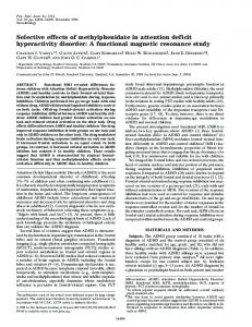
Figure 6. Right: single spatial-temporal dipole solutions for the activity in the response locked ERPs in the 0–50 ms postresponse interval, superimposed for targets (dipole 1), conjunction errors (dipole 2), and feature errors (dipole 3). Left: topographical map (contour step 1 µV) showing the scalp-distribution of the grand-average response-locked ERPs for target stimuli 40 ms post-response. The upper map shows the distribution as actually measured, the lower map shows the distribution as predicted by the dipole model.
3.91% for the target hits, feature errors, and conjunction errors, respectively.
Discussion We will first discuss the ERP-effects of attention in the correctly detected and rejected experimental trials, and second the ERP-effects in trials in which the participants made erroneous responses.
Correct Trials In the present experiment the participants were instructed to search for the occurrence of a colored target letter in a subsequently presented search array consisting of three different letters in three different colors. The color and identity of the target element varied randomly from trial to trial. This task can be conceived of as a transient multifeature selective attention task, i.e., the participants were cued on a trial-by-trial basis to attend to a combination of letter identity and color. A major difference between this study and previous multifeature selective attention studies is that subjects had to search for the target among distractors. Therefore, they not only had to detect the relevant features, but also had to decide whether these features were combined in the same display element (since the relevant features could also be present in different display elements, as in the color + letter present trials). Another difference between the present experiment and most earlier ERP-research on visual selective attention was that stimuli varied along five values (dimensions) of the two stimulus features (color and letHogrefe & Huber Publishers
ter identity) instead of varying only along two feature values. We found that when the search array contained the target element, the ERPs showed a prominent early posterior, contralaterally maximal negativity (i.e., occipital SN; onset at about 150–200 ms) relative to the situation in which there were no relevant features present (i.e., the nontarget ERPs). In addition, target ERPs elicited an anterior early positivity (i.e., FSP; onset at about 200 ms) relative to the nontarget ERPs. Very similar ERP-effects have been obtained in a large number of previous studies on sustained nonspatial visual selective attention (Hillyard & Münte, 1984; Kenemans et al., 1993, 1995, 2002; Wijers, Mulder, Okita, & Mulder, 1989; Wijers, Mulder, Okita, Mulder, & Scheffers, 1989; Wijers et al., 2002). First, this confirms that ERP-effects of selective attention in a phasic cuing situation are very comparable to the effects obtained in sustained attention conditions, as had already been shown for both spatial attention (Harter & Anllo-Vento, 1991; Luck et al., 1994; Mangun & Hillyard, 1991; Nobre et al., 2000) and, less so, for nonspatial attention (e.g., Eimer, 1995). Second, apparently the ERP effects of attending to a particular feature dimension are not much affected by the number of feature dimensions to be ignored (one in most previous studies and four in the present experiment). We might cautiously interpret this as showing that the differences between the ERPs to relevant and irrelevant stimuli are driven more by the facilitative processing of relevant stimuli than by the inhibition of irrelevant stimuli, since one would probably expect that inhibition would be harder the more irrelevant dimensions there are. Of course, a direct manipulation of the number of irrelevant dimensions could lead to much firmer conclusions. AlJournal of Psychophysiology 2005; Vol. 19(3):xxx–xxx
12
A.A. Wijers & M.A.S. Boksem: ERPs in an Illusory Conjunction Task
together, the present results show that the allocation of attention to nonspatial information is a flexible and dynamic process. Furthermore, it was found that the presence of a relevant color in the search display (color present trials) produced much more pronounced and earlier ERP effects than the presence of a relevant letter (letter present trials). Displays containing a letter in the relevant color elicited an early (onset slightly earlier than 200 ms) posterior contralateral selection negativity and a medial anterior selection positivity with a similar onset. Displays containing a letter with the relevant (target) identity elicited no posterior negativity and a small and delayed (as compared to the color effect) frontal positivity (see Figure 3). When both relevant attributes were present, but not combined in the same display element (color + letter present trials), this resulted in a posterior negativity which was identical to the effect when only the relevant color was present, and in a frontal positivity which seemed to consist of the summation of the effects of a relevant color only and a relevant letter only (see Figure 3). Therefore, the frontal positivity, but not the posterior negativity, showed a pattern of results indicating that color and letter identity were independently attended to (Hansen & Hillyard, 1983; Hillyard & Hansen, 1986). The dissociation between the patterns of results for the frontal selection positivity and the posterior selection negativity supports earlier suggestions that these effects are functionally separable and reflect different sources of neural activity (Rugg et al., 1987; Smid et al., 1999; Wijers, Mulder, Okita, & Mulder, 1989; Wijers, Mulder, Okita, Mulder, & Scheffers, 1989). More specifically, it has been hypothesized that SN and FSP reflect independent and parallel activation of posterior and anterior brain regions, respectively. SN is thought to reflect selective processing in the posterior visual system (selection-forperception), whereas FSP is thought to reflect (pre)frontal or subcortical selective processing (selection-for-action, Rugg et al., 1987; Smid et al., 1999). Of particular interest was the difference between ERPs elicited by the color + letter present trials, in which both relevant attributes were present but on different items, and the target trials in which both relevant attributes were combined in the same display element. For both stimulus types an FSP was found (relative to the
nontarget ERPs), but in the target ERPs this positivity was prominently enhanced after 250 ms (relative to the color + letter present ERPs; see Figure 3). This suggests that the later phase of the anterior positivity reflects the proper binding of the two relevant attributes to the same spatial object. Whereas the early phase of the anterior positivity reflects independent attributes selections, the later phase of the effect seems to reflect the selection of the spatially integrated attributes, i.e., this might reflect a form of object-based attention (Driver & Baylis, 1998). Alternatively, the fact that FSP was so dramatically increased in the target ERPs as compared to the color + letter ERPs might support the notion that FSP is associated with selection-for-action, enabling selective coupling of relevant stimuli to motor responses (Smid et al., 1999).
Error Trials In this experiment we succeeded in replicating the pattern of performance results that has been interpreted in earlier studies as evidence for the occurrence of illusory conjunctions (see introduction). Whereas participants made feature errors only about 10% of the time, they made many more conjunction errors (slightly less than 25%). However, the numbers of feature and conjunction errors that are supportive of the existence of “true illusory conjunctions” is dependent upon the particular theoretical model and parameters that one adopts for explaining these errors (Ashby et al., 1996; Donk, 1999) 1. Therefore, one of the questions in the present investigation was whether the ERPs in conjunction error trials and in feature error trials would differ, since these error types reflect qualitatively different processes according to FIT. We failed to demonstrate any convincing differences, however. It could be the case that illusory conjunction errors consist of a mixture of several different sources of error: feature errors, location errors, true illusory conjunctions, and others (Ashby et al., 1996; Donk, 1999; Navon & Ehrlich, 1995; Tsal, 1989). If the proportion of true illusory conjunctions is small relative to other error types, this could explain our nil result. It could also be the case that the number of participants was too small to reliably detect subtle differences. However, early ERP-
1 Actually, by straightforward commonsense considerations, one could argue that the number of conjunction errors should be larger than twice the number of feature errors (Navon & Ehrlich, 1995). In the present experiment this difference was not significant (F1,10 = 2.38, p = .15). However, an interaction was obtained between Visual Field and Error Type (F1,10 = 18.2, p < 0.005). This signified that the number of conjunction errors exceeded twice the number of feature errors for stimuli in the left visual field (F1,10 = 7.1, p < 0.05) but not for the right visual field (F1,10 = 0.1, p > 0.75). The percentages of conjunction errors and twice the feature error percentages were 24.7 and 18.5 for the left visual field and 21.8 and 21.1 for the right visual field. This might be an interesting observation, since it has been argued that the left hemisphere is specialized for object-based attention (Driver & Baylis, 1998), and, therefore, may be less likely to commit feature integration errors. Unfortunately, preliminary statistical analyses for the ERP results showed that Visual Field did not interact with other effects in an informative way. Therefore, the present paper reported the results pooled over both visual fields. Journal of Psychophysiology 2005; Vol. 19(3):xxx–xxx
Hogrefe & Huber Publishers
A.A. Wijers & M.A.S. Boksem: ERPs in an Illusory Conjunction Task
effects of selective attention are among the smallest effects investigated, and still the present experiment apparently had enough statistical power to prove these effects highly significant. Comparing the ERPs evoked in target hit and conjunction error trials, it was found that that targets elicited a prominent, parietally maximal P300, whereas the P300 was largely reduced in conjunction error trials. This suggests that in conjunction error trials participants responded with a low level of confidence (e.g., Sutton et al., 1982). This was also supported by the finding that the RTs in the false alarm trials were much slower than in the target hit trials. Altogether these results suggest that target hits and conjunction errors mainly differ with respect to decision-related processes. In the present experiment, the probability of the targets was only 20%. As is well-know from previous research, and was also found in the present experiment, low-probability events elicit prominent P300 components. Could it be that the low target probability had unwanted side-effects? For instance, if the subjects somehow expected that targets would be presented with a probability of 50%, this could explain why they made so many false-alarm responses. This seems unlikely, however. Subjects received extensive training, and were given feedback on misses and false alarms at the end of each of the 40-stimulus series. Therefore, we believe that the participants must have figured out the probabilistic structure of the stimulus sequences. The major result we report on the P300 is the larger P300 in target hit trials than in conjunction error trials. It also seems unlikely that this effect is contaminated by probability effects, since the number of target hits was much larger than the number of conjunction errors. Therefore, this probability difference would instead oppose the result we obtained. In the response locked averages, a fronto-central negativity, peaking at about 50 ms after the button-release response, was visible in the ERPs in error-trials (both feature and conjunction errors). This phenomenon, the ERN, was first described more than a decade ago (Falkenstein et al., 1990, 1991; Gehring et al., 1993, 1995), and is presently the topic of extensive research. The ERN usually peaks at about 100 ms after response-onset as measured by EMG onset (Coles et al., 2001). Since we used response button release as the synchronization point instead of EMG-onset, the somewhat shorter peak latency of 50 ms in the present experiment is to be expected. Surprisingly, an ERN of similar amplitude was also found in the response-locked correct target hit ERPs. Source localization yielded almost identical solutions for both error types and for the hit trials. The dipole-solutions consisted of a source in a deep medial frontal brain area. These results are well in accordance with previous ERP studies (Dehaene et al., 1994; Gehring & WilloughHogrefe & Huber Publishers
13
by, 2002; Van Veen & Carter, 2002b) and fMRI (Carter et al., 1998; Kiehl et al., 2000; Van Veen & Carter, 2002a), and support the suggestion that the ERN is generated from the ventral bank of the anterior cingulate cortex (Coles et al., 2001). Initially it was found that the ERN was only elicited by error trials and not by correct trials, and its functional significance was specifically related to error-processing, e.g., that it is associated with the error signal provided by a comparator system which compares a representation of the correct response with a representation of the actually executed response (Coles et al., 2001). However, many later studies found ERN-like activity to be present on correct trials also (e.g., Falkenstein et al., 2000; Luu et al., 2000; Vidal et al., 2000). Coles et al. (2001) propose two reasons why this can be the case. First, responselocked averages might contain artifacts from stimuluslocked negativities, which were not removed from the response-locked average. This can especially be the case with RTs that are fast and show low variability, and when the stimulus-locked averages show prominent negative waves. In our experiment, however, RTs were rather slow (more than 600 ms), and no clear negative components were present in the stimulus-locked averages, especially for the frontal ERPs, were the ERN was as large as for the central electrodes (compare Figures 1 and 5). Second, there might be error processing in correct trials. For instance, since most tasks involve speeded response requirements, trials with slow RTs might be considered as erroneous. Another example might be tasks in which the stimuli are degraded. In this case the representation of the appropriate or correct response will be compromised, and error processing may occur (Coles et al., 2001). We suggest that in typical studies on the topic of illusory conjunctions, in order to provoke enough conjunction errors, stimuli are presented in such a way that they are suboptimally perceived (e.g., in a double task situation or with a fast presentation rate). Therefore, the representation of the correct response (a GO versus a NOGO response) is also suboptimal. This might result in a state of continuous response-conflict (Braver et al., 2001; Van Veen & Carter, 2002a, b), both on target trials and on false alarm trials. The fact that the ERN was elicited both in correct hit target trials and in false alarm trials supports our earlier suggestion that feature errors and conjunction errors alike result from a vulnerable response decision process, in which there is ample conflict about whether or not to respond. Although the occasional occurrence of a (small) ERN in correct trials now seems to be well accepted, this experiment provided (as far as we know) the first demonstration of an equally sized ERN in correct and incorrect trials in healthy subjects. This seems to occur in rather Journal of Psychophysiology 2005; Vol. 19(3):xxx–xxx
14
A.A. Wijers & M.A.S. Boksem: ERPs in an Illusory Conjunction Task
restricted experimental circumstances, as in the conditions of an illusory conjunction paradigm, in which there is a large response uncertainty on each and every trial. An equally sized ERN on correct and incorrect response trials has been reported for schizophrenic subjects, which was interpreted as a response-monitoring dysfunction in these patients (Alain et al., 2002; Mathalon et al., 2002). However, in these patients the absence of a difference between correct and incorrect trials seems to be largely due to a decrease in ERN for incorrect trials, whereas in the present experiment the absence of a difference seems to be due to the substantial ERN for correct (hit) trials. Note that an apparent dissociation was present in the P300 and ERN results. Whereas a decreased P300 amplitude (and an increased RT) seemed to suggest that subjects responded less confidently in the false alarm trials than in the correct hit trials, the ERN-data suggested that there was an equal conflict for hits and false alarms. One could hypothesize that the P300 and ERN reflect different forms of conflict, perceptual conflict and response conflict, respectively. However, it is hard to imagine why different states of perceptual conflict could lead to equal states of response conflict. An alternative would be that the ERN (at least the aspect of it as obtained in the present experiment) reflects yet another functional process, for instance a state of general cognitive effort (or control), being present throughout an experimental run, independent of the particular trial being presented. Acknowledgments This research has been made possible by a fellowship from the Royal Netherlands Academy of Arts and Sciences. We wish to thank Joop Clots, Eise Hoekstra, Edwin Kiers, Jaap Ruiter, and Jan Smit for technical support. We thank Mieke Donk, Anton van Boxtel, and an anonymous reviewer for their comments on an earlier draft of this article.
References Alain, C., McNeely, H.E., He, Y., Christensen, B.K., & West, R. (2002). Neurophysiological evidence of error-monitoring deficits in patients with schizophrenia. Cerebral Cortex, 12, 840– 846. Ashby, F.G., Prinzmetal, W., Ivry, R., & Maddox, W.T. (1996). A formal theory of feature binding in object perception. Journal of Experimental Psychology: General, 111, 60–100. Braver, T.S., Barch, D.M., Gray, J.R., Molfese, D.L., & Snyder, A. (2001). Anterior cingulate cortex and response conflict: Effects of frequency, inhibition, and errors. Cerebral Cortex, 11, 825– 836. Journal of Psychophysiology 2005; Vol. 19(3):xxx–xxx
Carter, C.S., Braver, T.S., Barch, S.D.M., Botvinick, M.M., Noll, D., & Cohen, J.D. (1998). Anterior cingulate cortex, error detection, and the online monitoring of performance. Science, 280, 747–749. Coles, M.G.H., Scheffers, M.K., & Holroyd, C.B. (2001). Why is there an ERN/NE on correct trials? Response representations, stimulus-related components, and the theory of error-processing, Biological Psychology, 56, 173–189. Craver-Lemley, C., Arterberry, M.E., & Reeves, A. (1999). “Illusory” conjunctions: The conjoining of features of visual and imagined stimuli. Journal of Experimental Psychology: Human Perception and Performance, 25, 1036–1049. Dehaene, S., Posner, M.I., & Tucker, D.M. (1994). Localization of a neural system for error detection and compensation. Psychological Science, 5, 303–305. Donk, M. (1999). Illusory conjunctions are an illusion: The effects of target-nontarget similarity on conjunction and feature errors. Journal of Experimental Psychology: Human Perception and Performance, 25, 1207–1233. Driver, J., & Baylis, C. (1998). Attention and visual object segmentation. In R. Parasuraman (Ed.), The attentive brain (pp. 299–325). Cambridge, MA: MIT Press. Eimer, M. (1995). Event-related potential correlates of transient attention shifts to color and location. Biological Psychology, 41, 167–182. Falkenstein, M., Hohnsbein, J., Hoormann, J., & Blanke, L. (1990). Effects of errors in choice reaction tasks on the ERP under focused and divided attention. In C.H.M. Brunia, A.W.K. Gaillard, & A. Kok (Eds.), Psychophysiological brain research (pp. 192–195). Tilburg: Tilburg University Press. Falkenstein, M., Hohnsbein, J., Hoormann, J., & Blanke, L. (1991). Effects of crossmodal divided attention on late ERP components: II. Error processing in choice reaction tasks. Electroencephalography and Clinical Neurophysiology, 78, 447– 455. Falkenstein, M., Hoormann, J., Christ, S., & Hohnsbein, J. (2000). ERP components on reaction time errors and their functional significance: A tutorial. Biological Psychology, 52, 87–107. Gehring, W.J., Coles, M.G., Meyer, D.E., & Donchin, E. (1995). A brain potential manifestation of error-related processing. In G. Karmos, M. Molnar, V. Csepe, I. Czigler, & J.E. Desmedt (Eds.), Perspectives of event-related potentials research (EEG Journal Supplement 44, pp. 261–272). Amsterdam: Elsevier. Gehring, W.J., Goss, B., Coles, M.G., Meyer, D.E., & Donchin, E. (1993). A neural system for error detection and compensation. Psychological Science, 4, 385–390. Gehring, W.J., & Willoughby, A.R. (2002). The medial frontal cortex and the rapid processing of monetary gains and losses. Science, 295, 2279–2282. Hansen, J.C., & Hillyard, S.A. (1983). Selective attention to multidimensional auditory stimuli. Journal of Experimental Psychology: Human Perception and Performance, 9, 1–19. Harter, M.R., & Anllo-Vento, L. (1991). Visual-spatial attention: Preparation and selection in children and adults. In C.H.M. Brunia, G. Mulder, G., & M.N. Verbaten (Eds.), Event-related potentials of the brain (pp. 1832–94[??]). Amsterdam: Elsevier. Hillyard, S.A., & Hansen, J.C. (1986). Attention: Electrophysiological approaches. In M.G.H. Coles, E. Donchin, & S.W. Porges (Eds.), Psychophysiology: Systems, processes, and applications (pp. 227–243). New York: Guilford. Hogrefe & Huber Publishers
A.A. Wijers & M.A.S. Boksem: ERPs in an Illusory Conjunction Task
Hillyard, S.A., & Münte, T.F. (1984). Selective attention to color and location: An analysis with event-related brain potential. Perception and Psychophysics, 36, 185–198. Ivry, R.B., & Prinzmetal, W. (1991). Effect of feature similarity on illusory conjunctions. Perception and Psychophysics, 49, 105–116. Kenemans, J.L., Kok, A., & Smulders, F.T.Y. (1993). Event-related potentials to conjunctions of spatial frequency and orientation as a function of stimulus parameters and response requirements. Electroencephalography and Clinical Neurophysiology, 88, 51–63. Kenemans, J.L., Lijfijt, M., Camfferman, G., & Verbaten, M.N. (2002). Split-second sequential selective activation in human secondary visual cortex. Journal of Cognitive Neuroscience, 14, 48–61. Kenemans, J.L., Smulders, F.T.Y., & Kok, A. (1995). Selective processing of two-dimensional visual stimuli in young and old subjects: Electrophysiological analysis. Psychophysiology, 32, 108–120. Khurana, B. (1988). Visual structure and the integration of form and color information. Journal of Experimental Psychology: Human Perception and Performance, 24, 1766–1785. Kiehl, K.A., Liddle, P.F., & Hopfinger, J.B. (2000). Error processing and the rostral anterior cingulate: an event-related fMRI study. Psychophysiology, 37, 216–223. Lange, J.J., Wijers, A.A., Mulder, L.J.M., & Mulder, G. (1999). ERP effects of spatial attention and display search with unilateral and bilateral stimulus displays. Biological Psychology, 50, 203–233. Luck, S.J., & Girelli, M. (1998). Electrophysiological approaches to the study of selective attention in the human brain. In R. Parasuraman (Ed.), The attentive brain (pp. 71–94). Cambridge, MA: MIT Press. Luck, S.J., Hillyard, S.A., Mouloua, M., Woldorff, M.G., Clark, V.P., & Hawkins, H.L. (1994). Effects of spatial cuing on luminance detectability: Psychophysical and electrophysiological evidence for early selection. Journal of Experimental Psychology: Human Perception and Performance, 20, 887–904. Luu, P., Flaisch, T., & Tucker, D.M. (2000). Medial frontal cortex in action monitoring. Journal of Neuroscience, 20, 464–469. Mangun, G.R., & Hillyard, S.A. (1991). Modulation of sensoryevoked brain potentials provide evidence for changes in perceptual processing during visual-spatial priming. Journal of Experimental Psychology: Human Perception and Performance, 17, 1057–1074. Mathalon, D.H., Fedor, M., Faustman, W.O., Gray, M., Asakari, N., & Ford, M.J. (2002). Response-monitoring dysfunction in schizophrenia: An event-related brain potential study. Journal of Abnormal Psychology, 111, 22–41. Navon, D., & Ehrlich, B. (1995). Illusory conjunctions: Does inattention really matter? Cognitive Psychology, 29, 59–83. Nobre, A.C., Sebestyen, G.N., & Miniussi, C. (2000). The dynamics of shifting attention revealed by event-related potentials. Neuropsychologia, 38, 964–974. Perrin, F., Pernier, J., Bertrand, O., & Echalier, J.F. (1989). Spherical splines for scalp potential and current density mapping. Electroencephalography and Clinical Neurophysiology, 72, 184–187. Posner, M.I., Snyder, C.R.R., & Davidson, B.J. (1980). Attention and the detection of signals. Journal of Experimental Psychology: General, 109, 160–174. Hogrefe & Huber Publishers
15
Previc, F.H., & Harter, M.R. (1982). Electrophysiological and behavioral indicants of selective attention to multifeature gratings. Perception and Psychophysics, 32, 465–472. Prinzmetal, W., Henderson, D., & Ivry, R. (1995). Loosening the constraints on illusory conjunctions: Assessing the roles of exposure duration and attention. Journal of Experimental Psychology: Human Perception and Performance, 21, 1362– 1375. Prinzmetal, W., Hoffman, H., & Vest, K. (1991). Automatic processes in word perception: An analysis from illusory conjunctions. Journal of Experimental Psychology: Human Perception and Performance, 17, 902–923. Rugg, M.D., Milner, A.D., Lines, C.R., & Phalp, R. (1987). Modulation of visual event-related potentials by spatial and nonspatial visual selective attention. Neuropsychologia, 25, 85–96. Scherg, M. (1990). Fundamentals of dipole source potential analysis. In F. Grandori, M. Hoke, & G.L. Romani (Eds.), Auditory evoked magnetic fields and electric potentials. Advances in audiology, 6 (pp. 40–69). Basel: Karger. Smid, H.G.O.M., Jakob, A., & Heinze, H.J. (1999). An event-related brain potential study of visual selective attention to conjunctions of color and shape. Psychophysiology, 36, 264–279. Sutton, S., Ruchkin, D.S., Munson, R., & Kietzman, M.L. (1982). Event-related potentials in a two-interval forced-choice detection task. Perception and Psychophysics, 32, 360–374. Treisman, A.M., & Gelade, G. (1980). Afeature-integration theory of attention. Cognitive Psychology, 12, 97–136. Treisman, A.M., & Gormican, S. (1988). Feature analysis in early vision: Evidence from search asymmetries. Psychological Review, 95, 15–48. Treisman, A.M., & Sato, S. (1990). Conjunction search revisited. Journal of Experimental Psychology: Human Perception and Performance, 15, 394–400. Treisman, A., & Schmidt, H. (1982). Illusory conjunctions in the perception of objects. Cognitive Psychology, 14, 107–141. Treisman, A., & Souther, J. (1986). Illusory words: The roles of attention and of top-down constraints in conjoining letters to form words. Journal of Experimental Psychology: Human Perception and Performance, 12, 3–17. Tsal, Y. (1989). Do illusory conjunctions support the feature integration theory? A critical review of theory and findings. Journal of Experimental Psychology: Human Perception and Performance, 15, 394–[??]100. Tsal, Y., Meiran, N., & Lavie, N. (1994). The role of attention in illusory conjunctions. Perception and Psychophysics, 55, 350–358. Ungerleider, L.G., & Haxby, J.V. (1994). “What” and “where” in the human brain. Current Opinions in Neurobiology, 4, 157– 165. Van Veen, V., & Carter, C.S. (2002a).The anterior cingulate as a conflict monitor: FMRI and ERP studies. Physiology and Behavior, 77, 477–482. Van Veen, V., & Carter, C.S. (2002b). The timing of action-monitoring processes in the anterior cingulate cortex. Journal of Cognitive Neuroscience, 14, 593–602. Vidal, F., Hasbroucq, T., Grapperon, J., & Bonnet, M. (2000). Is the “error negativity” specific to errors? Biological Psychology, 51, 109–128. Wijers, A.A., Lamain, W., Slopsema, S., Mulder, G., & Mulder, L.J.M. (1989). An electrophysiological investigation of the spatial distribution of attention to colored stimuli in focused Journal of Psychophysiology 2005; Vol. 19(3):xxx–xxx
16
A.A. Wijers & M.A.S. Boksem: ERPs in an Illusory Conjunction Task
and divided attention conditions. Biological Psychology, 29, 213–245. Wijers, A.A., Mulder, G., Okita, T., & Mulder, L.J.M. (1989). An ERP-study on memory search and selective attention to lettersize and conjunctions of lettersize and color. Psychophysiology, 26, 529–547. Wijers, A.A., Mulder, G., Okita, T., Mulder, L.J.M., & Scheffers, M.K. (1989). Attention to colour: An ERP-analysis of selection, controlled search, and motor activation. Psychophysiology, 26, 89–109. Wijers, A.A., Van Besouw, N.J.P., & Mulder, G. (2002). Selective attention to a facial feature with and without facial context: An ERP-study. International Journal of Psychophysiology, 44, 13–35.
Journal of Psychophysiology 2005; Vol. 19(3):xxx–xxx
Accepted for publication: March 16, 2005 Address for correspondence Albertus A. Wijers Experimental and Work Psychology University of Groningen Grote Kruisstraat 2/1 NL-9712 TS Groningen The Netherlands Tel. +31 50 353-6466 Fax +31 50 363-6304 E-mail
[email protected]
Hogrefe & Huber Publishers