The microscale cell patterning inside the microchannel was successful ... several applications such as analyses of chemicals released from a single cell and.
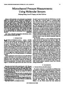
SELECTIVE CONTROL OF CELL ATTACHMENT IN MICROCHANNEL USING PHOTOCHEMICAL REACTION Kihoon Jang, Kae Sato, Tomohiro Konno, Kazuhiko Ishihara, Takehiko Kitamori The University of Tokyo, JAPAN ABSTRACT In this paper, we employed photochemical reaction for controlling the cell adhesion inside the microchannel. The microscale cell patterning inside the microchannel was successful, which was difficult in the glass microchip before. Using our method, cell micropatterning can be performed directly inside the microchannel without separating the substrate and cover of the microchip for surface patterning. KEYWORDS: Cell adhesion, Microchannel, Micropatterning, INTRODUCTION Interest of microchip-based cellular biochemical analysis systems has been increased to utilize them as a powerful tool for cell-based assays and basic biomedical researches. Cell culture in the microfluidic device has several advantages, because they provide three-dimensional small culture space, which reproduces an artificial minimum living tissue, and flow culture condition, which reproduces the vascularlike flow. In related developments, micropatterning of cells can be used in tissue engineering and cell-based fundamental studies. It is possible to control the number of cells and the distance between cells for highly efficient biological analyses. We thought that the microscale cell patterning inside the microchannel would be a powerful tool for the microchip-based cellular analysis systems. It will open the way for several applications such as analyses of chemicals released from a single cell and communication between different cells. EXPERIMENTAL We developed a UV-sensitive surface modification method, in which an exposure to UV changes from hydrophilic to moderately hydrophobic cell adhering surface (Figure 1). The technique is based on a combining 2methacryloyloxyethyl phosphorylcholine (MPC) containing polymer [1] which is known as a resistant barrier for cell adherence and nitrobenzyl group photolabile linker for selective cell patterning.
Figure 1. Preparation of cell-adhesive surface by photochemical reaction on MPC modified surface.
Twelfth International Conference on Miniaturized Systems for Chemistry and Life Sciences October 12 - 16, 2008, San Diego, California, USA 978-0-9798064-1-4/µTAS2008/$20©2008CBMS
901
Figure 2. Schematic representation of selective surface control of cell attachment in microchannel. (a) UV (365 nm, 150 mW/cm2) is illuminated through the photomask to MPC modified microchannel. (b) Rinse with deionized water, sterilize with ethanol, wash with deionized water. (c) Introduce cell suspension at a concentration of 8 × 10⁶ cells/ml and incubate for 2 hr (d) After 2 hr of incubation, fresh medium is connected to microsyringe pump at the flow rate of 0.2 µl/min. Figure 2 shows the schematic representation of cell patterning on the UVsensitive surface. The surface modification method was based on our previoius work [2]. Experimental procedures were as follows: the cleaned microchannel was treated with 3-aminopropyl–triethoxysilane (APTS), and then nitrobenzyl group containing photolabile linker was introduced and reacted for 2 h. Next, MPC was grafted by EDC coupling for 12 h, which made non-fouling surface. UV (365 nm) irradiation induced cleaving reaction of MPC which made the surface moderately hydrophobic, cells attached to the UV illuminated area. . RESULT AND DISCUSSION Figure 4a shows the continuous-perfusion microfluidic cell culture system, which was based on our previous work [3]. MC-3T3 E1 cells were used for cell micropatterning. After, the preparing the MPC modification of microchannel, UV was illuminated through the photomask having 200 µm wide stripes with black 300 µm separations. Figure 3b shows the result of the cells cultured on surface between UV irradiated and not irradiated in microchannel (width 400 µm, depth 100 µm). No cells were attached to MPC modified surface. However, cells localized selectively on UV exposed area. Cells were localized to UV exposed area where MPC had removed and size of the patterned cells were coincidence with that of photomask. These results show that this technique can be applied directly in microchannel for insitu surface patterning. Long-term stability of micro-patterned cells was confirmed in microchannel with flow condition for 14 days of cultures. After preparation of surface, endothelial cell (EC) suspension (cell concentration of 8×10⁶ cells/mL) was introduced into microchannel and ECs were micro-patterned (width 200 µm stripe) in mirochannel (width 800 µm, maximum depth 200 µm). These micro-patterned cells were cultured for 14 days at the flow rate of 0.2 µl/min. Micro-patterned cells were maintained in safe from day 1 (Figure 3c) to day 14 (Figure 3d) without outgrowing of the pattern cells. Twelfth International Conference on Miniaturized Systems for Chemistry and Life Sciences October 12 - 16, 2008, San Diego, California, USA
902
Even though in the case of microfluidic system, MPC modified surface still have a resistant to cell adherence for 2 weeks of cultures. Which showed that the property of MPC have not decreased in microfluidic cell culture conditions. (a)
Microchannel
Medium
Inlet
Chipholder
(b) Capillary
Outlet
(c)
(d)
Figure 3. Cell micropatterning in microchip system (scale bar = 200 µm). (a) Continuous microfluidic cell culture system. (b) Phase contrast images of MC3T3 E1 cells micro-patterned after 1 day of culture in microchannel (width 400 µm, depth 100 µm). Stability of the micro patterned ECs in microchannel (width 800 µm, depth 150 µm) (c, d). Phase contrast image of micropatterned ECs (c) After 1 day of culture. (d) After 14 days of culture. CONCLUSION In conclusion, we have developed a simple, straigthforward surface modification method for regulating the cell adherence inside the microchannel. The strategy was based on combining MPC polymer and photolabile linker. By using photochemical reaction, MPC was efficiently removed and MC3T3E1 cells were patterned on UV illuminated area in microchannel. We are currently extending the presented strategy to different cell types side-by-side in a single channel for cell-cell interaction studies. ACKNOWLEDGEMENTS This work was supported by the JSPS Core-to Core Program. REFERENCES [1] Y. Iwasaki, K. Ishihara, Anal Bioanal Chem, 381, 534-546 (2005). [2] K.H. Jang, K. Sato, K. Tomohiro, K. Ishihara, T. Kitamori, The principles of manuscript writing, Proc. Micro Total Analysis System 2007, The Chemical and Biological Microsystems Society, pp. 1628-1630 (2007). [3] K.H. Jang, K. Sato, K. Igawa, U. Chung, T. Kitamori, Anal Bioanal Chem, 390, 825-832 (2008).
Twelfth International Conference on Miniaturized Systems for Chemistry and Life Sciences October 12 - 16, 2008, San Diego, California, USA
903