CSIRO PUBLISHING
Functional Plant Biology, 2007, 34, 449–456
www.publish.csiro.au/journals/fpb
Selenium-induced oxidative stress in coffee cell suspension cultures ˜ B , Salete A. GaziolaB , Paulo MazzaferaC , Peter J. LeaD Rui A. Gomes-JuniorA , Priscila L. Gratao B,E and Ricardo A. Azevedo A
Centro de Estudos Superiores de Balsas, Universidade Estadual do Maranh˜ao, 65800-000, Balsas, MA, Brazil. B Departamento de Gen´etica, Escola Superior de Agricultura Luiz de Queiroz, Universidade de S˜ao Paulo, 13 418-900 Piracicaba, SP, Brazil. C Departamento de Fisiologia Vegetal, Instituto de Biologia, CP 6109, Universidade Estadual de Campinas, 13 083-970 Campinas, SP, Brazil. D Department of Biological Sciences, University of Lancaster, Lancaster, LA1 4YQ, UK. E Corresponding author. Email:
[email protected]
Abstract. Selenium (Se) is an essential element for humans and animals that is required for key antioxidant reactions, but can be toxic at high concentrations. We have investigated the effect of Se in the form of selenite on coffee cell suspension cultures over a 12-day period. The antioxidant defence systems were induced in coffee cells grown in the presence of 0.05 and 0.5 mM sodium selenite (Na2 SeO3 ). Lipid peroxidation and alterations in antioxidant enzymes were the main responses observed, including a severe reduction in ascorbate peroxidase activity, even at 0.05 mM sodium selenite. Ten superoxide dismutase (SOD) isoenzymes were detected and the two major Mn-SOD isoenzymes (bands V and VI) responded more to 0.05 mM selenite. SOD band V exhibited a general decrease in activity after 12 h of treatment with 0.05 mM selenite, whereas band VI exhibited the opposite behavior and increased in activity. An extra isoenzyme of glutathione reductase (GR) was induced in the presence of selenite, which confirmed our previous results obtained with Cd and Ni indicating that this GR isoenzyme may have the potential to be a marker for oxidative stress in coffee. Additional keywords: Coffea arabica, glutathione reductase, oxidative stress, selenium, superoxide dismutase.
Introduction The increase in contamination of the global environment is a matter of great concern, since anthropogenic activities are the main source of the pollution that is causing such contamination (Azevedo and Azevedo 2006). Toxic elements, including heavy metals have a particularly strong effect on plants and their productivity, although some metals and metalloids are essential for plant growth in small quantities (Grat˜ao et al. 2005). The considerable accumulation of metals and other toxic elements in some plant species has allowed the identification of hyperaccumulator species which may eventually be used in phytoremediation to replace wherever possible, traditional engineering approaches (LeDuc and Terry 2005; Lynch and Moffat 2005). Selenium (Se) can be considered a metalloid and occurs in several different oxidation states as selenide (Se2− ), elemental selenium (Se0 ), selenite (Se4+ ), and selenate (Se6+ ) and has chemical properties very similar to sulfur (Brown and Shrift 1982). It is now well established that Se is an essential element for humans, animals and bacteria (Hartikainen 2005). However, there is no definitive evidence so far that Se is an essential nutrient for higher plants. The Se containing amino acids found in animal proteins are not present in equivalent © CSIRO 2007
proteins in higher plants, although they are present in algae (Hatfield et al. 2006). The presence of Se in soil worldwide is very heterogeneous and can vary dramatically from Se-deficient to Se-toxic (Hartikainen 2005). The metabolic role and regulation of Se, particularly in plants, is poorly understood (Agalou et al. 2006). Most plant species cannot tolerate high concentrations of Se in their tissues and contain less than 25 µg g−1 DW even when grown on high-Se soils (White et al. 2004). Although a study on a range of different accession lines of Arabidopsis thaliana L. indicated that some lines could accumulate 1000 µg g−1 DW in the roots when grown on 20 µM selenite (Zhang et al. 2007). However, some plants (e.g, Astragalus bisulcatus A.Gray and Stanleya pinnata Britton) are able to accumulate very high concentrations of Se, up to 10 000 µg g−1 DW, in the periphery of the leaves (Freeman et al. 2006). There has recently been considerable interest in attempting to improve the Se tolerance and accumulation capacity of higher plants by the overexpression of key enzymes involved in selenium and sulfur metabolism (Van Huysen et al. 2004; Van Hoewyk et al. 2005; LeDuc et al. 2006). There have been a number of reports that toxic elements can induce oxidative stress in plants (Vit´oria et al. 2001; Anza et al. 2005; Canc¸ado et al. 2005; di Toppi et al. 2005;
10.1071/FP07010
1445-4408/07/050449
450
Functional Plant Biology
Garcia et al. 2006; Hassan 2006; Gajewska and Sklodowska 2007; Groppa et al. 2007; Saeidi-Sar et al. 2007). Seedlings of Trigonella foenum-graecum L. exhibited oxidative stress during Se deficiency and changes in the antioxidant enzyme activity, particularly of superoxide dismutase, catalase and peroxidase, in response to Se supplementation were identified (Santosh et al. 1999; Sreekala et al. 1999). The addition of low concentrations of selenate to ryegrass decreased lipid peroxidation, with small increases in glutathione peroxidase. However, high concentrations of selenate were toxic and there was a large increase in lipid peroxidation, α-tocopherol and in particular glutathione peroxidase activity (Hartikainen et al. 2000; Hartikainen 2005). Cartes et al. (2005) showed that selenite was more efficient than selenate at causing the increase in glutathione peroxidase activity in ryegrass. However, when additional sulfur was applied to the selenite grown ryegrass, the shoot Se concentration was reduced along with glutathione peroxidase activity, while lipid peroxidation increased (Cartes et al. 2006). Coffee is a very important crop and is extensively used worldwide as a beverage and apart from some reports (Praxedes et al. 2006; Ronchi et al. 2006), very little has been published on the response of this plant to abiotic stresses. Meija et al. (2003) described the Se volatile species in green and roasted coffee beans and the brewed coffee drink. Selenite treatment of coffee seedlings resulted in lower dry mass accumulation, lower leaf area, thicker leaves, decrease of chlorophyll and increase in caffeine and soluble sugars (Mazzafera 1998). Nitrogen metabolism was also affected as nitrate reductase activity was strongly reduced by selenite treatment (Mazzafera 1998). Selenite may also be interfering with nitrate influx into the roots (Lea and Azevedo 2006) and uptake by the cells, because nitrate membrane transporters are competitively inhibited by nitrite in plants (King et al. 1993). As part of an ongoing project in our laboratories, the antioxidative response of coffee to toxic elements is under investigation (Gomes-Junior et al. 2006a, 2006b). In this paper we report the antioxidant responses of the same coffee cell cultures subjected to two concentrations of Se added as selenite. Further research is being carried out to analyse coffee plants in the field and the effects of Cd, Ni and Se on plant growth and beverage quality. Materials and methods Cell suspension Leaf segments from leaves of the third and fourth leaf pairs of Coffea arabica L. cv. Catua´ı Vermelho were used to produce the cell cultures. The explants were maintained in solid CIM medium (callus inducing medium – Neuenschwander and Baumann 1992) containing Murashige and Skoog salts, and supplemented with 10 mg L−1 thiamineHCl, 100 mg L−1 inositol, 30 g L−1 sucrose, 4 mg L−1 kinetin and 1 mg L−1 2,4-dichlorophenoxyacetic acid, at pH 5.8. After 9–10 weeks in the dark, friable calli were selected and transferred to liquid medium (30 mL in 250 mL Erlenmeyer flasks, at 20g in the dark, 25 ± 2◦ C). Large aggregates were eliminated during weekly culture maintenance, when half the volume of the flask was transferred to a new flask containing 15 mL medium.
R. A. Gomes-Junior et al.
Treatment of the cells and cell growth Coffee cells maintained in liquid medium for 7 days were suction–dried and washed with distilled-deionised sterile water and transferred to the liquid CIM medium at a density of 4 g cells per 50 mL. Preliminary analyses on the effect of several sodium selenite (Na2 SeO3 ) concentrations (0 to 5 mM) on the growth of the cell culture were performed and two selenite concentrations (0.05 and 0.5 mM) were chosen for the main experiments. Sodium selenite was added to a final concentration of 0 (control), 0.05 and 0.5 mM and the cells were grown for 12 days (288 h) under the same conditions, harvested and stored as described by (GomesJunior et al. 2006a, 2006b). Se accumulation Samples of coffee cells were dried at 60◦ C for 14 days and quantitative Se analysis of the cells was carried out using energy disperse X-ray fluorescence (EDXRF) with the radioisotopic excitation technique as described previously (Gomes-Junior et al. 2006a). Lipid peroxidation Lipid peroxidation in the coffee cells was determined by measuring the concentration of thiobarbituric acid-reacting substances (TBARS) as described previously (Gomes-Junior et al. 2006a). Antioxidant enzyme analyses The following steps were carried out at 4◦ C unless otherwise stated. The coffee cells were homogenised (2 : 1 buffer volume/fresh weight) in a mortar with a pestle with 100 mM potassium phosphate buffer (pH 7.5) containing 1 mM EDTA, 3 mM DTT and 5% (w/v) insoluble polyvinylpyrrolidone (PVPP). The homogenate was centrifuged at 10 000g for 30 min and the supernatant was kept stored in separate aliquots at −80◦ C, before superoxide dismutase (SOD, EC 1.15.1.1), guaiacol peroxidase (GOPX, EC 1.11.1.7), catalase (CAT, EC 1.11.1.6), ascorbate peroxidase (APX, EC 1.11.1.11) and glutathione reductase (GR, EC 1.8.5.1) analyses. Catalase, GR, APX and GOPX activities were determined as described by Gomes-Junior et al. (2006a). Activity staining following non-denaturing PAGE was also carried out for CAT, GR and SOD. Electrophoresis buffers and gels were prepared as described previously (Gomes-Junior et al. 2006a) except that SDS was excluded. Electrophoresis was carried out under nondenaturing condition in 9% polyacrylamide gels for CAT, GR and SOD activity staining. A constant current of 30 mA gel−1 was applied for 8 h (gels to be stained for CAT activity), 4 h (gels to be stained for GR activity) or 3 h (gels to be stained for SOD activity) at 4◦ C. Equal amounts of protein (20 µg) were loaded on to each lane. After electrophoretic separation, SOD, CAT and GR activity staining was carried out as described previously (Gomes-Junior et al. 2006a). The concentration of protein was determined by the method of Bradford (Bradford 1976) using BSA as a standard. Statistical analysis Data were statistically analysed within the experiment and the results expressed as mean and standard error of the mean
Selenium effects in coffee cells
Functional Plant Biology
(± s.e.m.) of three independent replicates of cell growth, TBARS, CAT, GR, GOPX, APX activities, and Se accumulation measurements.
8
451
(A)
7
Results Fresh weight
Growth of Coffea arabica cell cultures Preliminary experiments with increasing concentrations of selenite were carried out to determine the doses that cause inhibition of growth (data not shown). Concentrations higher than 1 mM showed a strong toxic effect on the cells, which exhibited a deep brown colouration after a few hours of treatment. Even at 0.05 and 0.5 mM selenite concentrations, the fresh weight of the cell cultures showed a strong inhibition of growth during the period of the experiment (Fig. 1A).
6 5 4 3
2 0
48
96
Se accumulation
144
192
240
288
192
240
288
192
240
288
Time (h)
There was a massive accumulation of Se in the 0.5 mM selenite treated cells, which was over 10-fold higher than that detected in the 0.05 mM selenite treated cells (Fig. 1B). There was a linear increase in Se concentration in the 0.5 mM selenite treated cells between 48 and 192 h, whereupon the concentration stabilised. However, in the 0.05 mM selenite treated cells, the Se cell concentration remained unaltered after 96 h of growth.
3000
(B)
Se concentration
2500
Lipid peroxidation A visual examination of the coffee cells after 12 days of growth indicated that the cells treated with selenite were pigmented, with those grown in 0.5 mM exhibiting a deep brown colour (Fig. 2). Evidence of Se-induced lipid peroxidation and consequently oxidative stress, was obtained with the increase in TBARS in the cells subject to exposure to both concentrations of selenite (Fig. 1C).
2000 1500 1000 500 0 0
48
96
144
Time (h)
SOD activity 10
(C)
9
TBARS content
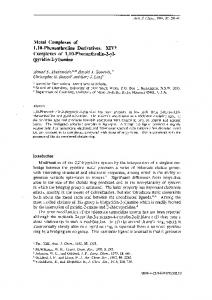
Bands of SOD enzyme activity representing the presence of isoenzymes, were observed following the staining of native PAGE gels, after the separation of proteins extracted from the cultured cells (Fig. 3A). In a previous paper we have classified SOD isoenzymes from coffee cell suspension cultures (GomesJunior et al. 2006a). On the basis of this classification, SOD isoenzymes I, II, III, IV, V and VI were classified as Mn-SOD, whereas the SOD isoenzymes VII, VIII and IX were classified as Fe-SOD. Cu/Zn-SOD activity was not observed following native PAGE separation of coffee cell culture extracts. The activity staining showed two major Mn-SOD isoenzymes (bands V and VI) in coffee cells, with band V exhibiting a general decrease in activity after 24 h of treatment with 0.05 mM selenite, whereas band VI exhibited the opposite behaviour. Therefore, after 288 h of treatment at 0.05 mM selenite, band V had almost disappeared, whereas band VI exhibited its highest activity essentially accounting for all the Mn-SOD activity (lane 6, Fig. 3A). The other Mn-SOD isoenzymes were not detected after 12 h treatment at both selenite concentrations. Following treatment with 0.5 mM selenite, the Mn-SOD bands I, II, III, IV were not detected and all Mn-SOD activity was because of bands V and VI which did not clearly exhibit any major alterations during the whole time length of the experiment. A curious result was the detection of an extra SOD band (numbered band X, also
8 7 6 5 4 3 0
48
96
144
Time (h) Fig. 1. (A) Cell growth (g fresh weight); (B) Se accumulation (µg g−1 dry weight); (C) thiobarbituric acid-reacting substances (TBARS) content (nmol g−1 fresh weight) in cultured coffee cells grown for a 288 h period in three concentrations of sodium selenite (Na2 SeO3 ). (䊏) Control-zero Se, ( ) 0.05 mM Na2 SeO3 and (N ) 0.5 mM Na2 SeO3 . Values are the means of four replicates ± s.e.m. for cell growth, means of two replicates ± s.e.m. for Se accumulation and means of three replicates ± s.e.m. for TBARS content.
◦
452
Functional Plant Biology
R. A. Gomes-Junior et al.
I II III IV
(A)
V VI VII VIII IX X
(B)
0
0.05 mM Na2SeO3
I
0.5 mM
Fig. 2. Coffee cells suspension cultures grown for 288 h in zero Se, 0.05 mM Na2 SeO3 and 0.5 mM Na2 SeO3 .
classified as Fe-SOD, data not shown), which appeared in the 0.5 mM treatment after 192 h (lane 9 and 10, Fig. 3A).
(C)
CAT activity Catalase activity staining after the separation of extracts by native PAGE (Fig. 3B) revealed the presence of only one CAT isoenzyme in coffee cell suspension cultures. Due to the diffuse appearance of the bands, it is difficult to see the relationship between the band intensity with the spectrophotometer data (Fig. 4A). However, it is possible to see that the peak observed after 12 h coincides with an increased intensity in the corresponding lanes 3 (0.05 mM) and 7 (0.5 mM), and that overall the general trend is the same. Catalase activity determined with the spectrophotometer assay (Fig. 4A) showed a varying effect of selenite at the two concentrations. At a concentration of 0.05 mM, selenite induced a different behaviour in CAT activity, which was strongly inhibited between 48–96 h of treatment, but then continuously increased until the end of the experiment. In contrast, at 0.5 mM selenite induced a 3-fold increase in activity when compared with the control after 12 h of exposure, which was followed by a rapid decrease at 24 h of exposure, and again another peak of activity at 96 h. GR activity Following native PAGE, GR activity staining showed the presence of at least four GR isoenzymes in coffee cells (bands I, II, III and IV) that were differentially affected by selenite treatment (Fig. 3C). Following exposure to 0.05 mM selenite, there was an increase in the activity of GR isoenzyme I. Initially there was a similar effect after 24 h treatment with 0.5 mM selenite, but it can be clearly seen that after 192 h of treatment, isoenzymes I and II were totally inhibited (Fig. 3C). It is also interesting to note that at both selenite concentrations after 12 h of exposure, band IV was clearly and definitely present but disappeared after 24 h to return as a less visible band after longer periods (Fig. 3C).
I II III IV
1
2
3
4
5
6
7
8
9
10
Fig. 3. Activity staining for (A) superoxide dismutase (SOD), (B) catalase (CAT) and (C) glutathione reductase (GR) following native PAGE of extracts of cultured coffee cells. Lane 1, bovine SOD, bovine liver CAT and Saccharomyces cerevisiae GR standards for (A), (B) and (C), respectively; lane 2, control (zero Se) after 96 h; lane 3, 12 h, lane 4, 24 h; lane 5, 192 h and lane 6, 288 h of growth in 0.05 mM Na2 SeO3 ; lane 7, 12 h; lane 8, 24 h; lane 9, 192 h; and lane 10, 288 h of growth in 0.5 mM Na2 SeO3 .
Following treatment with 0.05 mM selenite, there was a peak of GR activity at the 24 h point, after this time the activity remained essentially constant without any major changes, but was always higher when compared to the control (Fig. 4B). Initially there was some variation in the GR activity in the cells treated with 0.5 mM selenite, but after 96 h there was clearly a continuous decrease in activity (Fig. 4B). Although a densitometry analysis was not carried out, it is possible to see that overall the total stained GR activity was higher in the 0.05 mM selenite treatment, as also observed when activity was determined using the spectrophotometer assay. APX activity Ascorbate peroxidase activity was also determined in the extracts of the selenite treated coffee cells and exhibited some interesting trends (Fig. 4C). APX activity did not vary
Selenium effects in coffee cells
Functional Plant Biology
453
2.5
70
(A)
(B)
60 50
GR specific activity
CAT specific activity
2.0
40 30 20
1.5
1.0
0.5 10
0.0
0 0
48
96
144
192
240
288
0
48
96
Time (h)
(C)
240
288
192
240
288
(D) 1.2
GOPX specific activity
1.0
APX specific activity
192
1.4
1.2
0.8
0.6
0.4
0.2
0.0
144
Time (h)
1.0 0.8 0.6 0.4 0.2
0
48
96
144
192
240
288
0.0
0
48
Time (h)
96
144
Time (h)
Fig. 4. Specific activity of (A) catalase (CAT) (µmol min−1 mg−1 protein); (B) glutathione reductase (GR) (µmol min−1 mg−1 protein); (C) ascorbate peroxidase (APX) (µmol min−1 mg−1 protein) and (D) GOPX (u) in coffee cells grown for a 288 h period in three concentrations of Na2 SeO3 . (䊏) Control zero-Se, ( ) 0.05 mM Na2 SeO3 and (N ) 0.5 mM Na2 SeO3 .Values are the means of three replicates ± s.e.m.
◦
significantly in the control treatment during the whole time of the experiment. However, both selenite concentrations tested induced a dramatic increase in APX activity after 24 h, which was then followed by a sharp drop in activity and a later steady increase (Fig. 4C). GOPX activity As with the other enzyme activities determined in these experiments, both selenite treatments induced a rapid increase in GOPX activity in extracts of coffee cells, which was followed by a considerable reduction and later recovery of enzyme activity (Fig. 4D). Discussion Selenium is toxic to most plants and other organisms when present at higher than trace concentrations, and the toxic effects have been related to the chemical similarity of Se to S (Brown and Shrift 1982). Mazzafera (1998) demonstrated that at high selenite concentrations (1 mM), coffee plants exhibited lower
dry matter accumulation in roots and leaves. Selenite additions generally did not affect plant biomass, but still resulted in accumulations of Se in above ground tissues of sorgrass to concentrations potentially toxic to animals (Carlson et al. 1991). In ryegrass, Se concentration in the shoots increased with the application of both selenite and selenate, However, the highest shoot Se concentrations were obtained in selenate-treated plants but for both sources of Se, there was a significant positive correlation between the shoot and soil Se concentrations (Cartes et al. 2005). At both concentrations used we were able to show in this work that selenite is toxic for the coffee cells (Fig. 1A) and can be rapidly accumulated (Fig. 1B), particularly if a high concentration is used. This effect could be directly related to the rate of cell growth. A comparison between selenite exposed cells with control coffee cells showed a marked increase in TBARS indicating that lipid peroxidation was taking place in Se-treated cells (Fig. 1C). Both concentrations were toxic to coffee cells, clearly indicating a Se-induced oxidative stress. Cell oxidative stress levels can be
454
Functional Plant Biology
. determined by the amounts of O2 − , H2 O2 and .OH radicals (Foyer and Noctor 2005). However, reactions involving reactive oxygen species (ROS) are an inherent feature of plant cells and contribute to a process of oxidative deterioration that may result in cell death, but the addition of selenite to the culture medium induced the production of extra ROS in the cell system. H2 O2 can be directly metabolised by peroxidases, particularly those in the apoplast (Zoller et al. 2003), and by CAT in the peroxisome (Igamberdiev and Lea 2002). The mitochondria can also be very susceptible to oxidative induced inhibition of function (Bartoli et al. 2004). In the case of coffee cells growing in the dark, the effects of oxidative stress may have had a direct effect on the mitochondria, because the Mn-SOD isoenzymes, which are located in the mitochondria (Azevedo et al. 1998) were the ones strongly affected by selenite treatment (Fig. 3A). The response of SOD to toxic element stress varies considerably depending on plant species, tissue, stage of development, toxin utilised and exposure time. Significant increases in antioxidant enzyme activity, including SOD, CAT and glutathione peroxidase, was positively correlated with Se content in different plants species such as soybean (Djanaguiraman et al. 2005), lettuce (Xue et al. 2001) and Trigonella foenum-graecum (Sreekala et al. 1999). In contrast, decreases in SOD activity in ryegrass and lettuce have also been detected, which could be explained by the antioxidative function of Se (Hartikainen et al. 1997). As a matter of fact, plant antioxidant responses to Se seems to vary considerably depending on tissue, developmental stage and plant species, as has been shown for other abiotic stresses (Grat˜ao et al. 2005). For instance, total SOD activity was shown to be decreased in young ryegrass tissue (Hartikainen et al. 2000), but increased in older ryegrass tissue (Hartikainen et al. 2000), in senescing lettuce plants (Xue et al. 2001) and in soybean leaves (Djanaguiraman et al. 2005). However, in another study with potato, the accumulation of the mRNA transcript of chloroplastic CuZn-SOD increased, whereas that of Mn-SOD was unaltered in response to Se supplementation (Sepp¨anen et al. 2003). We did not determine total SOD activity, but instead we have directly identified alterations in specific isoenzymes through the activitystaining assay on non-denaturing PAGE, which indicated that two main bands appeared to respond to selenite (Fig. 3A). In two previous reports, we tested Cd and Ni on the growth and antioxidant response of coffee cells and the response observed in this report is similar to those induced by Ni (Gomes-Junior et al. 2006a). In cells treated with low concentrations of Se and Ni, there was a decrease in activity of SOD band V and an increase in the activity of SOD band VI (Fig. 3A), whereas, the SOD band V accounted for the great majority of SOD activity in Cd-treated and control coffee cells (Gomes-Junior et al. 2006b). The changes in CAT activity during the time course of the experiment varied with the concentration of selenite applied to the coffee cells (Figs 3B, 4A). Exposure of the Trigonella foenum-graecum to selenite caused a decrease in CAT activity (Sreekala et al. 1999), whereas in wheat, CAT activity decreased with increasing Se content, while the lowest dose of selenite caused a 10% increase in CAT activity (Nowak et al. 2004). The number of CAT isoenzymes varies among plant species but only one CAT isoenzyme was observed in cultured coffee cells.
R. A. Gomes-Junior et al.
Such a result confirms our previous works with the same cells subjected to growth in the presence of heavy metals in which one CAT isoenzyme was identified. Furthermore, the response of CAT to selenite was similar to that observed for Cd and Ni treatments, with an increase in activity particularly early in the treatment (Gomes-Junior et al. 2006a, 2006b), which suggest that the alterations are result of oxidative stress and not Se specifically. In the case of coffee cells, in certain periods of the treatment CAT activity was reduced in the presence of selenite but other ROS-scavenging enzymes, such as APX and GOPX, were upregulated particularly when compared to the control cells (Fig. 4). In previous work on coffee cells, Ni induced an early increase in APX activity (Gomes-Junior et al. 2006a), whereas the response to Cd was varied, dependent on the concentration used (Gomes-Junior et al. 2006b). The role of the GPOX in plant metabolism is not clear although recent work has indicated that apoplastic peroxidases are key enzymes in manganese toxicity-induced processes (Fecht-Christoffers et al. 2006). The analysis of GPOX was not particularly helpful in understanding Se-induced oxidative stress in coffee cells, because there was no clear trend of activity change in response to the selenite (Fig. 4D). When this enzyme was examined in coffee cells subjected to Cd (Gomes-Junior et al. 2006b) and Ni (Gomes-Junior et al. 2006a), only a slight increase in activity was observed. The flavoprotein GR catalyses the NADPH-dependent reduction of oxidised GSSG to the reduced form GSH (Mullineaux and Creissen 1997). The lowest selenite concentration induced a significant increase in GR activity in coffee cells (Figs 3C, 4B). Another interesting result was the isoenzyme pattern observed after 0.5 mM selenite treatment for 192 h, which resulted in a complete inhibition of GR isoenzymes I and II. Studies with Trigonella foenun-graecum have shown a decline in GR activity by 50–60% in both the mitochondrial and soluble fractions of selenite-treated plants (Santosh et al. 1999). However, the most important result was that related to GR isoenzyme IV, which clearly appeared after 12-h of exposure in the selenite-treated cells. This result was almost identical to data obtained with the same cell suspension cultures, which had been exposed to Cd (Gomes-Junior et al. 2006b) and Ni (Gomes-Junior et al. 2006a). GR isoenzyme IV was barely present in control cells, but was strongly induced in the metal-treated cells, indicating that the response by this enzyme is not specific for one toxin. Thus in coffee cells, the increase in the activity of GR isoenzyme IV may be a general response to toxic element induced oxidative stress or perhaps to different types of stress. If such result can be confirmed with other forms of stress, this isoenzyme could be a potential molecular maker for oxidative stress. GSH also plays an important role within the cell system, in relation to the synthesis of phytochelatins (PC). Thus, the response of GR activity in coffee cells to selenite may not only be related to the enzymatic antioxidant system, but may also be reflecting alterations in the pool of GSH to form PC. Our results have shown that selenite caused an inhibition of growth and a rapid increase in lipid peroxidation in coffee cells, leading to a range of antioxidant responses that varied
Selenium effects in coffee cells
depending on the concentration of selenite used. It appeared that a high concentration of selenite caused severe damage to the cell defence system which was not be able to respond sufficiently to avoid oxidative stress. A lower selenite concentration induced a clearer response by the coffee antioxidant system, particularly the GR and SOD isoenzymes responses. Furthermore, we have been able to identify a general trend for all the enzymes that responded very quickly to the addition of selenite, because the enzyme activities exhibited a very sharp increase early in the treatment (12–24 h), which was followed by reduced levels of activities during the rest of the experiment. The effects of toxic elements on coffee is now being carried out in the field, where we may confirm the antioxidant responses observed in cell suspension cultures, for Cd, Ni and Se, as well as the effect they have on the coffee growth and beverage quality. Acknowledgements This work was financed by the Fundac¸a˜ o de Amparo a` Pesquisa do Estado de S˜ao Paulo (FAPESP, Grant no. 04/08444-6) and Conselho Nacional de Desenvolvimento Cient´ıfico e Tecnol´ogico (CNPq-Brazil, Grants no. 471814/2003-2 and 452944/2006-6). The authors also thank Dr Virgilio Nascimento Filho for technical support. RAA and PM thank CNPq for the researches fellowships.
References Agalou A, Spaink HP, Roussis A (2006) Novel interaction of seleniumbinding protein with glyceraldehyde-3-phosphate dehydrogenase and fructose-bisphosphate aldolase of Arabidopsis thaliana. Functional Plant Biology 33, 847–856. doi: 10.1071/FP05312 Anza M, Riga P, Garbisu C (2005) Time course of antioxidant responses of Capsicum annuum subjected to a progressive magnesium deficiency. Annals of Applied Biology 146, 123–134. doi: 10.1111/j.17447348.2005.04023.x Azevedo JA, Azevedo RA (2006) Heavy metals and oxidative stress: where do we go from here? Communications in Biometry and Crop Science 1, 135–138. Azevedo RA, Alas RM, Smith RJ, Lea PJ (1998) Response of antioxidant enzymes to transfer from elevated carbon dioxide to air and ozone fumigation, in the leaves and roots of wild-type and a catalasedeficient mutant of barley. Physiologia Plantarum 104, 280–292. doi: 10.1034/j.1399-3054.1998.1040217.x Bartoli CG, Gomez F, Martinez DE, Guianet JJ (2004) Mitochondria are the main target for oxidative damage in leaves of wheat (Triticum aestivum L.). Journal of Experimental Botany 55, 1663–1669. doi: 10.1093/jxb/erh199 Bradford MM (1976) A rapid and sensitive method for the quantitation of microgram quantities of protein utilizing the principle of proteindye binding. Analytical Biochemistry 72, 248–254. doi: 10.1016/00032697(76)90527-3 Brown TA, Shrift A (1982) Selenium: toxicity and tolerance in higher plants. Biological Reviews of the Cambridge Philosophical Society 57, 59–84. Canc¸ado GMA, De Rosa VE, Fernandez JH, Maron LG, Jorge RA, Menossi M (2005) Glutathione S-transferase and aluminum toxicity in maize. Functional Plant Biology 32, 1045–1055. doi: 10.1071/FP05158 Carlson CL, Adriano DC, Dixon PM (1991) Effects of soil-applied selenium on the growth and selenium content of a forage species. Journal of Environmental Quality 20, 363–368. Cartes P, Gianfreda L, Mora ML (2005) Uptake of selenium and its antioxidant activity in ryegrass when applied as selenate and selenite forms. Plant and Soil 276, 359–367. doi: 10.1007/s11104-005-5691-9
Functional Plant Biology
455
Cartes P, Shene C, Mora MD (2006) Selenium distribution in ryegrass and its antioxidant role as affected by sulfur fertilization. Plant and Soil 285, 187–195. doi: 10.1007/s11104-006-9004-8 di Toppi LS, Marabottini R, Vattuone Z, Musetti R, Favali MA, Sorgona A, Badiani M (2005) Cell wall immobilisation and antioxidant status of Xanthoria parietina thalli exposed to cadmium. Functional Plant Biology 32, 611–618. doi: 10.1071/FP04237 Djanaguiraman M, Devi D, Shanker AK, Sheeba JA, Bangarusamy U (2005) Selenium – an antioxidative protectant in soybean during senescence. Plant and Soil 272, 77–86. doi: 10.1007/s11104-004-4039-1 Fecht-Christoffers MM, Fuhrs H, Braun H-P, Horst WJ (2006) The role of hydrogen peroxide-producing and hydrogen peroxide-consuming peroxidases in the leaf apoplast of cowpea in manganese tolerance. Plant Physiology 140, 1451–1463. doi: 10.1104/pp.105.070474 Foyer CH, Noctor G (2005) Redox homeostasis and antioxidant signaling: a metabolic interface between stress perception and physiological responses. The Plant Cell 17, 1866–1875. doi: 10.1105/ tpc.105.033589 Freeman JL, Zhang LH, Marcus MA, Fakra S, McGrath SP, Pilon-Smits EAH (2006) Spatial imaging, speciation, and quantification of selenium in the hyperaccumulator plants Astragalus bisulcatus and Stanleya pinnata. Plant Physiology 142, 124–134. doi: 10.1104/pp.106.081158 Gajewska E, Sklodowska M (2007) Effect of nickel on ROS content and antioxidative enzyme activities in wheat leaves. Biometals 20, 27–36. doi: 10.1007/s10534-006-9011-5 Garcia JS, Grat˜ao PL, Azevedo RA, Arruda MAZ (2006) Metal contamination effects on sunflower (Helianthus annuus L.) growth and protein expression in leaves during development. Journal of Agricultural and Food Chemistry 54, 8623–8630. doi: 10.1021/jf061593l Gomes-Junior RA, Moldes CA, Delite FS, Grat˜ao PL, Mazzafera P, Lea PJ, Azevedo RA (2006a) Nickel elicits a fast antioxidant response in Coffea arabica cells. Plant Physiology and Biochemistry 44, 420–429. doi: 10.1016/j.plaphy.2006.06.002 Gomes-Junior RA, Moldes CA, Delite FS, Pompeu GB, Grat˜ao PL, Mazzafera P, Lea PJ, Azevedo RA (2006b) Antioxidant metabolism of coffee cell suspension cultures in response to cadmium. Chemosphere 65, 1330–1337. doi: 10.1016/j.chemosphere.2006.04.056 Grat˜ao PL, Polle A, Lea PJ, Azevedo RA (2005) Making the life of heavy metal-stressed plants a little easier. Functional Plant Biology 32, 481–494. doi: 10.1071/FP05016 Groppa MD, Ianuzzo MP, Tomaro ML, Benavides MP (2007) Polyamine metabolism in sunflower plants under long-term cadmium or copper stress. Amino Acids 32, 265–275. doi: 10.1007/s00726006-0343-9 Hartikainen H (2005) Biogeochemistry of selenium and its impact on food chain quality and human health. Journal of Trace Elements in Medicine and Biology 18, 309–318. doi: 10.1016/j.jtemb.2005.02.009 Hartikainen H, Ekholm P, Piironen V, Xue TL, Koivu T, Tli-Halla M (1997) Quality of the ryegrass and lettuce yields as affected by selenium fertilization. Agricultural and Food Science in Finland 6, 381–387. Hartikainen H, Xue TL, Piironen V (2000) Selenium as an antioxidant and pro-oxidant in ryegrass. Plant and Soil 225, 193–200. doi: 10.1023/A:1026512921026 Hassan IA (2006) Physiological and biochemical response of potato (Solanum tuberosum L. cv. Kara) to O-3 and antioxidant chemicals: possible roles of antioxidant enzymes. Annals of Applied Biology 148, 197–206. doi: 10.1111/j.1744-7348.2006.00058.x Hatfield DL, Carlson BA, Xu XM, Mix H, Gladyshev VN (2006) Selenocysteine incorporation machinery and the role of selenoproteins in development and health. Progress in Nucleic Acid Research and Molecular Biology 81, 97–142. Igamberdiev AU, Lea PJ (2002) The role of peroxisomes in the integration of metabolism and evolutionary diversity of photosynthetic organism. Phytochemistry 60, 651–674. doi: 10.1016/S0031-9422(02)00179-6
456
Functional Plant Biology
R. A. Gomes-Junior et al.
King BJ, Siddiqi MY, Ruth TJ, Warner RL, Glass ADM (1993) Feedback regulation of nitrate influx in barley roots by nitrate, nitrite, and ammonium. Plant Physiology 102, 1279–1286. Lea PJ, Azevedo RA (2006) Nitrogen use efficiency. 1. Uptake of nitrogen from the soil. Annals of Applied Biology 149, 243–247. doi: 10.1111/j.1744-7348.2006.00101.x LeDuc DL, AbdelSamie M, Montes-Bayon M, Wu CP, Reisinger SJ, Terry N (2006) Overexpressing both ATP sulfurylase and selenocysteine methyltransferase enhances selenium phytoremediation traits in Indian mustard. Environmental Pollution (Barking, Essex : 1987) 144, 70–76. doi: 10.1016/j.envpol.2006.01.008 LeDuc DL, Terry N (2005) Phytoremediation of toxic elements in soil and water. Journal of Industrial Microbiology & Biotechnology 32, 514–520. doi: 10.1007/s10295-005-0227-0 Lynch JM, Moffat AJ (2005) Bioremediation – prospects for the future application of innovative applied biological research. Annals of Applied Biology 146, 217–221. doi: 10.1111/j.1744-7348.2005.040115.x Mazzafera P (1998) Growth and biochemical alterations in coffee due to selenite toxicity. Plant and Soil 201, 189–196. doi: 10.1023/ A:1004328717851 Meija J, Bryson JM, Vonderheide AP, Montes-Bayon M, Caruso JA (2003) Studies of selenium-containing volatiles in roasted coffee. Journal of Agricultural and Food Chemistry 51, 5116–5122. doi: 10.1021/jf034210h Mullineaux PM, Creissen GP (1997) Glutathione reductase: regulation and role in oxidative stress. In ‘Oxidative stress and the molecular biology antioxidant defences’. (Ed. JG Scandalios) pp. 667–713. (Cold Spring Harbor Laboratory Press, Cold Spring Harbour: New York, NY) Neuenschwander B, Baumann TW (1992) A novel type of somatic embryogenesis in Coffea arabica. Plant Cell Reports 10, 608–612. doi: 10.1007/BF00232380 Nowak J, Kaklewski K, Ligocki M (2004) Influence of selenium on oxidoreductive enzymes activity in soil and in plants. Soil Biology & Biochemistry 36, 1553–1558. doi: 10.1016/j.soilbio.2004.07.002 Praxedes SC, DaMatta FM, Loureiro ME, Ferrao MAG, Cordeiro AT (2006) Effects of long-term soil drought on photosynthesis and carbohydrate metabolism in mature robusta coffee (Coffea canephora Pierre var. kouillou) leaves. Environmental and Experimental Botany 56, 263–273. doi: 10.1016/j.envexpbot.2005.02.008 Ronchi CP, DaMatta FM, Batista KD, Moraes GABK, Loureiro ME, Ducatti C (2006) Growth and photosynthetic down-regulation in Coffea arabica in response to restricted root volume. Functional Plant Biology 33, 1013–1023. doi: 10.1071/FP06147 Saeidi-Sar S, Khavari-Nejad RA, Fahimi H, Ghorbanli M, Majd A (2007) Interactive effects of gibberellin A(3) and ascorbic acid on lipid peroxidation and antioxidant enzyme activities in Glycine max seedlings under nickel stress. Russian Journal of Plant Physiology: a Comprehensive Russian Journal on Modern Phytophysiology 54, 74–79. doi: 10.1134/S1021443707010116
Santosh TR, Sreekala M, Lalitha K (1999) Oxidative stress during selenium deficiency in seedlings of Trigonella foenum-graecum and mitigation by mimosine – Part II. Glutathione metabolism. Biological Trace Element Research 70, 209–222. Sepp¨anen M, Turakainen M, Hartikainen H (2003) Selenium effects on oxidative stress in potato. Plant Science 165, 311–319. doi: 10.1016/ S0168-9452(03)00085-2 Sreekala M, Santosh TR, Lalitha K (1999) Oxidative stress during selenium deficiency in seedlings of Trigonella foenum-graecum and mitigation by mimosine – Part I. Hydroperoxide metabolism. Biological Trace Element Research 70, 193–207. Van Hoewyk D, Garifullina GF, Ackley AR, Abdel-Ghany SE, Marcus MA et al. (2005) Overexpression of AtCpNifS enhances selenium tolerance and accumulation in Arabidopsis. Plant Physiology 139, 1518–1528. doi: 10.1104/pp.105.068684 Van-Huysen T, Terry N, Pilon-Smits EAH (2004) Exploring the selenium phytoremediation potential of transgenic Indian mustard overexpressing ATP sulfurylase or cystathionine-γ-synthase. International Journal of Phytoremediation 6, 111–118. doi: 10.1080/16226510490454786 Vit´oria AP, Lea PJ, Azevedo RA (2001) Antioxidant enzymes responses to cadmium in radish tissues. Phytochemistry 57, 701–710. doi: 10.1016/ S0031-9422(01)00130-3 White PJ, Bowen HC, Parmaguru P, Fritz M, Spracklen WP et al. (2004) Interactions between selenium and sulphur nutrition in Arabidopsis thaliana. Journal of Experimental Botany 55, 1927–1937. doi: 10.1093/jxb/erh192 Xue TL, Hartikainen H, Piironen V (2001) Antioxidative and growthpromoting effect of selenium on senescing lettuce. Plant and Soil 237, 55–61. doi: 10.1023/A:1013369804867 Zhang L, Ackley AR, Pilon-Smits EAH (2007) Variation in selenium tolerance and accumulation among 19 Arabidopsis thaliana accessions. Journal of Plant Physiology 164, 327–336. doi: 10.1016/ j.jplph.2006.01.008 Zoller T, Skroppa T, Johnsen O, Polle A (2003) Apoplastic peroxidases in needles of Norway spruce (Picea abies) progenies from different crossing environments. Forstwissenschaftliches Centralblatt 122, 153–159. doi: 10.1046/j.1439-0337.2003.00153.x
Manuscript received 17 January 2007, accepted 28 March 2007
http://www.publish.csiro.au/journals/fpb