The speciation of selenium in the soluble fraction of the soybean protein was ..... Sittipout, P. Chantiratikul, Hydroponic cultivation of selenium-enriched kale.
Microchemical Journal 123 (2015) 70–75
Contents lists available at ScienceDirect
Microchemical Journal journal homepage: www.elsevier.com/locate/microc
Selenium speciation in soybean by high performance liquid chromatography coupled to electrospray ionization–tandem mass spectrometry (HPLC–ESI–MS/MS) Mei Tie a, Baorui Li a, Xiaohong Zhuang a, Jie Han a, Li Liu a, Yuanyuan Hu a, Huawei Li b,⁎ a b
College of Environment, Liaoning University, Shenyang, China College of Chemistry and Life Sciences, Shenyang Normal University, Shenyang, China
a r t i c l e
i n f o
Article history: Received 20 March 2015 Received in revised form 14 May 2015 Accepted 19 May 2015 Available online 27 May 2015 Keywords: HPLC–ESI–MS/MS Se-enriched soybean Speciation
a b s t r a c t Selenium enriched soybean was cultivated in experimental fields for 4 months in which the manure-based growth medium contained different concentrations (0, 50, 100, 150, 200 mg/kg) of sodium selenite. Total selenium in the soybean increased across the different concentrations of selenium used, reaching a concentration of 1118.0 μg/kg at the maximum concentration of selenium used. The concentration of Se-protein in the soybean was 447.2 μg/kg. The speciation of selenium in the soluble fraction of the soybean protein was determined by enzymatic hydrolysis, followed by high performance liquid chromatography coupled to electrospray ionization–tandem mass spectrometry (HPLC–ESI–MS/MS), performed under optimized parameters. Se-methyselenocysteine (Se-MeSeCys) and selenomethionine (Se-Met) were monitored by selecting the multiple reaction monitoring (MRM) mode. The results showed that inorganic selenium was absorbed by the soybean and biotransformed mainly into Se-MeSeCys. Se-MeSeCys represented 66.4% of the selenium in Se-protein, and 29.2% of the total selenium in the soybean. The result showed that HPLC–ESI–MS/MS was a sensitive and accurate method for the identification and quantification for the speciation of selenium in soybean. © 2015 Elsevier B.V. All rights reserved.
1. Introduction Selenium is an essential trace element for human health. It fulfills the role of an antioxidant, and also enhances the immunity of the body [1,2]. However, different chemical forms of selenium determine different metabolic processes, bioavailabilities and toxicities [3,4]. The inorganic forms are mostly selenite and selenate, while the known organic forms are selenoamino acids, selenopeptides and selenoproteins [5]. Compared with inorganic selenium, organic selenium has higher bioavailability and can be more readily absorbed and is less toxic [6,7]. The main source of selenium for human is food [8,9]. Due to the differences in the level of selenium in the soil in different regions, not all the cultivated foods contain similar levels of selenium. Thus dietary supplement is usually used to address the problem associated with selenium deficiency in the diet [10]. The labels of selenium-enriched food and medicine available in the market only contain information on the total selenium, and do not provide specific information on the different forms of selenium. Those products may bring potential and unpredictable risks to consumers, so the study of selenium speciation is essential. The most common selenium supplements are selenium-enriched yeast, in which the primary selenium is selenomethionine (Se-Met). Synthesis ⁎ Corresponding author.
http://dx.doi.org/10.1016/j.microc.2015.05.017 0026-265X/© 2015 Elsevier B.V. All rights reserved.
of Se-Met occurs through the sulfur metabolic pathway, and Se-Met is incorporated non-specifically and randomly into proteins in place of Met [11]. On the other hand, many edible plants can absorb inorganic selenium and convert it into organic selenium, including Allium sativum (garlic), Allium schoenoprasum. The major form of selenium reported for these vegetables is Se-methyselenocysteine (Se-MeSeCys), which is a more effective inhibitor than Se-Met for inhibiting the formation of tumor, but excessive intake of Se-Met may cause an undesirable accumulation of selenium in the tissues [12–14]. In order to maintain the existing chemical speciation of selenium in a specimen before speciation analysis, it is important to prepare the samples properly. The most commonly used methods are hot water extraction [15] and acid hydrolysis [16]. Though these two methods can extract inorganic selenium and water soluble organic selenium, the water extraction method often leads to a low level of selenoamino acids content being detected because a large portion of the selenium is bound to the protein structure [17]. For acid hydrolysis, the process is uncontrollable and may cause the destruction of selenoamino acids. Due to the amounts of selenium bound to protein chains, enzymatic hydrolysis should be used to break the peptide bonds between amino acids within the protein structures before analysis. The quantitative and qualitative determinations of selenium speciation from the literature are mainly based on high performance liquid chromatography (HPLC) with inductively-coupled plasma mass
M. Tie et al. / Microchemical Journal 123 (2015) 70–75
spectrometry as a detector (ICP-MS) [18–21]. Various HPLC methods have been used, with size-exclusion (SEC-HPLC) and ion-pair reversed-phase (IP-RP-HPLC) being the most commonly used methods. However, HPLC–ICP-MS cannot provide structural information for the identification of known and unknown compounds in selenium-enriched plants. HPLC coupled to electrospray ionization tandem mass spectrometry technique (HPLC–ESI–MS/MS) has become a more popular method for use in the speciation of selenium [15,22]. This method is more sensitive and accurate than the other methods because it utilizes the multiple reaction-monitoring technology (MRM) that is applied to mass spectrometry detection [23]. This method selects the precursor ion m/z and the m/z of one of its successive product ions, making it a sensitive and accurate method. The purpose of the present study was to develop a method of identification and quantification for the speciation of selenium in soybean based on HPLC–ESI–MS/MS. 2. Materials and methods 2.1. Materials and chemicals All chemicals used were of analytical grade. Standards of sodium selenite (99%), seleno-L-methionine (Se-Met), Se-methy-selenocysteine (Se-MeSeCys) were purchased from Sigma. All standard stock solutions for HPLC–ESI–MS/MS analysis were prepared to have a concentration of 100 μg/L by appropriate dilution with deionized water (18.2 MΩ). Seleno-L-methionine and Se-methy-selenocysteine solutions were stored at − 20 °C except for sodium selenite, which was stored at 4 °C. Acetone, acetonitrile, formic acids, proteinase K, tripsin, Tris aminomethane hydrochloride, and nitric acid (69%, v/v) were purchased from Sigma.
71
Table 2 HPLC–ESI–MS/MS parameters. HPLC Column Mobile phase Mobile phase flow Chromatographic sample volume Mass spectrometer Ionization method Mode of operation Nebulizer gas Desolvation gas Capillary voltage Source temperature
Agilent 1100 ZORBAX 300A SB-C18 (100 mm × 2.1 mm i.d., 3.5 μm) 30% A: water +70% B: acetonitrile; both A and B contained 0.1% formic acid 200 μL/min 10 μL Agilent 6410 triple quadrupole mass spectrometer ESI Positive N2 N2 3845 V 300 °C
(NH4)2PO4 and (NH4)2SO4 plus with different concentrations (0, 50, 100, 150, 200 mg/kg) of sodium selenite. The manure was added to the fields before the seeds were planted. The seeds were planted in June (2012) and harvested in October (2012). The harvested soybeans were frozen at −84 °C overnight, and then freeze-dried. The dried soybeans were homogenized to a powder and stored at room temperature (RT) in a vacuum dryer until analysis. 2.4. Extraction procedure
An Analyst 200 atomic absorption spectrometer including the AS-200 autosampler (Varian, USA) were used for measuring total selenium. The optimum operating conditions for GFAAS are given in Table 1. An Agilent 1100 liquid chromatography coupled to an Agilent 6410 ESI-triple quadrupole mass spectrometry was used to detect the presence of selenoamino acids. An Agilent Zorbax 300A SB-C18 column (100 mm × 2.1 mm i.d. 3.5 μm) was used with a flow rate of 200 μL/min. Positive ion multiple reaction monitoring (MRM) was used to measure the intensity of the eluting selenoamino acid derivatives. The HPLC–ESI– MS/MS operating conditions are given in Table 2.
For the speciation of selenoamino acids in soybean, approximately 3 g soybean powder was weighed and dispensed in a plastic tube, and 45 mL of 30 mM Tris–HCl (pH = 7.5) was then added to the sample. The sample was placed in a water bath oscillator set at 40 °C for 4 h. The extract was then centrifuged at 4000 ×g for 30 min and the supernatant was transferred to a new tube. To precipitate the protein in the supernatant, four volumes of acetone was added to the supernatant and the mixture was allowed to stand at − 20 °C for 8 h, followed by centrifugation at 10,000 ×g/4 °C for 20 min. The supernatant was discarded and the pellet was dried over a stream of N2. Approximately 0.2 g of the precipitated protein was dissolved in 5 mL of 30 mM Tris– HCl (pH = 7.5) plus 10 mg of trypsin, and the sample was incubated at 50 °C for 4 h with continuous agitation. After this, 10 mg of proteinase K was added to the sample, followed by incubation at 50 °C for 4 h with continuous agitation. The sample was centrifuged at 5000 ×g for 20 min and the supernatant was filtered through a 0.45-μm PVDF filter. The filtrate was subjected to HPLC–ESI–MS/MS to identify and quantify the different selenium amino acids.
2.3. Cultivation and preparation of Se-enriched soybean
2.5. Determination of total selenium and protein-Se
Soybean seeds were purchased from a local food market. The seeds were planted in five experimental fields. The area of each field was 3 × 4 m2, and 240 seeds were planted per field. The manure used to grow the soybeans contained (NH4)2PO4 and (NH4)2SO4 only or
Approximately 0.5 g dry soybean powder was weighed into a tube, and 5 mL of nitric acids (69%, v/v) and 1 mL hydrogen peroxide (30%, v/v) were added to the tube. The tube was placed in the microwave (model MDS 2002A) using a three step procedure as follows: first, the pressure was ramped to 0.5 MPa and held for 2 min. It was then increased to 1.0 MPa and held for 2 min before it was increased to 1.5 MPa and held for 5 min. Digestion performed without soybean powder was used as a blank control. After digestion, the solution was diluted with deionized water up to 25 mL and the selenium content was determined by GFAAS (Table 1). Solid soluble protein (0.1 g) was digested with 2 mL of nitric acid (69%, v/v) inside the microwave under the same conditions as mentioned above. The calibration and equation, and the limit of detection were examined. The calibration curve for selenium was linear up to a concentration of 50 μg/L, with a correlation coefficient (r) of 0.9996. The limit of detection (LOD) was calculated as the concentration that gave a signal equal to three times the standard deviation of the blank solution. The instrument LOD of 0.8 μg/L was obtained for the standard blank solution.
2.2. Instrument and apparatus
Table 1 Optimum operating condition for GFAAS. Element
Selenium
Lamp current (mA) Wavelength (nm) Slit (nm) Measurement mode Chemical modifier Graphite furnace Dry temp (°C) Pyrolysis temp (°C) Atomization temp (°C) Cleaning temp (°C) Sample volume (μL)
7.0 196.0 1.0 Peak area 1 g/L Ni(NO3)2 120 (ramp 15 s, hold 30 s) 800 (ramp 10 s, hold 15 s) 2300 (ramp 0 s, hold 3 s) 2500 (ramp 1 s, hold 3 s) 20
72
M. Tie et al. / Microchemical Journal 123 (2015) 70–75
3. Results and discussion
Table 4 Total Se and Se-proteins of soybean cultivated under different concentrations of added selenium.
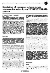
3.1. Total selenium and protein-Se in soybean The presence of added sodium selenite in the manure appeared to provide a positive benefit for the soybeans. Soybeans cultivated in manure containing 50–150 mg/kg sodium selenite showed increased germination rate and growth, compared to those cultivated in the absence of added sodium selenite. However, when the concentration of sodium selenium was increased to 200 mg/kg both germination rate and growth decreased (Table 3). Likewise, Lintschinger has previously shown that high concentration of selenium in the soil can result in lower germination rate and growth for the selenium-enriched sprouts [24]. Total selenium in the soybean also increased across the different concentrations of sodium selenite used, reaching 1118.0 μg/kg at the maximum concentration of sodium selenite used (Table 4). Thus it appeared that the absorption of selenium by soybean still increased at the concentration (200 mg/kg) that already started to inhibit its germination rate and growth. The presence of inorganic selenium in the manure also increased the incorporation of selenium into protein. Compared to the control, the selenium concentration obtained for the soluble protein fraction of soybean cultivated in manure containing 50–200 mg/kg clearly increased, reaching a maximum of 447.2 μg/kg, but the proportion of proteinconjugated selenium to total selenium decreased from 53.2% for the control to 39% for soybean cultivated in the presence of 200 mg/kg sodium selenite (Table 4). This indicated that as the concentration of inorganic selenium in the soil was increased, it not only stimulated the incorporation of selenium into selenoproteins, but also increased the accumulation of non protein-conjugated selenium. 3.2. Optimization of HPLC/MS parameters In order to optimize the parameters for mass spectrometry, solution containing 30% water/70% acetonitrile (v/v) and 0.1% formic acids was used as the mobile phase in the positive ion mode. Capillary, fragmenting voltage and collision energy were optimized by injecting each standard solution into the mass spectrometer to obtain the characteristic MRM ion pairs that could be used as the basis for quantitative analysis. MRM mode was determined by selecting the precursor ion m/z and one of its daughter ion m/z with high specificity and sensitivity. A capillary of 3800 V was optimal. Optimal fragmenting voltage and collision energies varied for each selenoamino acid (Table 5). A fragmenting voltage that is too low would lead to incomplete desolvation, and one that is too high would result in more product ions before the sample enter the first quadrupole, producing less intense [M + H]+ signals in the MS spectrum, and lower signals for the product ions in MS–MS spectrum. Se-MeSeCys and Se-Met were investigated through intense [M + H] + signals to obtain optimal fragmenting voltage. The precursor ion of Se-MeSeCys was measured at m/z of 184 and Se-Met at m/z of 198. The ions produced from Se-MeSeCys and Se-Met are shown in Fig. 1. For Se-MeSeCys, the product ions were found at m/z 167: loss of NH3; m/z 149: loss of NH3 and H2O; m/z 139: loss of NH3 and CO; and m/z Table 3 Germination rate and output (dry weight) of soybean cultivated in the presence of different concentrations of sodium selenium (sodium selenite) in solution. Selenium concentration in solutions (mg/kg)
Germination rate (%)
Output (kg, dw)
0 50 100 150 200
39.8 45.1 56.7 57.9 31.3
1.9 2.2 2.75 3.55 1.73
Selenium concentration in solutions (mg/kg)
Total-Se (μg/kg)
Protein-Se (μg/kg)
0 50 100 150 200
123.3 660.7 778.1 885.9 1118.0
65.4 202.3 280.1 390.5 447.2
73: loss of NH3 and SeCH+ 3 . For Se-Met, four significant product ions were found at m/z: 181, loss of NH3; m/z 153: loss of CO and H2O; m/z 135: loss of CO, NH3 and H2O; and m/z 109: corresponding to the ion CH3SeCH+ 2 . By comparing the two selenoamino acids, it was clear that Se-MeSeCys (m/z 184) and Se-Met (m/z 198) were characterized by typical fragments produced after collision induced dissociation. The two [M + H]+ species each lost an ammonia (m/z 17) that corresponded to the [M + H–NH3]+ species. Identification of the standards (Se-MeSeCys and Se-Met) was performed in order to determine the retention times of the compounds of interest in the chromatogram (Fig. 2). Description of the structure of the main ions is given in Table 6. 3.3. Limit of detection and quantification The characteristic MRM ion pairs 184–167 (Se-MeSeCys) and 198– 181 (Se-Met) were selected to determine the linear range and detection and quantification limits of the method. In order to check the linearity of the standards, solutions of Se-MeSeCys and Se-Met were prepared with concentrations ranging from 0 to 100 μg/L. The detection limit for each compound was calculated as three times the noise of the base line, while the quantification limit was calculated as ten times the noise of the base line. The observed value of the relative standard deviation (RSD) of precision for each compound was calculated from the analysis of 11 replicates at the 100 μg/L level. The result of the above analytical performance is given in Table 7. According to the data, the method had a broad linear range, with high sensitivity and good precision. 3.4. Identification and quantification of selenium-conjugated soluble proteins HPLC–MS was applied to study the distribution and accumulation of selenium in the soybean. Formic acid (0.1%) was added to the mobile phase to enhance the formation of ion. The identification of selenoamino acids was performed by comparing the MS spectra of the sample with those of standards at the corresponding retention times. Fig. 3 shows the chromatograms and precursor ion mass spectra of soybean extract. The two peaks corresponding to tR = Se-MeSeCys and tR = Se-Met. The two individual retention times were assigned a 70-V fragmenting voltage (Se-MeSeCys) and a 50-V fragmenting voltage (Se-Met). The mass spectra of the precursor ions revealed m/z values of 184 in the case of Se-MeSeCys, and 198 in the case of Se-Met, indicating that the two compounds could be Se-MeSeCys and Se-Met. All species were monitored by selecting the MRM mode. A significant increase in the content of selenoamino acid was observed for soybeans cultivated in the presence of added selenium compared to the control. For Se-MeSeCys, the content of
Table 5 Fragmenting voltage and collision energies used in MS/MS mode to give optimum fragmentation of precursor ions into product ions. Compounds
Precursor ion
Product ion
Fragmenting voltage [V]
Collision energy [V]
Se-MeSeCys Se-Met
184 198
167 181
70 50
20 10
M. Tie et al. / Microchemical Journal 123 (2015) 70–75
73
Fig. 1. Product ions of Se-MeSeCys and Se-Met.
Fig. 2. Chromatograms of Se-MeSeCys and Se-Met standard solution (100 μg/L).
Table 6 MS/MS fragmentation pattern of organic selenium species. Compounds Se-MeSeCys
Se-Met
Precursor ion
Product ion
selenium increased with increasing concentrations of sodium selenite added to the soil, peaking at 592.2 μg/kg when the concentration in the manure was 150 mg/kg. Above 150 mg/kg, the content of selenium in the soybean extract started to decrease. Se-MeSeCys therefore accounted for 66.4% of the selenium in Se-protein and 29.2% of the total selenium in the soybean. However, the proportion of Se-Met in the control soybean was not determined, but the content of Se-Met in soybean cultivated in the presence of selenium increased linearly with the concentration of selenium in the manure (Fig. 4), and it accounted for 5.8% of the selenium in Se-protein and 2.4% of the total selenium in the soybean. The most obvious result obtained in the present study was the much higher level of Se-MeSeCys compared to Se-Met (Table 8). This result was different from those reported in some other studies. Maseko found that SeCys is the main selenoamino acid in Agaricus bisporus when it is grown in compost enriched with sodium selenite [5].
74
M. Tie et al. / Microchemical Journal 123 (2015) 70–75
Table 7 Limits of detection and quantification for selenium compounds. Compound
Correlation
LOD (μg/L)
LOQ (μg/L)
Precision (%)
Se-MeSeCys Se-Met
0.9993 0.9998
0.25 0.5
0.9 1.7
1.7 1.4
Maneetong demonstrated that the concentration of Se-Met is higher than that of other forms of organic selenium in selenium-enriched kale [11]. Our results appeared to contrast the results reported by other investigators [14,25], and the reason might be due to the different species of plants and extraction methods used, as well as the source of selenium and cultivation conditions.
Fig. 3. Chromatograms and mass spectra for selenoamino acids of selenium-enriched soybean (100 mg/kg solution) after enzymatic hydrolysis.
Fig. 4. MRM chromatograms of Se-MeSeCys and Se-Met for selenium-enriched soybeans.
M. Tie et al. / Microchemical Journal 123 (2015) 70–75 Table 8 Results of the speciation analysis of soybean. Se concentration in manure (mg/kg)
0 50 100 150 200
Selenoamino acids concentration in soybeans (μg/kg) Se-MeSeCys
Se-Met
57.0 258.5 373.3 592.2 472.6
– 29.8 38.4 42.1 64.8
4. Conclusions A HPLC–ESI–MS-based method for the speciation of selenium in soybean was described. For Se-MeSeCys and Se-Met, the fragmenting voltage and collision energy were optimized. All species were monitored by the selected MRM mode in which the precursor ions and product ions were selected: MRM of ion pairs with m/z values of 184–167 were characteristic of Se-MeSeCys while those with m/z values of 198–181 were characteristic of Se-Met. The result showed that Se-MeSeCys was the main selenoamino acid in the soluble fraction of soybean protein, and accounted for 66.4% of the selenium in Se-protein. Se-Met was detected at a much lower concentration than Se-MeSeCys. Thus, soybean could be a valuable dietary source of Se-MeSeCys, the species associated with cancer-preventive properties. Further study is in progress to study the speciation of organic selenium using other extraction and purification methods. Acknowledgments This work was supported by Liaoning Scientific Research Fund 2011205001. The authors thank Dr Alan K Chang (Liaoning University) for his valuable contribution to the writing of the manuscript. References [1] Y. Mehdi, J.-L. Hornick, L. Istasse, I. Dufrasne, Selenium in the environment, metabolism and involvement in body functions, Molecules 18 (2013) 3292–3311. [2] H. Zeng, J.J. Cao, G.F. Combs, Selenium in bone health: roles in antioxidant protection and cell proliferation, Nutrients 5 (2013) 97–110. [3] D. Umysová, M. Vítová, I. Doušková, K. Bišová, M. Hlavová, M. Čížková, J. Machát, J. Doucha, V. Zachleder, Bioaccumulation and toxicity of selenium compounds in the green alga Scenedesmus quadricauda, BMC Plant Biol. 9 (2009) 58. [4] B. Guerrero, M. Llugany, O. Palacios, M. Valiente, Dual effects of different selenium species on wheat, Plant Physiol. Biochem. 83 (2014) 300–307. [5] T. Maseko, D.L. Callahan, F.R. Dunshea, A. Doronila, S.D. Kolev, K. Ng, Chemical characterisation and speciation of organic selenium in cultivated selenium-enriched Agaricus bisporus, Food Chem. 141 (2013) 3681–3687. [6] M. Kieliszek, S. Błażejak, Selenium: significance, and outlook for supplementation, Nutrition 29 (2013) 713–718.
75
[7] A. Kouba, J. Velíšek, A. Stará, J. Masojídek, P. Kozák, Supplementation with sodium selenite and selenium-enriched microalgae biomass show varying effects on blood enzymes activities, antioxidant response, and accumulation in common barbel (Barbus barbus), Biomed. Res. Int. 2014 (2014). [8] S.J. Fairweather-Tait, Y. Bao, M.R. Broadley, R. Collings, D. Ford, J.E. Hesketh, R. Hurst, Selenium in human health and disease, Antioxid. Redox Signal. 14 (2011) 1337–1383. [9] C. Thiry, A. Ruttens, L. De Temmerman, Y.-J. Schneider, L. Pussemier, Current knowledge in species-related bioavailability of selenium in food, Food Chem. 130 (2012) 767–784. [10] S.O. Knowles, N.D. Grace, Parenteral selenomethionine for production of seleniumrich foods, Google Patents, 2014. [11] S. Maneetong, S. Chookhampaeng, A. Chantiratikul, O. Chinrasri, W. Thosaikham, R. Sittipout, P. Chantiratikul, Hydroponic cultivation of selenium-enriched kale (Brassica oleracea var. alboglabra L.) seedling and speciation of selenium with HPLC–ICP-MS, Microchem. J. 108 (2013) 87–91. [12] J.E. Spallholz, B.J. Shriver, T.W. Reid, Dimethyldiselenide and methylseleninic acid generate superoxide in an in vitro chemiluminescence assay in the presence of glutathione: implications for the anticarcinogenic activity of L-selenomethionine and L-Se-methylselenocysteine, Nutr. Cancer 40 (2001) 34–41. [13] D. Medina, H. Thompson, H. Ganther, C. Ip, Se-methylselenocysteine: a new compound for chemoprevention of breast cancer, Nutr. Cancer 40 (2001) 12–17. [14] K. Wróbel, K. Wróbel, S.S. Kannamkumarath, J.A. Caruso, I.A. Wysocka, E. Bulska, J. Świa̧tek, M. Wierzbicka, HPLC–ICP-MS speciation of selenium in enriched onion leaves—a potential dietary source of Se-methylselenocysteine, Food Chem. 86 (2004) 617–623. [15] F. Gosetti, P. Frascarolo, S. Polati, C. Medana, V. Gianotti, P. Palma, R. Aigotti, C. Baiocchi, M. Gennaro, Speciation of selenium in diet supplements by HPLC–MS/ MS methods, Food Chem. 105 (2007) 1738–1747. [16] M. Tie, B. Li, Y. Liu, J. Han, T. Sun, H. Li, HPLC–ICP-MS analysis of selenium speciation in selenium-enriched Cordyceps militaris, RSC Adv. 4 (2014) 62071–62075. [17] J. Zembrzuska, H. Matusiewicz, H. Polkowska-Motrenko, E. Chajduk, Simultaneous quantitation and identification of organic and inorganic selenium in diet supplements by liquid chromatography with tandem mass spectrometry, Food Chem. 142 (2014) 178–187. [18] B. Chen, M. He, X. Mao, R. Cui, D. Pang, B. Hu, Ionic liquids improved reversed-phase HPLC on-line coupled with ICP-MS for selenium speciation, Talanta 83 (2011) 724–731. [19] M. Roman, Development and Applications of New Analytical Methodologies Based on HPLC–ICP-MS for Trace Speciation Analysis of Selenium in Biological Samples, 2011. [20] W. Thosaikham, K. Jitmanee, R. Sittipout, S. Maneetong, A. Chantiratikul, P. Chantiratikul, Evaluation of selenium species in selenium-enriched pakchoi (Brassica chinensis Jusl var parachinensis (Bailey) Tsen & Lee) using mixed ion-pair reversed phase HPLC–ICP-MS, Food Chem. 145 (2014) 736–742. [21] M. Tie, Y.-z. Fang, T.-b. Sun, C. Li, J. Fei, H. Li, S. Zang, Application of HPLC–ICP-MS in speciation analysis of selenium in selenized Flammulina velutipes, Chem. J. Chin. Univ. 28 (2007) 635. [22] Y. Lu, A. Rumpler, K.A. Francesconi, S.A. Pergantis, Quantitative selenium speciation in human urine by using liquid chromatography-electrospray tandem mass spectrometry, Anal. Chim. Acta 731 (2012) 49–59. [23] E. Dumont, Y. Ogra, F. Vanhaecke, K.T. Suzuki, R. Cornelis, Liquid chromatography– mass spectrometry (LC–MS): a powerful combination for selenium speciation in garlic (Allium sativum), Anal. Bioanal. Chem. 384 (2006) 1196–1206. [24] J. Lintschinger, N. Fuchs, J. Moser, D. Kuehnelt, W. Goessler, Selenium-enriched sprouts. A raw material for fortified cereal-based diets, J. Agric. Food Chem. 48 (2000) 5362–5368. [25] M. Kotrebai, M. Birringer, J.F. Tyson, E. Block, P.C. Uden, Selenium speciation in enriched and natural samples by HPLC–ICP-MS and HPLC–ESI–MS with perfluorinated carboxylic acid ion-pairing agents presented at SAC 99, Dublin, Ireland, July 25–30, 1999, Analyst 125 (2000) 71–78.