an Internet portal for browsing/querying online medical databases, and applied .... All actions supported by. ILive are based on the categorization of images into classes ... diagnostic queries, such as looking for tumors in X-ray images. Our goal is to design a ..... Semantic Based Medical Image Indexing and Retrievalâ, Tech.
SEMANTIC BASED CATEGORIZATION, BROWSING AND RETRIEVAL IN MEDICAL IMAGE DATABASES Aleksandra Mojsilovic and Jose Gomes IBM T. J. Watson Research Center, 30 Saw Mill River Road, Hawthorne, NY 10532 ABSTRACT Content-based retrieval (CBIR) methods in medical databases have been designed to support specific tasks, such as retrieval of digital mammograms or 3D MRI images. These methods cannot be transferred to other medical applications since different imaging modalities require different types of processing. To enable content-based queries in diverse collections of medical images, the retrieval system must be familiar with the current image class prior to the query processing. We describe a novel approach for the automatic categorization of medical images according to their modalities. We propose a semantically based set of visual features, their relevance and organization for capturing the semantics of different imaging modalities. The features are used in conjunction with a new categorization metric, enabling “intelligent” annotation, browsing/searching of medical databases. Our algorithm provides basic semantic knowledge about the image, and may serve as a front-end to the domain specific medical image analysis methods. To demonstrate the effectiveness of our approach, we have designed and implemented an Internet portal for browsing/querying online medical databases, and applied it to a large number of images. Our results demonstrate that accurate categorization can be achieved by exploiting the important visual properties of each modality. 1. INTRODUCTION Medical information systems with advanced browsing capabilities play an increasingly important role medical training, research, and diagnostics. So far, the utilization of online medical data was limited by the lack of effective search methods and the text-based searches have been the dominating approach for the medical database management. Since images represent an essential component of the diagnosis, follow-up and research, it is natural to use medical images to support browsing and querying of medical databases. Existing CBIR systems depend on visual attributes, such as color, texture and shape, to classify and search for similar images. This approach provides excellent results when constrained to a single application domain, however, no matter how sophisticated they are, color, texture and shape features alone do not adequately model image semantics and thus have many limitations when applied to broad content image databases. This problem becomes even more apparent when dealing with semantics of medical images. For this reason, CBIR methods in medical applications have been designed to support specific medical tasks, such as retrieval of tumor shapes in mammograms [1], computed tomographies of the lung [2], 3D MRI images in neurology [3]-[5], or pathology [6]. However, these methods are task-specific and cannot be transferred to other medical applications since different imaging modalities require different processing methods. Therefore, to enable content-based queries for research and diagnostic purposes, the information retrieval system must be familiar with the current image class prior to the query processing. Hence the categorization of medical images into different imaging modalities is required to support further queries.
This paper describes methods and a system for automatic categorization of medical images according to their modalities. We propose a semantically based set of visual features, their relevance and organization for capturing the semantics of different imaging modalities. The features are used in conjunction with the categorization metric, to allow for the “intelligent” annotation, browsing and searching of medical databases. Our method provides a basic semantic knowledge about the analyzed images, and may serve as a front-end for detailed, domain specific medical image analysis. 2. SEMANTIC IMAGE CATEGORIZATION Our previous work on image semantic modeling was focused on overcoming the limitations of traditionally used features for image retrieval, understanding the important semantic categories that drive our visual perception, and studying human similarity judgments to extract meaningful, discriminating features of these semantic categories [7]. This work demonstrated that for images that represent a semantically well-defined set, combinations of the low level global and local features, such as color composition, lines, region color, boundaries, shape and texture, frequency information, blob information, etc., can be used to capture image semantics. Given such a set, and given a balanced set of image features, the semantic image categorization problem can be defined through the following two steps. 1) for each semantic category ci , determine a feature vector f (c i ) that discriminates that category from other images, and 2) given the feature vectors for all the categories, categorize image x by finding:
min{ dist ( f ( x | ci ), f (ci )) } i
where: f ( x | ci ) is a feature set for image x, assuming that it belongs to the category ci , and dist (⋅) is some distance metric between the feature vectors f ( x | ci ) and f (c i ) . In this paper we expand this approach and apply it for automatic categorization, browsing and retrieval in medical image databases. Our work is based on the hypothesis that different types of imaging modalities, as well as different classes of medical images within the same modality, represent semantically well-defined sets. Given a medical image database comprising of different imaging modalities (X-ray, MRI, ultrasound, microscopy, photography, medical diagrams, etc.) the goal of our work is to: 1) design versatile global and regional image features as a basis for the semantic descriptors, 2) develop feature combinations that capture the visual characteristics of each modality, 3) implement an image categorization metric, which will enable intelligent browsing and navigation of the image repository. Since images within the same modality represent a semantically well-defined set, image classification and retrieval share the same goal. Therefore, our classification method can be further used as a similarity metric for searching for similar images. To demonstrate the effectiveness of this approach we have designed and implemented an Internet portal for browsing the online medical databases.
3. THE OVERVIEW OF THE SYSTEM
ILive, Interactive Livesciences Visualization and Exploration system is an Internet portal developed as a part of the ongoing project for semantic based image modeling. ILive contains three logical parts that operate independently as follows: 1) A distributed World Wide Web crawler. Numerous ”tiny spiders”', starting independently from arbitrary home nodes, cover the web no further than a certain predefined distance. The metric used by the spiders to measure how far they are from their home node reflects their preference for staying within the same physical server, which limits the depth of the search. Thus, each spider works within a domain around its home node and completes its traversal in minutes on the average WWW client (i.e. any personal computer). The spiders send the URLs of the found images back to the common server. A server receives all the packets and eliminates redundancies. 2) A server back-end. This part computes the image features. The image features, along with their URLs, and the URLs of the document referencing them, are stored in a repository. 3) A server front-end. The front-end of the search engine runs on the HTTP server and searches the repository according to the queries of the users. Users interacts with ILive through a graphical user interface, typical for the Internet search engines. All actions supported by ILive are based on the categorization of images into classes representing different imaging modalities, and their subclasses. Each imaging modality is modeled with a different combination of features. The feature selection and extraction process is further described in Section 4. Images are assigned to classes using the categorization method (described in Section 5). Additional image classes and subclasses can be added at any time. The queries in ILive can be formulated in two ways. The first way is using functions, such as X-rays() or Tissues(). All the functions are listed on the front page in the ILive web directory. The functions implement our automatic image categorization algorithm, to retrieve all the images from the one of the predefined image classes. Another level of interaction can be achieved by combining these functions with the low level features using the Boolean comparison and arithmetic operators. As we will show later, since our features are related to the human perception of semantic content, it is possible to use “natural language” to query a database. The web can be also searched using combinations of functions, features and text keywords. 4. FEATURE SELECTION AND EXTRACTION FOR SEMANTIC CATEGORIZATION OF MEDICAL IMAGES
Even an untrained subject can distinguish between certain imaging modalities, since they have entirely different visual appearance. For example, we make a distinction between an Xray and tissue photograph, due to entirely different color properties. We can also set apart an ultrasound, X-ray, and MRI image, due to substantial difference in their grayscale contrast. Therefore, at the first level of semantics we need global features to support this level of knowledge. However, medical knowledge further arises from anatomical and physiological information. Therefore, at the second level of semantics, regional features are required to support semantic queries. For example, we need regional features to distinguish between different organs and body parts. On the deeper level, these features may be used for diagnostic queries, such as looking for tumors in X-ray images. Our goal is to design a feature set that is linked to human perception of image content. Such a set allows for an efficient categorization metric, as well as intuitive queries. Consequently, the features in our set are linked to some of the semantic attributes we use to describe image appearance. Some examples of these
attributes are “background is black”, “there is one round object in the center”, “image is monochromatic”, “there is a human in the picture”, etc. For each image we therefore extract a set of semantically relevant global and local features through the following processing steps. The input image is first subjected to the following three types of segmentation. The first one is texture segmentation, followed by color segmentation, and foreground/background segmentation. Texture segmentation has two goals. The first goal is to check if the image is uniform in the texture domain (in which case it most likely represents a tissue). The second goal of texture segmentation is to provide a texture map, which will be used later to provide additional information about specific image regions. The original image is then subjected to color segmentation. Each relevant region from the color segmentation is assigned the structure containing the information about its size, boundary, texture (from the texture map), mean color and colorname (for example red, light pink, black, dark gray, etc.). In the third step, the texture and color maps are combined to achieve the foreground/background segmentation, i.e. to determine if there is a dominant object (or objects) in the image. For each relevant object we compute simple shape features (boundary, eccentricity, moments, symmetry features etc.), as well as color and texture properties. In the current implementation, texture map is generated by computing a set of directional edge maps, followed by a region growing procedure. For the color segmentation we use the meanshift algorithm [8]. For each extracted region, the novel color naming procedure is carried by comparing the average region color with the set of standard colors described in the ICCS NBS Color dictionary [9], using the L2 norm in the Lab color space. Another important semantic feature is the presence of humans in the image, i.e. skin. We designed a novel algorithm for detection of skin regions. This is a geometric method where image pixels are interpreted as points in the 4D-Euclidian space. The coordinates of a pixel in this space are the Lab color coordinates and the measure of the color variation. The later is totally encoded in the spatial Jacobian of (L,a,b), but we used only its Euclidean norm n. To build a skin color model in this space, we used “skin pixels” collected from the training set of 260 images. The manifold of the skin color is then reconstructed via the 4D anisotropic diffusion. This diffusion is achieved through the resolution of a dedicated Partial Differential Equation. The skin regions are identified by computing the distance between each relevant region and the skin manifold. All the regional features are then combined to provide global descriptors. These include number of regions, number of blobs, number of regions with specific color, measures of local and global contrast. Furthermore, the color names from all relevant regions are combined into a color name histogram to determine a color appearance of the image. The color name histogram generates descriptions such as grayscale, monochromatic, flesh, pink overtones, graphic colors, etc. In many cases these descriptions only are capable of capturing image semantics. For example, due to bright colors, medical diagrams are often characterized with “graphic colors”. Similarly, certain stainings in histology create “monochromatic appearance” with “pink or purple overtones”, while the photographs of the internal tissues have color composition described as “flesh”. 5. IMAGE CATEGORIZATION AND RESULTS
Initial image categories were established according to the most common medical imaging modalities. These include, X-rays (with different subclasses according to the body parts), MRI, histological stainings (again divided according to the different types of stainings), micrographs, photographs of internal organs,
photographs of internal tissues, etc. For each semantic category we have determined a feature combination that best captures the semantics of that category. The feature selection was conducted on a training set of 200 images. In modeling the semantics of each category, we have followed our previous perceptual findings [7]. Namely, within a certain category, not all the features are equally important. For example, all histological images obtained with the same staining process have the same color composition. This feature is thus considered as required feature for the particular class. On the other hand, most of the images from this category (but not all of them) have uniform texture properties, or a large number of small/medium irregularly distributed blobs. Therefore, texture properties, blob number and blob size are considered as frequently occurring features for this category. According to our model, each semantic category ci, is “uniquely” described by the set of features S ci . Ideally, these features can be used to separate ci from other categories in the set. To test if the input image x belongs to the category ci, we will therefore use only the features that are important for this category. This is done according to the following scheme. The semantics of each category ci, is captured by the following four sets: 1), 2) The set of features, S ci , that are representative for the category, and their values, V ci
S ci
c c = { {RF j i } j =1,...,M , {FOk i }k =1,..., N } i i c
V ci = {V j i } j =1,...,M + N i i
(1) (2)
where: RF and FO are the required and frequently occurring features for the category ci. For the image x, the values involved in testing if x ∈ c i are then c
S ci ( x) = {S ji ( x)} j =1,...,M + N = i i c c { {RF j i ( x)} j =1,...,M , {FOk i ( x)}k =1,..., N } i
(3)
i
3) The set of operators, or functions, O ci , describing how S ci (x) will be compared to V ci . c c c c c Oc = {O ji ( S ci ( x) ji , V j i , P j i ) | O j i ∈ [0,1]} j =1,...,M + N (4) i i i c
Pc = {P j i } j =1,...,M + N i i i
where :
(5)
is the set of parameters involved in the comparison. The comparison is then performed according to the following metric: sim ( x, ci ) = [
1 M i ci c c c [ Π O ( RF j i ( x), V j i , P j i ) ] ⋅ N i j =1 j
Ni + M i
Σ
j = M i +1
(6)
c c c c O j i ( FO ji− M ( x), V j i , P j i ) ] i
sim ( x, c i ) > 0
x ∈ ci .
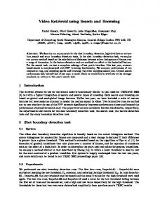
Note that, according to (6), to classify image into a semantic category, all the required and at least one of the frequently occurring features for that category have to be present. Fig. 1 shows images shows a typical screen from ILive, with images classified into “Stainings/Papanicolau” category. The query was performed on a 2000 images “crawled” from the medical databases registered on the WWW. In addition semantic categorization into preselected imaging modalities, ILIve allows users to compose their own queries, by typing in the query window. Since the feature set is semantically based, the users can write queries in the same way they would describe images. For
example, to find X-ray images of skull, the user may compose the following query: texture = no AND composition = grayscale AND
Tcmin < contrast < Tcmax AND number of relevant objects = 1 AND eccentricity < Tcmin AND y-symmetry > Tsymy AND Tomin