Students can view this process and explore the created structures, which helps them in ..... free exploration, but could lead students to unproductive learn- ing [DHB01]. ... circular cross-sections, and provide an on-the-fly reconstruction of the vessel surface ... and motion parallax, the virtual ray allows an accurate definition of.
Semi-Immersive 3D Sketching of Vascular Structures for Medical Education Patrick Saalfeld, Aleksandar Stojnic, Bernhard Preim, Steffen Oeltze-Jafra
The definite version of this article is available at http://diglib.eg.org/.
To cite this version: Patrick Saalfeld, Aleksandar Stojnic, Bernhard Preim, Steffen Oeltze-Jafra Semi-Immersive 3D Sketching of Vascular Structures for Medical Education Proc. of Eurographics Workshop on Visual Computing for Biology and Medicine (EG VCBM), pp. 123-132, 2016
Semi-Immersive 3D Sketching of Vascular Structures for Medical Education P. Saalfeld, A. Stojnic, B. Preim and S. Oeltze-Jafra Visualization Group, Faculty of Computer Science, University of Magdeburg, Germany
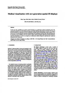
Figure 1: Arbitrarily complex vascular systems can be created by sketching in 3D space. The images show a consecutive sketching of branching vessels with a balloon-like pathology. The created surface model can be enriched with a plausible, real-time blood flow simulation. Abstract We present a semi-immersive 3D User Interface to sketch complex vascular structures and vessel pathologies by drawing centerlines in 3D. Our framework comprises on-the-fly reconstruction of the corresponding vessel surface and subsequent local surface compression and expansion. Additionally, we allow the enrichment with an illustrative, plausible blood flow visualization. Our framework is designed for medical educators and students to support anatomy and pathology education. Anatomy educators can realize the step-by-step process of creating and explaining complex spatial relationships of interlinked vascular structures and blood flow behavior. Students can view this process and explore the created structures, which helps them in reproducing and memorizing them. To create a surface model based on the sketched centerlines, we employ implicit surfaces. This allows for easy adding, editing, and removing vessel branches and achieve continuous surfaces with smooth transitions at branchings. The blood flow can be interactively added and is realized with a topology-aware particle simulation. We qualitatively evaluated our framework and demonstrate the applicability and usability of our approach. Categories and Subject Descriptors (according to ACM CCS): I.3.7 [Computer Graphics]: Three-Dimensional Graphics and Realism—Virtual reality H.1.2 [GENERAL]: User/Machine—Human factors
1. Introduction The anatomy and pathology curriculum is a key part in the education of medical students. They need a comprehensive knowledge to perform future tasks as a physician [DHB01]. An important subject of medical education is the vascular system and its diseases, which is reasoned by the high amount of deaths associated with them. Cardiovascular diseases are responsible for 17.5 million deaths world-
wide [MPN∗ 11], i.e., every third person’s death, which makes them a key threat to maintain health in the twenty-first century [RO15]. Education, however, is challenging for the students and educators as well [PCP10]. Students need a comprehensive understanding of spatial relationships, shapes, and names of medical structures, and educators need appropriate tools to convey these. One way is using sketches drawn by educators, e.g., on chalkboards.
P. Saalfeld et al. / Semi-Immersive 3D Sketching of Vascular Structures for Medical Education
Here, complex vascular systems are drawn by incrementally adding new structures and explaining their functionality. Thereby, students learn successively the functionality of small parts towards a broader understanding of the whole system. In parallel, they are educated to reproduce these sketches. This common step-by-step approach is helpful to convey medical knowledge [PCP10]. However, 2D sketches have limitations. Besides results depending on artistic talent, it is challenging to represent 3D shapes and spatial relationships. Furthermore, a sketched image only conveys one viewing direction and may hide important structures.
flowing through it. A stenosis could lead to an undersupply of surrounding structures. Besides the narrowing of the vessel, a bulging called aneurysm may occur. The bulge itself could press surrounding structures, which is especially critical in the brain. Additionally, the bulged vessel can rupture, which leads to a bleeding into the surrounding tissue. In the brain, these critical incidents have a mortality rate of 40-60% [BCB∗ 94].
Therefore, we present a framework to sketch vessels and pathologies as 3D surface models. This introduces the challenge of depth-aware sketching. We tackle this by using a 3D User Interface (3DUI), i.e., the zSpace. This semi-immersive system consists of a stereoscopic display with head tracking. For interaction, a six degrees of freedom (6DoF) stylus is used, which lets users define the centerline of the vascular structures. These technical components allow for an easy, natural sketching and viewing in a semiimmersive environment. To create the actual vessel geometry, we use implicit surfaces. This enables the user to create editable complex vasculature with smooth transitions at branchings in real time. Additionally, the sketched vascular system can be enriched with an illustrative, plausible blood flow visualization, which is realized with a particle system. The blood flow adapts dynamically to topological changes by interpreting the vascular system as a graph. The qualitative evaluation of our framework with computer scientists shows the applicability and usability of our approach as well as the suitability of the zSpace. Our work contributes to the field of medical education by combining methods from sketch-based modeling and 3DUIs to create 3D vascular systems.
The following description is based on discussions with our clinical partners. Medical students learn about the vascular system in a variety of courses and media along their academic studies. In lectures and seminars, educators present vascular systems through sketches on blackboards, images, and videos. In parallel, students acquire medical knowledge with anatomic atlases and textbooks. Here, an integration of computer-assisted methods is useful. Besides lectures and seminars, preparation courses are given in which students are presented a body donor. Here, their task is to uncover tissue, vessels and organs. During this process, educators explain general anatomic knowledge accompanied by peculiarities and diseases of the body donor and their cause.
The paper is organized as follows: the next section gives a brief overview of the medical and mathematical background comprising an introduction to the cardiovascular system and vascular pathologies, the medical curriculum with a focus on blood vessels, and implicit surfaces. In Section 3, related work in the domains of implicit surfaces for vascular modeling, sketching in immersive environments, and computer-aided medical education is presented. Section 4 describes our semi-immersive 3D sketching approach from a technical and user interaction point of view motivating decisions based on medical education goals. Our evaluation is summarized in Section 5 and Section 6 concludes the paper. 2. Medical and Mathematical Background First, the medical knowledge about the vascular system including its physiology, pathologies and blood flow is summarized. Then, the medical curriculum and implicit surfaces are introduced. 2.1. Cardiovascular System The cardiovascular system comprises arteries, which transport blood from the heart to peripheral capillaries of the body. Veins, on the other hand, carry back blood from tissue parts [She12]. A common disease of arteries is arteriosclerosis, which changes the vessel morphology by depositing fat, thrombi and lime inside the vessel wall [KBY13]. This could result in a narrowing or occlusion of the vessel (stenosis) which has a critical impact on blood
2.2. Anatomy Education of the Vascular System
An example of a specific course is the gross anatomy lecture, where the foundation about general physiology of all body parts is imparted. The vascular system is described from its central part, the heart, to the peripheral capillaries of the body. Clinical reference is demonstrated by describing possible diseases, patients as well as examination methods. During the first years, the clinical reference is mainly added to motivate the students in learning. In the later years, dedicated courses are offered with topics on medical conditions, their reasons, and diagnosis. Thus, to support medical students and educators in medical education, our framework should fulfill the following two requirements: (1) allow them to illustrate a wide variety of vascular configurations in different body areas and (2) illustrate the most common vascular diseases, i.e., stenoses and aneurysms. 2.3. Implicit Surfaces in a Nutshell An implicit surface is defined in Euclidean space as the zero set of an implicit equation F(x, y, z) = 0. For instance, all points p(x, y, z) satisfying the equation x2 + y2 + z2 − r2 = 0 are on the surface of a sphere with radius r. Implicit surfaces have been introduced to computer graphics as Blobby Molecules for visualizing electron density fields of hydrogen atoms [Bli82]. There, each atom (“blob”) is associated with a scalar field function — a three-dimensional Gaussian function modeling the electron density, which falls off from the atom’s center into the vicinity. An implicit surface is constructed based on an isovalue representing the density of interest. In overlapping scalar fields of multiple atoms, surface construction yields a smooth, organic looking blending of the atoms’ individual surfaces. Since construction based on the infinitively defined Gaussian function requires a consideration of all atoms at each point in space, bounded field functions were soon introduced under the concepts of Metaballs [NHK∗ 85] and Soft Objects [WMW86]. For a point p in
P. Saalfeld et al. / Semi-Immersive 3D Sketching of Vascular Structures for Medical Education
space, the field function F(p) for Metaballs is defined as: � � �2 � d w 1 − 3 : 0 ≤ d ≤ 3r r � �2 F(p) = 3w d : 3r ≤ d ≤ r 2 1− r 0 :r≤d
(1)
with d being the distance from p to the center of the Metaball, w being the weight of the Metaball, and r being its radius of influence. In our application, all Metaballs contribute equally and thus, w equals 1. The contribution of multiple Metaballs M at p is computed as the sum of their individual function values Fi (p). The implicit surface is then defined based on an isovalue Iso as the zero set of:
∑
Fi (p) − Iso = 0
(2)
mi ∈M
The definition of scalar field functions was later extended by J. Bloomenthal and K. Shoemaker from points to line segments, planar curves, and polygons based on a convolution of these primitives with a three-dimensional Gaussian filter [BS91]. This extension paved the way for skeleton-based surface modeling and is referred to as convolution surfaces. While it matches our centerline-based sketching approach, we decided to distribute Metaballs along the centerline, using an existing implementation. This allowed us to focus early on interaction techniques and sketching aspects. 3. Related Work In this section, we survey work on sketching in immersive environments, computer-aided education, and implicit surfaces for vascular modeling. 3.1. Sketching in Immersive Environments With sketch-based interfaces (SBIs), it is possible to visualize and communicate ideas without the necessity to draw accurately [JS11]. The advantages of sketching as a reality-based interaction are demonstrated by several works, e.g., Xu et al. [XLJA02] and Naya et al. [NCAC07]. They show that users prefer sketching over classical desktop metaphors, such as WIMP-based interaction (Windows, Icon, Menu, Pointer), and are more efficient with it. In medicine, sketches are employed for a wide range of applications. For example, they are used as an intuitive interaction technique for medical reports [RS06] or to segment structures in 2D image data [HMTH13]. In clinical practice, sketching is also an easy supplement for patient education or the interchange with the medical staff. Sketch-based interfaces for modeling (SBIMs) use drawings and transform them into 3D branching vessels [PCP10] or 2D vascular structures [SBP∗ 15]. Apart from that, sketches in immersive environments can be fully immersive, e.g., with a head-mounted display (HMD). Here, users benefit from depth cues conveyed by stereopsis and binocular disparity and, additionally, from immersion. 6DoF input devices allow the user to sketch directly in 3D space. An example of an artistic application is Lift-Off [JK16]. Here, artists import 2D sketches
and interactively create 2D curves which are lifted into 3D space. In contrast to full immersion, semi-immersive environments constrain stereopsis and binocular disparity to the frontal visual field, e.g., a computer display. This so-called fish tank VR [WAB93] is used in this paper and has the same spatial benefits regarding depth cues. An example for semi-immersive environments used in medical applications is described by Saalfeld et al. [SGP15]. Here, 3D free-form sketches are used to annotate existing vascular structures, e.g., to illustrate treatment methods and plan therapies. To create vascular systems, two major approaches would be possible: sketching the boundaries of vessels or their centerline. Systems, such as Teddy [IMT99], use the former approach. This gives the user higher freedom of the resulting shape, but is also more time-intensive and complicated. Other systems to sketch plants [SLSS06] use a centerline approach. They are not as flexible, but allow a fast creation of structures. Using the boundary approach to sketch vessels, the user would mostly draw two parallel lines, since vessels have a tubular shape. If an educator draws inaccurately, the resulting vessel diameter would be uneven and could be misinterpreted by the students as a pathology. Therefore, we choose the centerline approach which is faster, prevents unequal diameter variations and is therefore easier to handle for the user. 3.2. Computer-Aided Medical Education Software has been supporting medical education for a long time. Already in 1967, Starkweather described benefits due to computer assistance, which helps students explore clinical problems at their own pace [Sta67]. In a literature study, Letterie [Let03] compared articles from 1988 to 2000 dealing with computer-assisted learning in a wide variety of medical fields. The majority of analyzed articles (96%) describe a positive impact of computer-assisted instructions. The used media comprise online technologies, multimedia work stations, but also VR. Computers can be integrated differently into an existing curriculum by augmenting, enhancing, or replacing traditional teaching methods [DHB01]. Our developed framework augments the curriculum of vascular system education by representing vessels spatially in a more tangible way. The form of computer assistance ranges from very structured, with e.g., digital flashcard systems, to open exploratory approaches. There is a trade-off between these approaches. The structured approach is beneficial for teaching facts but does not allow students to detour from planned learning paths. On the other hand, exploratory systems, such as medical 3D puzzles [RPDS00] or tools to elaborate vessel variations [SKJ∗ 16], encourage an independent free exploration, but could lead students to unproductive learning [DHB01]. Therefore, consideration of students, educators and their needs is important. An example for a commercially available system for computer-aided education is VOXEL-MAN (Hamburg, Germany). Different versions can be used, e.g., as a digital anatomical learning atlas [GHL∗ 06] or as a surgery trainer [TPH∗ 10]. 3.3. Implicit Surfaces for Vascular Modeling Vascular modeling techniques have been surveyed and classified into explicit and implicit with respect to the type of geometric re-
P. Saalfeld et al. / Semi-Immersive 3D Sketching of Vascular Structures for Medical Education
construction [Hon14]. Explicit modeling requires a careful, casespecific connection of sets of polygons or parametric patches to ensure smooth surface transitions at branchings. Implicit modeling achieves smoothness without specific constructional effort by blending the surfaces of individual branches. Since these surfaces describe a volume, no inner polygons are created, benefiting a semitransparent display and virtual angioscopy applications [HLT11]. Oeltze et al. [OP05] applied convolution surfaces [BS91] for modeling vascular structures represented by a vessel skeleton and associated circular cross-sections. Specific emphasis was put on faithfully representing the local radius information, preventing blending artifacts, and speed of the surface reconstruction. Schumann et al. [SOB∗ 07] employed multi-level partition unity (MPU) implicits [OBA∗ 05] for reconstructing the vascular surface from a segmentation mask, thereby facilitating arbitrarily-shaped crosssections. In follow-up work, Schumann et al. [SNB∗ 08] focused on an application of their method in Computation Fluid Dynamics (CFD) simulations of blood flow. They presented a post-processing step enhancing the triangle quality of the surface mesh and reducing the number of triangles. Wu et al. [WWL∗ 10] avoided such postprocessing by a curvature-dependent, quality-aware reconstruction of the vascular surface yielding less triangles in straight regions. Similar to Schumann et al. [SOB∗ 07], they generated the surface based on the boundary voxels of a segmentation mask. In followup work, they quantitatively compared both approaches regarding, e.g., triangle quality, number and surface smoothness [WHM13]. Hong et al. [HLT12] presented an alternative method for generating high-quality vascular surface meshes based on boundary voxel interpolation by swept bivariate piecewise algebraic splines and polynomial blending at ramifications. At about the same time, Kretschmer et al. [KBT∗ 12] proposed the connection of interpolating free-form contours along the centerline and the combination of surface branches by means of Boolean operations. Later, Kretschmer et al. [KGPS13] presented a surface reconstruction technique similar to the one of Hong et al. [HLT12], but significantly faster, facilitating a modification of incorrect vessel segmentations at interactive frame rates. Hong et al. [HLL∗ 15] in turn improved their approach guaranteeing an arbitrary level of geometric continuity as compared to the C1 continuity of the surfaces reconstructed by Kretschmer et al. [KGPS13]. The approach of Yureidini et al. [YKD∗ 12] is closest to ours, since it is based on distributing Blobby Molecules along a given vessel skeleton. Blobs are locally adapted to points on the boundary of a segmentation mask. The resulting local surfaces are stored in a tree for fast contact management in simulated procedures of interventional radiology. In contrast to Yureidini et al., we let the user sketch a vessel skeleton, make the simplifying assumption of circular cross-sections, and provide an on-the-fly reconstruction of the vessel surface during sketching.
Figure 2: The image shows the zSpace 100 used for our framework. The stereoscopic display with head-tracked glasses and 6DoF stylus for input allows for sketching vascular centerlines in 3D.
data is described. Finally, the augmentation of the vascular model with simulated blood flow is presented. 4.1. 3D User Interface In the following, our 3DUI according to the classification of Bowman et al. [BKLP04] is described, comprising input and output device, as well as the general interaction technique. The semiimmersive sketching is implemented on the 3DUI zSpace 100 (zSpace Inc., San Francisco, U.S.A.). It uses a full HD display with an update rate of 120Hz. The stereoscopic effect is achieved with circular polarized rendered images, which are filtered by glasses worn by the user. These glasses are tracked passively through infrared (IR) markers and allow 6DoF head tracking. This combination of stereopsis (binocular parallax) and head tracking (motion parallax) allows users to spatially explore medical structures. For user input, a stylus with three buttons is used. It is tracked actively by IR light and allows 6DoF input. The interaction technique is based on the ray-based interaction, i.e., the orientation of the stylus is virtually extended into the scene. The visible virtual ray is constrained to a specific length. Together with the binocular and motion parallax, the virtual ray allows an accurate definition of 3D points and, thus, sketching 3D lines. Furthermore, the user can translate and rotate the whole vascular structure with a dedicated stylus button. After pointing the stylus to the vessel, the movement and rotation delta of the physical stylus is transformed into the 3D
4. Semi-Immersive 3D Sketching of Vascular Structures This section introduces our 3DUI for semi-immersive sketching, and elaborates on the centerline drawing and the real-time vessel surface reconstruction. Furthermore, the possibility to explore computer-generated vascular surfaces derived from real patient-
Figure 3: A sketched centerline and the resulting surface model by using raw input data (left) and input processed with resampling and smoothing (right) is depicted.
P. Saalfeld et al. / Semi-Immersive 3D Sketching of Vascular Structures for Medical Education
(a)
(b)
(c)
(d)
(e)
Figure 4: The images show different possibilities to create and edit centerlines. In (a), a centerline is sketched. (b-c) shows the oversketching functionality to correct existing lines. (d) depicts a branching of centerlines by sketching on existing lines. Here, the user is supported by snapping. In (e), the deletion is illustrated. The user erases existing centerlines by sketching over them with an eraser metaphor.
environment and applied to the surface mesh. The zSpace and stylus are shown in Figure 2. Our framework controlling input and output, as well as providing a graphical user interface (GUI) was developed with the game engine Unity (Unity Technologies, San Francisco, USA) 4.2. Sketching Vascular Systems 4.2.1. Creating and Editing Center Lines The zSpace provides 3D positions of the tracked stylus. These points result from sampling the 3D movement of the virtually extended stylus and represent the basis for our centerlines. Due to tracking inaccuracies and hand tremble, these points are irregularly sampled and noisy. However, our surface reconstruction requires points with equal distance. Therefore, the points are resampled first and secondly smoothed by a Gaussian kernel (Fig. 3). An overview of our interaction techniques to generate centerlines is depicted in Figure 4. With the resampled and smoothed input data, centerlines can be sketched and edited in the following ways. Create. The centerline is sketched directly and a uniform vessel surface is created around it in real-time. The creation of a centerline starts by pressing a button on the stylus. Then, the user sketches the line by moving the stylus in 3D space (Fig. 4a). Each sample point is stored as a control point. The control points are connected to create the line. Edit. The created line can be edited by two techniques: oversketching and erasing, both triggered with dedicated buttons on the stylus. To oversketch an existing line, the user has to start and finish drawing at a control point, which is supported by snapping. After finishing, the old line is replaced with the new one (Fig. 4b). For deleting line segments, an eraser metaphor is implemented. Around the stylus tip, a sphere is created and every control point which touches this sphere is deleted (Fig. 4e). The sphere has a smaller diameter than the distance between two control points to allow the user to delete precisely. Ramify. To build complex vessel trees with bifurcations or trifurcations, new lines can be attached to old ones. The user starts sketching on an existing control point and is supported by snapping (Fig. 4d). Hereby, not only trees can be created, but also cycles, which is important to create systems such as the Circle of Willis. 4.2.2. Surface Creation with Metaballs and Marching Cubes While sketching the centerline, the vessel surface is created around it in real-time. To obtain a continuous surface with smooth
branches, we use implicit surfaces, i.e., Metaballs (Eq. 1). In our framework, the implementation Skinned Metaballs [Met] is used. The implicit surface is created out of a list of Metaballs, where each Metaball can be defined by a 3D position and its radius of influence r. We create a Metaball for every control point of our centerline with a fixed scalar for its radius. If control points are close enough, the Metaballs blend into each other, forming a vascular structure with smooth surface transitions at branchings. Then, the 3D space is discretized to a grid of cells. The necessary distance between control points, the grid size, and the uniform radius r were empirically determined such that a smooth surface without gaps, dents, and bulges was generated. For every cell influenced by Metaballs, the value F(p) is calculated (recall Eq. 1 and Eq. 2). To polygonize the grid, the Marching Cubes algorithm of the Skinned Metaballs [Met] implementation was applied. The quality of the resulting mesh and the computational complexity depend on the amount of Metaballs as well as the cell width. Since new Metaballs are generated with every new control point, the cell size has to be low enough to ensure real-time performance even for large vascular structures. Figure 5 shows different settings for the cell sizes, the resulting triangle count, and the necessary time to calculate the surface model. We decided to use a medium surface quality to ensure real-time performance. However, the user can set a low cell size and thus, achieve a high mesh quality. This reduces the render time to a non-interactive frame rate during sketching, but the vessel can still be inspected at interactive frame rates. 4.2.3. Creating Stenoses and Aneurysms The illustration of pathologies is important in medical education to understand their implications for vessel morphology changes, their influence on the blood flow behavior, and, finally, possible risks for a patient. An educator can sketch vascular systems parts, which are commonly affected by diseases, and point out differences in their appearance. The most important vascular diseases caused by vessel lumen and wall changes are stenoses and aneurysms. Therefore, we implemented possibilities to create these pathologies. As described before, the initially sketched vessels have a constant diameter. To create a narrowed vessel part representing a stenosis, this diameter has to be decreased, which can be realized by reducing the size of the underlying Metaballs. This is achieved by first, selecting control points similar to the eraser metaphor. The user draws over control points, which are then highlighted. Now, the user can push the stylus forward, which decreases the size of the selected Metaballs and thus creates a stenosis (Fig. 6a). With
P. Saalfeld et al. / Semi-Immersive 3D Sketching of Vascular Structures for Medical Education
Figure 7: In our two-pass shader, the back faces and front faces are rendered separately. The amount of how much the front vessel wall exposes the inner vessel wall can be adjusted to occlude (left) or reveal the inner vessel wall (right).
(a)
(a) low (b) medium (c) high
(b)
Grid Size 0.38 0.2 0.05
(c)
Time to Calculate 2ms 15ms 768ms
#Triangles 852 3948 66276
Figure 5: Different cell sizes used to discretize the 3D space on a grid are shown. The calculation time and triangle count are stated as well. For real-time sketching, we use the medium setting.
ergy conservation is used, e.g., less light is reflected than received, specular light minimizes the diffuse amount of light, and a Fresnel effect is added (the surface becomes more reflective at grazing angles). This realism supports depth perception and the understanding of spatial relationships [PBC∗ 16], which are hard to convey with, e.g., chalk board drawings.
4.2.4. Rendering Vessels
The created vascular surface consists of single face geometry. We create a two-pass shader to give the appearance of an inner and outer vessel wall. In the first render pass, only the back faces are sent to the rendering pipeline. Their normals are flipped in the vertex shader to make them visible. After that, the faces are rendered with physically-based shading. In the second render pass, the remaining front faces are rendered with an addition. The dot product between the normals and the viewing direction is calculated. The result represents how much the face normal is pointing to the user. With a higher value, the transparency is increased. The strength of this effect can be adjusted, i.e., the extent to which the outer vessel wall exposes the inner vessel wall and blood flow (Fig. 7).
The models are rendered with physically-based shading [PH10], a shading technique introduced in Unity 5. This technique simulates the behavior of light more realistically. For example, the idea of en-
4.3. Exploration of Real-Patient Data
the same technique, but a pulling the stylus backward, the Metaball size increases, which results in fusiform aneurysms (Fig. 6b). However, many aneurysms have a saccular shape, i.e., are only bulged on one side. For these, a combination of attaching centerlines and adjusting the diameter of the associated Metaballs can be applied (Fig. 6c).
The possibility to import vessel surfaces derived from real-patient data allows users to inspect rare conditions and is faster compared to sketch very complex structures. As a basis for this, we use the file format of MeVisLab (MeVis Medical Solutions AG, Bremen, Germany), a framework with methods for medical image processing and visualization. In MeVisLab, vascular structures of real patient-data can be segmented. The segmentation mask can be skeletonized obtaining a centerline with associated diameter information [SPSP02].
(a)
(b)
(c)
Figure 6: Different pathologies created with our framework are depicted. In (a) a stenosis is shown, (b) depicts a fusiform aneurysm and (c) a saccular aneurysm.
We implement a parser to load this skeleton and transform it to our centerlines. The diameter information is used for the Metaball radius and, thus, allows for loading vascular systems with varying diameter. However, the quality of the resulting vascular system depends on the extracted centerline. If control points of the imported centerline are far apart from each other, gaps along the vessel can occur. If two centerlines run too close to each other, kissing artifacts are possible. An overview of different cases loaded with this approach is shown in Figure 8.
P. Saalfeld et al. / Semi-Immersive 3D Sketching of Vascular Structures for Medical Education
(a)
(a)
(b)
Figure 8: Real-patient data imported into our framework. In (a) the Circle of Willis is visualized. (b) shows a bronchial tree with an adapted rendering style. The surface models are created implicitly with Metaballs after importing a centerline.
4.4. Adding Plausible Blood Flow An illustration of the blood flow, which adapts dynamically to the sketched centerlines, could support students to understand relationships between pathological vessel changes and their influence on the blood flow. There are different possibilities to create a blood flow simulation and visualization. The most accurate but computationally expensive way is a hemodynamic simulation, i.e., reproducing the real-world behavior of blood. Since our goal is to illustrate blood flow in a plausible and informative way, the simulation does not necessarily have to be physically correct. Therefore, we choose a fast and simple approach based on a particle simulation. We employ the vessel surface and centerline for the blood flow creation. The surface is used in combination with a collision detection for each particle. If a particle hits the vessel wall, e.g. at a stenosis, it gets repelled and thus, directed closer to the vessel center. While this approach works well for stenoses, it leads to chaotic behavior in saccular aneurysms. Thus, we take advantage of the centerline and guide the blood flow along this line. To account for branching vessels, we interpret the vascular system as a graph similar to the work of Zhu et al. [ZIH∗ 11]. By traversing the graph, all possible paths are calculated. Now, each particle is assigned to one of these paths. From an interactive point of view, the user places particle emitters on a freely selectable control point. By pressing and holding the stylus button, the emitter is placed. Now while dragging, the user can determine the direction of the blood flow by dragging to a neighboring control point (Fig 9). To delete an existing particle emitter, the user simply starts dragging on the control point again into empty space.
(b)
(c)
Figure 9: The three images give an overview of the interaction to create blood flow. The user starts dragging on a control point. Now, an arrow gets visible which points to the direction of the blood flow. The user has to place the arrow on a neighboring control point, and thus, determine the start position and flow direction of the blood flow.
blood cells, i.e., erythrocytes from our emitter through the vascular system. We use a self-modeled 3D geometry, calculated texture coordinates for the model and applied a normal map to increase realism. Additionally, the 3D geometry of the particles allows us to visualize particle shadows on the inner surface (Fig. 9c). 4.5. Graphical User Interface In our framework, all control elements are part of the 3D world and are represented by geometry. An overview of control elements is depicted in Figure 10. The user can chose one of the three modes create, edit, and blood flow. The modes are activated by pressing virtual buttons with the stylus ray. For each mode, a textual description of the different functionalities of the three stylus buttons is shown. Additionally, an image of the stylus with labeled stylus buttons is shown. The labels contain the functions associated with the stylus buttons. This supports the self-description capability of the user interface and helps in learning control our framework. At the bottom of the user interface, a reset button can be pressed to clear the sketched vascular system and blood flow. Next to the reset button, a toggle button allows the users to switch between two types of mesh quality (recall Section 4.2.2). 5. Evaluation This section comprises our qualitative evaluation including goals, a description of participants, their tasks, and the procedure. The results are analyzed descriptively and ideas for improvement are discussed. 5.1. Goals
Modern approaches for blood flow visualization employ static representations, e.g., by representative integral curves [OLK∗ 14], or dynamically created ones, e.g., by propagating scalar values in a real-time blood flow simulation [SBP∗ 16].
The goal of our evaluation is to assess the usability and usefulness of our semi-immersive sketching approach. This provides the first step towards an integration into the medical curriculum. Therefore, aspects such as suitability for the tasks, error tolerance etc. are assessed using a questionnaire based on usability norm ISO 924111 [Prü97]. However, questions regarding customizability were neglected, since the application does not support it.
Since our framework should support education, we also generate a dynamic visualization showing blood as a moving fluid. For our particles we choose a realistic representation. We populate red
The integration of blood flow and the possibility to inspect patient-specific data were added after the evaluation and thus, were not considered.
4.4.1. Particles
P. Saalfeld et al. / Semi-Immersive 3D Sketching of Vascular Structures for Medical Education
medical students or educators, respectively. To attenuate a bias reasoned by a missing medical background, we first introduced every participant to the field of vascular systems and their pathologies. Secondly, our tasks were described very general, e.g., the description of the task to sketch a trifurcation was illustrated with an image and explained without using medical terms. 5.4. Procedure Starting with an introduction to the medical background, every function of our framework was described and shown. We ensure that each participant understood the medical background and different sketching functions. Then, the task-sheet was given to the participant. During solving the tasks, participants were asked to comment their actions (Think-aloud method [JØR90]). After finishing the tasks, the usability questionnaire was handed out. Figure 10: The user interface of our system is part of the 3D world, e.g., buttons are spatial objects. The user can change the sketching modes by pressing buttons with the virtual ray of the stylus.
5.2. Tasks We designed four tasks to assess the usability of our semiimmersive sketching approach. Here, participants had to create vascular configurations which were shown as a 2D sketch on a sheet of paper. The tasks were designed to represent the sketching of the most common vascular system parts and pathologies. Furthermore, they build up on each other to recreate the scenario of sketching a more complex vascular system. First, participants were asked to sketch a trifurcation. Here, we asked to sketch at least one branch into depth to be able to evaluate the 6DoF input and stereo output. After that, high mesh quality should be activated and the result should be inspected by changing the head position and take advantage of motion parallax. The second task was to activate the edit mode and delete one branch. On the remaining two branches, a fusiform aneurysm and stenosis should be created, respectively. As the third task, a new vessel should be sketched and corrected with the oversketching functionality. As the fourth, most complex task, a saccular aneurysm should be created. This task involves the creation of at least two branches (the second forming the saccular aneurysm), deflate the region around the aneurysm neck and inflating the aneurysm itself.
5.5. Results Observations and Think-aloud method. After the first drawing trials, the semi-immersive 3D sketching of centerlines was clear to all participants. However, participants differed in drawing speed, i.e., some sketched especially branching vessels very carefully. All participants were able to reproduce the general shape of the required vessel configurations. Additionally, the participants were able sketch into depth and thus, could take advantage of the 3DUI components. If participants made errors, they mostly did not use the oversketching or erasing functionality. They usually resetted the whole sketch and redrew completely. Since a mode change is necessary to edit the vessel, it is possible that this was too troublesome for the participants. Alternatively, this could be reasoned by a lack of training, which would allow more easy switching between modes. Therefore we plan to better communicate the functionalities to edit existing sketches. Editing for changing the vessel diameter was appreciated by all participants. One major problem was the functionality to toggle high mesh quality. Although participants used the functionality and were pleased with the improved vascular structure, almost all forgot to toggle it back. This resulted in low frame rates during the following sketching. Since real-time sketching is not possible with the high quality setting, we plan to solve this problem by automatically toggle the mesh quality if the users starts sketching again.
5.3. Participants
One participant stated that a functionality to abort sketching would be helpful or alternatively, a function to redo an executed operation.
We conducted the user study with nine participants (4 female, 5 male). Their age ranged from 18 to 34. All of them are computer scientists and one was involved in the project. Three participants had experience with ray-based interaction similar or equal to the interaction technique used in our framework. Nobody had a medical condition leading to problems perceiving stereoscopic content. Conducting the user study with computer scientists is a limitation regarding generalization and restricts the possibility to transfer the results to medical students and educators. However, a similar experience with 3DUIs can be excepted for computer scientists and
Usability Questionnaire. All questions were answered with a 7point Likert scale. The majority of usability questions were rated positively (Fig. 11). The questions regarding the Error Tolerance had the lowest average rating and a high variance (x = 1.11, σ = 0.45), where x is the average and σ the standard deviation. This matches the results of our observations, since participants often deleted the whole vascular system instead of correcting parts. The usability aspect Suitability for the Task had the highest values (x = 2.22, σ = 0.29), implicating our interaction techniques are appropriate to sketch vascular structures with the semi-immersive approach.
P. Saalfeld et al. / Semi-Immersive 3D Sketching of Vascular Structures for Medical Education
Mean Ratings for Usability
match our centerline-based sketching approach better. Additionally, we like to use our 3D sketching approach to illustrate treatment options for vascular pathologies, which could help understand the impact of these options to blood vessels and blood flow.
3
2 1
Acknowledgments
0 ST
CE
ET
SL
SD
CO
-1 Mean
-2 -3
Figure 11: Averaged results and standard deviations of the adapted usability questionnaire [Prü97]. ST Suitability for the Task, CE Conformity with User Expectations, ET Error Tolerance, SL Suitability for Learning, SD Self-descriptiveness, CO Controllability.
Regarding the overall rating, it can be assumed that semiimmersive sketching of centerlines is a usable possibility to create vascular systems. However, the results were obtained for computer scientist. It can be assumed that they have an affinity to new technology. Therefore, an evaluation with medical students is still necessary. 6. Discussion We presented a semi-immersive 3D sketching approach to create centerlines of vascular systems and its pathologies. The vascular surface is reconstructed on-the-fly with Metaballs, forming a smooth, continuous surface mesh. By allowing for the exploration of real-patient data and enrichment with a plausible blood flow simulation, our framework is able to convey different vascular phenomena and support medical education. Finally, we demonstrated the applicability of our interaction methods with the usability study. The presented framework has several limitations. The sketched vascular system is not tested for plausibility. Therefore, impossible vessel configurations and pathologies can be created, resulting in unrealistic blood flow. However, trying to prevent these errors needs to be done carefully. For example, preventing sketching cycles would render a creation of the Circle of Willis impossible. Another limitation is that the fast and simplistic centerline approach only allows for creating vessels with circular cross sections. A possibility to deform the cross section could show vessels and pathologies more realistically. Our next step is the integration of our approach into the medical curriculum. We collaborate with medical educators and students to define necessary functionalities to use our framework as an authoring system and create learning content. To establish our semi-immersive sketching approach in a lecture environment, the educator and students need to have access to their own zSpace. These need to be synchronized to visualize the same vascular structures on the educator’s and students’ devices. The head-tracking, however, can be decoupled, which allows students to analyze and explore vascular systems from their own perspective. From a technical point of view, we want to improve our reconstruction method by integrating convolution surfaces instead of Metaballs, since they
This work was partially funded by the German Federal Ministry of Economics and Technology under grant number ’ZF4028201BZ5’. References [BCB∗ 94] B EDERSON J. B., C ONNOLLY E. S., BATJER H. H., DACEY R. G., D ION J. E., D IRINGER M. N., D ULDNER J. E., H ARBAUGH R. E., PATEL A. B., ROSENWASSER R. H.: Guidelines for the Management of Aneurysmal Subarachnoid Hemorrhage. Stroke 40, 3 (1994), 994–1025. 2 [BKLP04] B OWMAN D. A., K RUIJFF E., L AV IOLA J. J., P OUPYREV I.: 3D User Interfaces: Theory and Practice. Addison Wesley Longman Publishing Co., Inc., Redwood City, CA, USA, 2004. 4 [Bli82] B LINN J. F.: A generalization of algebraic surface drawing. ACM transactions on graphics (TOG) 1, 3 (1982), 235–256. 2 [BS91] B LOOMENTHAL J., S HOEMAKE K.: Convolution surfaces. SIGGRAPH Comput. Graph. 25, 4 (1991), 251–256. 3, 4 [DHB01] D EV P., H OFFER E. P., BARNETT G. O.: Computers in Medical Education. In Medical informatics. Springer, 2001, pp. 610–637. 1, 3 [GHL∗ 06] G EHRMANN S., H OHNE K. H., L INHART W., P FLESSER B., P OMMERT A., R IEMER M., T IEDE U., W INDOLF J., S CHUMACHER U., RUEGER J. M.: A novel interactive anatomic atlas of the hand. Clin Anat 19, 3 (2006), 258–266. 3 [HLL∗ 15] H ONG Q., L I Y., L I Q., WANG B., YAO J., W U Q., S HE Y.: An implicit skeleton-based method for the geometry reconstruction of vasculatures. The Visual Computer (2015), 1–12. 4 [HLT11] H ONG Q., L I Q., T IAN J.: Virtual angioscopy based on implicit vasculatures. In Computational Science and Its Applications. Springer, 2011, pp. 592–603. 4 [HLT12] H ONG Q., L I Q., T IAN J.: Implicit reconstruction of vasculatures using bivariate piecewise algebraic splines. IEEE Transactions on Medical Imaging 31, 3 (2012), 543–553. 4 [HMTH13] H ECKEL F., M OLTZ J. H., T IETJEN C., H AHN H. K.: Sketch-Based Editing Tools for Tumour Segmentation in 3D Medical Images. Computer Graphics Forum 32, 8 (2013), 144–157. 3 [Hon14] H ONG Q.: A survey on the visualization and reconstruction of vasculatures. In Proc. of SPIE, International Conference on Graphic and Image Processing (2014), pp. 90690F–90690F–5. 4 [IMT99] I GARASHI T., M ATSUOKA S., TANAKA H.: Teddy: A Sketching Interface for 3D Freeform Design. In Proc. of Computer Graphics and Interactive Techniques (1999), ACM Press/Addison-Wesley Publishing Co., pp. 409–416. 3 [JK16] JACKSON B., K EEFE D. F.: Lift-Off: Using Reference Imagery and Freehand Sketching to Create 3D Models in VR. IEEE Trans Vis Comput Graph 22, 4 (2016), 1442–1451. 3 [JØR90] JØRGENSEN A. H.: Thinking-aloud in user interface design: a method promoting cognitive ergonomics. Ergonomics 33, 4 (1990), 501–507. 8 [JS11] J ORGE J., S AMAVATI F.: Sketch-based Interfaces and Modeling. Springer London, 2011. 3 [KBT∗ 12]
K RETSCHMER J., B ECK T., T IETJEN C., P REIM B., S TAM M.: Reliable adaptive modelling of vascular structures with non-circular cross-sections. Computer Graphics Forum 31, 3pt2 (2012), 1055–1064. 4 MINGER
P. Saalfeld et al. / Semi-Immersive 3D Sketching of Vascular Structures for Medical Education [KBY13] K UTIKHIN A., B RUSINA E., Y UZHALIN A. E.: A Hypothesis of Virus-Driven Atherosclerosis. In Viruses and Atherosclerosis, vol. 4 of SpringerBriefs in Immunology. Springer New York, 2013, pp. 1–3. 2 [KGPS13]
K RETSCHMER J., G ODENSCHWAGER C., P REIM B., S TAM MINGER M.: Interactive patient-specific vascular modeling with sweep surfaces. IEEE Transactions on Visualization and Computer Graphics 19, 12 (2013), 2828–2837. 4
[Let03] L ETTERIE G. S.: Medical education as a science: the quality of evidence for computer-assisted instruction. Am. J. Obstet. Gynecol. 188, 3 (2003), 849–853. 3 [Met] M ETABALL I MPLEMENTATION: Skinned metaballs from junk games. http://nkdtr.hatenablog.com/entry/2015/06/ 05/125027, last accessed on 13th August 2016. 5 [MPN∗ 11] M ENDIS S., P USKA P., N ORRVING B., O RGANIZATION W. H., F EDERATION W. H., O RGANIZATION W. S.: Global Atlas on Cardiovascular Disease Prevention and Control. Nonserial Publications Series. World Health Organization in collaboration with the World Heart Federation and the World Stroke Organization, 2011. 1 [NCAC07] NAYA F., C ONTERO M., A LEIXOS N., C OMPANY P.: ParSketch: A Sketch-based Interface for a 2D Parametric Geometry Editor. In Proc. of International Conference on Human-computer Interaction: Interaction Platforms and Techniques (2007), Springer-Verlag, pp. 115– 124. 3 N ISHIMURA H., H IRAI M., K AWAI T., K AWATA T., S HI I., O MURA K.: Object modeling by distribution function and a method of image generation. The Transactions of the Institute of Electronics and Communication Engineers of Japan 68, Part 4 (1985), 718– 725. 2
[NHK∗ 85]
RAKAWA
[OBA∗ 05] O HTAKE Y., B ELYAEV A., A LEXA M., T URK G., S EIDEL H.-P.: Multi-level partition of unity implicits. In SIGGRAPH 2005 Courses (2005), ACM, pp. 463–470. 4 [OLK∗ 14] O ELTZE S., L EHMANN D. J., K UHN A., JANIGA G., T HEISEL H., P REIM B.: Blood Flow Clustering and Applications in Virtual Stenting of Intracranial Aneurysms. IEEE Transactions on Visualization and Computer Graphics (TVCG) 20(5) (2014), 686–701. 7 [OP05] O ELTZE S., P REIM B.: Visualization of Vascular Structures: Method, Validation and Evaluation. IEEE Transactions on Medical Imaging 25(4), 4 (2005), 540–549. 4 P REIM B., BAER A., C UNNINGHAM D., I SENBERG T., ROPINSKI T.: A Survey of Perceptually Motivated 3D Visualization of Medical Image Data. Computer Graphics Forum 35, 3 (2016), 501–525. 6
[PBC∗ 16]
[PCP10] P IHUIT A., C ANI M.-P., PALOMBI O.: Sketch-Based Modeling of Vascular Systems: a First Step Towards Interactive Teaching of Anatomy. In Eurographics Workshop on Sketch-Based Interfaces and Modeling (2010). 1, 2, 3 [PH10] P HARR M., H UMPHREYS G.: Physically Based Rendering, Second Edition: From Theory To Implementation. Morgan Kaufmann Publishers Inc., 2010. 6 [Prü97] P RÜMPER J.: Der Benutzungsfragebogen ISONORM 9241/10: Ergebnisse zur Reliabilität und Validität. Vieweg+Teubner Verlag, 1997, pp. 253–262. 7, 9 [RO15] R EID C., OWEN A.: Handbook of Psychocardiology. Springer Singapore, 2015, ch. Epidemiology of Cardiovascular Disease, pp. 1–20. 1 [RPDS00] R ITTER F., P REIM B., D EUSSEN O., S TROTHOTTE T.: Using a 3D Puzzle as a Metaphor for Learning Spatial Relations. In Graphics Interface (2000), pp. 171–178. 3 [RS06] R EINER B., S IEGEL E.: Radiology Reporting: Returning to Our Image-Centric Roots. American Journal of Roentgenology 187 (2006), 1151–1155. 3 [SBP∗ 15] S AALFELD P., BAER A., P REIM U., P REIM B., L AWONN K.: Sketching 2D Vessels and Vascular Diseases with Integrated Blood Flow.
In Proc. of the International Conference on Computer Graphics Theory and Applications (GRAPP) (2015), pp. 379–390. 3 [SBP∗ 16] S AALFELD P., BAER A., P REIM U., P REIM B., L AWONN K.: A Sketch-Based Interface for 2D Illustration of Vascular Structures, Diseases, and Treatment Options with Real-Time Blood Flow. Springer International Publishing, 2016, ch. Computer Vision, Imaging and Computer Graphics Theory and Applications: International Joint Conference, Revised Selected Papers, pp. 19–40. 7 [SGP15] S AALFELD P., G LASSER S., P REIM B.: 3D User Interfaces for Interactive Annotation of Vascular Structures. In Proc. of Mensch und Computer (2015), pp. 255–258. 3 [She12] S HERWOOD L.: Human Physiology: From Cells to Systems. Cengage Learning, 2012. 2 [SKJ∗ 16] S MIT N. N., K RAIMA A. C., JANSMA D., D ERUITER M. C., E ISEMANN E., V ILANOVA A.: Varvis: Visualizing anatomical variation in branching structures. In EuroVis - Short Papers (2016). 3 [SLSS06] S TREIT L., L APIDES P., S OUSA M. C., S HARLIN E.: Modeling Plant Variations through 3D Interactive Sketches. In Eurographics Workshop on Sketch-Based Interfaces and Modeling (2006). 3 [SNB∗ 08] S CHUMANN C., N EUGEBAUER M., BADE R., P REIM B., P EITGEN H.-O.: Implicit vessel surface reconstruction for visualization and cfd simulation. International Journal of Computer Assisted Radiology and Surgery 2, 5 (2008), 275–286. 4 [SOB∗ 07] S CHUMANN C., O ELTZE S., BADE R., P REIM B., P EITGEN H.-O.: Model-free surface visualization of vascular trees. In IEEE Eurographics Symposium on Visualization (EuroVis) (2007), pp. 283–290. 4 [SPSP02] S ELLE D., P REIM B., S CHENK A., P EITGEN H.-O.: Analysis of Vasculature for Liver Surgery Planning. IEEE Transactions on Medical Imaging 21, 11 (2002), 1344–1357. 6 [Sta67] S TARKWEATHER J. A.: Computer-assisted learning in medical education. Can Med Assoc J 97, 12 (Sep 1967), 733–738. 3 [TPH∗ 10] T OLSDORFF B., P OMMERT A., H OHNE K. H., P ETERSIK A., P FLESSER B., T IEDE U., L EUWER R.: Virtual reality: a new paranasal sinus surgery simulator. Laryngoscope 120, 2 (2010), 420– 426. 3 [WAB93] WARE C., A RTHUR K., B OOTH K. S.: Fish Tank Virtual Reality. In Proc. of the Conference on Human Factors in Computing Systems (1993), pp. 37–42. 3 [WHM13] W U J., H U Q., M A X.: Comparative study of surface modeling methods for vascular structures. Computerized Medical Imaging and Graphics 37, 1 (2013), 4–14. 4 [WMW86] W YVILL B., M C P HEETERS C., W YVILL G.: Animating soft objects. The Visual Computer 2, 4 (1986), 235–242. 2 [WWL∗ 10] W U J., W EI M., L I Y., M A X., J IA F., H U Q.: Scaleadaptive surface modeling of vascular structures. BioMedical Engineering OnLine 9, 1 (2010), 1–16. 4 [XLJA02] X U X., L IU W., J IN X., A Z. S.: Sketch-based User Interface for Creative Tasks. In Proc. of Asia Pacific Conference on Computer Human Interaction (2002), pp. 560–570. 3 [YKD∗ 12] Y UREIDINI A., K ERRIEN E., D EQUIDT J., D URIEZ C., C OTIN S.: Local implicit modeling of blood vessels for interactive simulation. In Medical Image Computing and Computer-Assisted Intervention–MICCAI 2012. Springer, 2012, pp. 553–560. 4 [ZIH∗ 11] Z HU B., I WATA M., H ARAGUCHI R., A SHIHARA T., U METANI N., I GARASHI T., NAKAZAWA K.: Sketch-based Dynamic Illustration of Fluid Systems. In Proc. of the SIGGRAPH Asia Conference (2011), pp. 134:1–134:8. 7