The Journal of Immunology
Replication Initiation from a Novel Origin Identified in the Th2 Cytokine Cluster Locus Requires a Distant Conserved Noncoding Sequence1 Toshiro Hayashida,* Masako Oda,2† Kanako Ohsawa,† Atsumi Yamaguchi,* Takumi Hosozawa,* Richard M. Locksley,‡ Mauro Giacca,§ Hisao Masai,3† and Shoichiro Miyatake3* Lineage commitment of Th cells is associated with the establishment of specific transcriptional programs of cytokines. However, how Th cell differentiation affects the program of DNA replication has not been addressed. To gain insight into interplays between differentiation-induced transcription regulation and initiation of DNA replication, we took advantage of an in vitro differentiation system of naive T cells, in which one can manipulate their differentiation into Th1 or Th2 cells. We searched for replication origins in the murine IL-4/IL-13 locus and compared their profiles in the two Th cell lineages which were derived in vitro from the same precursor T cells. We identified a replication origin (oriIL-13) downstream from exon 4 of IL-13 and showed that this origin functions in both Th2 and Th1 cells. A distant regulatory element called CNS-1 (conserved noncoding sequence 1) in the IL-4/IL-13 intergenic region coincides with a Th2-specific DNase I-hypersensitive site and is required for efficient, coordinated expression of Th2 cytokines. Replication initiation from oriIL-13 is significantly reduced in Th1 and Th2 cells derived from CNS-1-deficient mice. However, the replication timing of this locus is consistently early during S phase in both Th1 and Th2 cells under either the wild-type or CNS-1 deletion background. Thus, the conserved noncoding element in the intergenic region regulates replication initiation from a distant replication origin in a manner independent from its effect on lineage-specific transcription but not the replication timing of the segment surrounding this origin. The Journal of Immunology, 2006, 176: 5446 –5454.
N
aive Th lymphocytes mature in the thymus and are released in the periphery to wait for an appropriate Ag. Once stimulated by the appropriate peptide-MHC complex, cells are activated and induced to proliferate, and initiate their differentiation into effector Th cells capable of producing the specific spectrum of cytokines that determine the immunological functions. Th1 effector cells produce IFN-␥ and other cytokines for self-defense against intracellular pathogens, whereas Th2 effector cells produce IL-4, IL-5, IL-13, and other cytokines to fight against extracellular pathogens. The IL-4, IL-5, and IL-13 genes are clustered within ⬃200-kb syntenic regions of the human chromosome 5 and murine chromosome 11 (1). Upon commitment to differentiation, the chromosomal regions of these genes are subject to epi*Cytokine Project and †Genome Dynamics Project, Tokyo Metropolitan Institute of Medical Science, Tokyo, Japan; ‡Howard Hughes Medical Institute and Department of Medicine, University of California San Francisco, CA 94143; and §International Centre for Genetic Engineering and Biotechnology, Trieste, Italy Received for publication March 31, 2005. Accepted for publication February 21, 2006. The costs of publication of this article were defrayed in part by the payment of page charges. This article must therefore be hereby marked advertisement in accordance with 18 U.S.C. Section 1734 solely to indicate this fact. 1 This work was supported in part by Grants-in-Aid from the Ministry of Education, Culture, Sports, Science, and Technology and by the Japan Health Science Foundation (to S.M.). This study was supported in part by Grants-in-Aid from the Japanese Ministry of Education, Culture, Sports, Science and Technology and by Astellas Foundation for Research on Metabolic Disorders (to H.M.). We also thank the Naito Foundation for supporting the short-term visit of M.G. to Japan. 2
genetic modifications (2– 4). Chromatin accessibility in the IL-4, IL-5, and IL-13 region detected by sensitivity to endonuclease is enhanced in Th2 cells but not in Th1 cells, while the opposite is the case with the chromosomal region for the IFN-␥ gene (5, 6). In naive T cells, the DNA surrounding the IL-4/IL-13 locus is highly methylated and extensive demethylation only takes place in Th2 cells (7). The Ag signal transiently induces acetylation of H3 and H4 histones over both the IL-4/IL-13 locus and IFN-␥ promoter regions in naive cells (8, 9). If activated naive cells are induced to differentiate into Th2 cells, acetylation of histones over the IL-4/ IL-13 locus is further enhanced while that of the IFN-␥ promoter is diminished, and the opposite pattern is observed in Th1 cells (10, 11). The maintenance of extensive acetylation of histones over a wide range of the chromosomal region is presumably required for the permissiveness of those cytokine genes in a specific cell lineage. Comparison of the syntenic region of the IL-4/IL-13 locus revealed the presence of conserved sequences (conserved noncoding sequences (CNS)4) in the intergenic and intronic regions. Th2-specific nuclease-sensitive sites had been mapped within these conserved segments (5, 6), and construction of mice lacking these conserved sequences revealed their functional significance for the expression of specific cytokine genes in Th2 effector cells (1, 12–15). Duplication of large linear chromosomes of eukaryotes depends on replication initiation from many origins, which facilitates efficient and highly coordinated DNA replication. There are 300 – 400 replication origins in the Saccharomyces cerevisiae genome, while
T.H. and M.O. contributed equally to this work.
3
Address correspondence and reprint requests to Dr. Hisao Masai, Genome Dynamics Project, Tokyo Metropolitan Institute of Medical Science, 3-18-22, Honkomagome, Bunkyo-ku, 113-8613 Tokyo, Japan. E-mail address:
[email protected], or Dr. Shoichiro Miyatake, Cytokine Project, Tokyo Metropolitan Institute of Medical Science, 3-18-22, Honkomagome, Bunkyo-ku, 113-8613 Tokyo, Japan. E-mail address:
[email protected] Copyright © 2006 by The American Association of Immunologists, Inc.
4 Abbreviations used in this paper: CNS, conserved noncoding sequence; ORC, origin recognition complex; ChIP, chromatin immunoprecipitation; PI, propidium iodide; HSS, hypersensitive site; ADA, adenosine deaminase.
0022-1767/06/$02.00
The Journal of Immunology the human genome contains an estimated 10,000 replication origins. Replication origins of S. cerevisiae carry one or more copies of the 11-bp consensus sequence and can be delineated within an ⬃150-bp segment, which harbors autonomously replicating sequence activity (16, 17). In Schizosaccharomyces pombe, origin activity requires a segment of 0.5–1 kb in length, which does not have recognizable consensus sequence elements. However, intergenic regions with distinctively high A⫹T content and 0.5–1.0 kb in length have a high probability of being origins (17, 18). In multicellular organisms, only a limited numbers of replication origins have been identified, and comparison of the structures of these origins reveals a high degree of diversity except that they are in most cases associated with clusters of AT-rich sequences without any hint of consensus sequences. Some origins are localized within a few kilobases of the next origin, whereas others are organized as clusters of multiple origins spreading over several hundreds of kb, designated as an initiation zone (19 –23). In some origins, chromosomal regions where DNA synthesis actually initiates and the sequence elements required for the initiation of replication, namely, replicator, are separated by a large segment of DNA (24). S. cerevisiae replication origins are composed of the consensus sequences essential for origin function and the auxiliary elements for increased efficiency of initiation. They are recognized by the hexameric origin recognition complex (ORC) in a sequence-specific manner (25). ORC serves as a platform to recruit other replication proteins to form a prereplicative complex. The ORC of S. pombe binds to AT-rich sequences of its origins through SpOrc4 containing AT-hook motifs (26, 27). ORCs of multicellular organisms, in contrast, bind to DNA in a sequence-independent manner, raising an important problem of how origins are selected in higher eukaryotes (28, 29). Furthermore, the processes of origin selection are developmentally regulated. In Xenopus and Drosophila, after the mid-blastula transition (when transcription begins), replication initiation sites switch from random locations to restricted locations (30, 31). These observations suggest that specification of origins in multicellular organisms require epigenetic modification of template DNA structures. In Drosophila follicle cells, eggshell (chorion) protein gene clusters are amplified to maximize the level of chorion gene expression (32). It has been shown that the origin activity found in this region is suppressed by deacetylation of histones in the flanking regions, while up-regulation of histone acetylation by tethering histone acetyltransferase near the origin enhances the efficiency of replication initiation in a cell-cycle-independent manner (33). When plasmid is injected into Xenopus eggs, replication initiates without preference for any sequences, but when a transcription factor is coinjected together with a template DNA carrying its binding site, replication initiates from the binding site where concomitant acetylation of bound histones was detected (34). These results suggest that histone modifications have roles in the specification of origins. In mammalian cells, the -globin gene cluster has been a subject for studying regulatory elements of gene expression, chromatin accessibility, epigenetic changes of chromatin structure, localization of origins, and replication timing. Although the organization of genes and regulatory elements are highly conserved, the distribution of origins differ significantly among species and the relationship of histone modification and origin specification is not clear (21, 35, 36). The inactive X chromosome is highly heterochromatinized and many genes are silenced. However, replication origins identified in the X chromosome can initiate replication in both inactive and active X chromosomes with similar efficiency (37). These examples indicate that epigenetic changes such as hi-
5447 stone modification and DNA methylation do not affect the origin specification. We have chosen the mouse IL-4/IL-13 locus to study how differentiation processes affect replication initiation in mammalian cells. The advantages of this system include the fact that Th1 and Th2 lineage cells can be directly induced from a primary culture of common precursor, CD4⫹ naive T cells isolated from animals, permitting the examination of differentiation effect on origin selection processes. Another advantage is the availability of mutant mice lacking CNS-1, known to be required for the expression of Th2 cytokine genes. We have identified a putative replication origin downstream from the IL-13 gene, which functions in both Th1 and Th2 cells. Interestingly, the activity of this origin depends on CNS-1, which is present 4 kb distant from it. We will discuss how the origin activity in the IL-4/IL-13 locus may be regulated by distant regulatory elements as well as by chromatin structures.
Materials and Methods Mice BALB/c mice and CNS-1-deficient mice were maintained in the pathogenfree animal care facility at Tokyo Metropolitan Institute of Medical Science (Tokyo, Japan) in accordance with institutional guidelines.
Cell cultures and in vitro differentiation CD4⫹ T cells were purified from the spleen of BALB/c mice by using anti-CD4 Ab-coated magnetic beads and an AutoMACS (Miltenyi Biotec). For Th1 differentiation, CD4⫹ T cells were stimulated with 0.1 g/ml anti-CD3⑀ (BD Biosciences) and irradiated splenocytes as APCs in RPMI 1640 medium containing 1.5 ng/ml rIL-12 (PeproTech) and IL-2 (PeproTech). After 7 days, the dead cells were removed and the remaining cells were restimulated with plate-bound anti-CD3⑀ and 1 g/ml anti-CD28 (37.5; BD Pharmingen). Cells were cultured for 3 days in Th1 differentiation medium in the presence of 20 M benzyloxycarbonyl-Val-AlaAsp(OMe)-fluoromethylketone (Enzyme Systems Products) to avoid apoptosis. Cells were pulse-labeled with 50 M BrdU for 30 min before harvest. For Th2 differentiation, CD4⫹ T cells were stimulated with 10 g/ml anti-CD3⑀ and APC in RPMI 1640 medium containing 10 ng/ml rIL-4 (PeproTech) and IL-2. Cells were cultured for 7–9 days and viable cells were then restimulated with plate-bound anti-CD3⑀ and anti-CD28 Abs. The cells were cultured in Th2 differentiation medium for 3 more days in the presence of benzyloxycarbonyl-Val-Ala-Asp(OMe)-fluoromethylketone. Harvested cells were washed twice with PBS, and kept at ⫺80°C for nascent strand preparation.
Preparation of nascent DNA Briefly, 1 ⫻ 108 cells were resuspended in 4 ml of lysis buffer (100 mM Tris-Cl (pH 8.5), 10 mM EDTA, 200 mM NaCl, 1% SDS, and 20 mg/ml protease K). After incubation at 55°C overnight, RNase was added at 100 g/ml and lysate was incubated for 1 h at 37°C. Genomic DNA was extracted with phenol-chloroform, precipitated with ethanol, and dissolved in TE (10 mM Tris-Cl (pH 7.5) and 1 mM EDTA). To 0.5-ml DNA samples (300 –500 g), 3.46 ml of water, 450 l of 1 M NaOH, 90 l of 0.5 M EDTA, and 3.18 g of Cs2SO4 were added. The mixture was transferred to a centrifugation tube and centrifuged at 54,000 rpm for 24 h at 20°C in the Beckman Vti65.2 rotor (Beckman Coulter). Fractions (10 drops each) were collected from the top of the gradient by Auto densi-flow (Labconco). Each fraction was replaced with TE by microcon (Millipore). DNA in each fraction was denatured and was transferred to nylon membrane (Hybond-N⫹; Amersham Biosciences) by a dot blot apparatus. Transferred membrane was incubated with TBST (20 mM Tris-HCl (pH 7.6), 137 mM NaCl, and 0.1% Tween 20) containing 5% skim milk at room temperature for 2 h. After washing with TBST, the membrane was incubated with 1 g/ml anti-BrdU Ab (3D4; BD Pharmingen) in TBST containing 1% skim milk at room temperature for 1 h, washed twice, and incubated with a second Ab conjugated with HRP at room temperature for 1 h. After washing, the membrane was reacted with detection reagent (Amersham Biosciences) and exposed to x-ray film. The intensities of signals were quantified by a densitometer and the fractions incorporating BrdU were pooled. DNA in the pooled fractions was ethanol-precipitated, dissolved in TNE (10 mM Tris-HCl (pH 8.0), 1 mM EDTA, and 100 mM NaCl) and sizefractionated on a 5–30% sucrose gradient by centrifugation at 26,000 rpm
5448
REPLICATION ORIGIN IN Th2 CYTOKINE LOCUS Table I. Oligonucleotides used in competitive PCR mapping of nascent DNAs Primer
AF AR CF CR #1F #1R #2F #2R #3F #3R #4F #4R #5F #5R #6F #6R #7F #7R #8F #8R #9F #9R #10F #10R #11F #11R
Sequence (5⬘33⬘)
Primer
Sequence (5⬘33⬘)
TGAGACTATCCTCCAGGTCT ATGGCTGCCTATGACCAACA CAATATGCCTCCTTCGTGTG GCTGTGAGCAGTAAACCTG AGCTCACACGTCCGCATTCT AGCTGGGCAGTTTGCAGGTT CCGCTAAAGGAAAGAGTCTG GGTGACAAGTACTTAGTGATC GAGTTTGAGGCTAGCCTGGA GTAGCAACGAAACAAGTTTCTG GATAGGAGGAGTGATGAGCA CAAGGGAGACCTGAGACTGT AGTGTCAGCCTGTCTGTCTG TTGAGGGACAGCTGTTCCAT GCTGCATTGCTCTTCCTGCA AGTATGTAGCAGGGAGGAGA ATTTGGGGCCAGGGCATGATG GCTATGCCTCTGAGCCACATATC GATTGAACTTGGGTCATCAG GGAAGTGAAGGGTGTCAGAA GGATAGTTGCTTCTCCTAGA GCTTTCATGTGTGTACCAAAC TCGGACACCTGTGACCTCTT AATCAGCACCTCTCTTCCAG CCAATTCTGAGTCCTACCAG AACACCGGACCTCCAACGTT
#12F #12R #13F #13R #14F #14R #15F #15R #16F #16R 3-4-1F 3-4-1R 3-4-2F 3-4-2R 3-4-3F 3-4-3R 3-4-4F 3-4-4R 4-5-1F 4-5-1R 4-5-2F 4-5-2R 4-5-3F 4-5-3R 4-5-4F 4-5-4R
TTATGTCCGCCGATGCTACCTG GATGGGCAAAGGCTTACTAAGGC GTGTCTCCTAGGTTACTCGT CCAGCAAGTTTTGACCTGCT CACTGTGTCAGAGCTCATGT GCCATAGACCCATATGCTTG CTGCTGTACTAGGGCTTGAA CCAGATCTCTGGGCTTCACA GTGCATGGTTAAAGTGAGAG GCATTGGTGTTTTGCCTGCA GGGCTCCCAACCTTCACCTGAACC GGTATCGGGGAGGCTGGAGACCTG AAGACCGTGAGTCCAGCCTGTGGC AATATTGCCAGGGACCCCAGAGCC AGCAGGCAGGCCTATCTGTC AGCAACCAGCCCTCAGCCAT GGCTAGCAGGAGTGGGTTCA ACCCACCTGGCCAGTTCAAC GGCCCGCAAGCTGGAGTGTTGGGA GCGTTGGCTAAGTTGCTGCCATGT AGAGTTTCTCTCTGTGGCCCTGCC AGCAAGCTGGTCAGTGTGGCGCAC TTCCTCCCAGTACATGCCTTGG TTAGGAGCACTGACTGAGCCAG AGGGCTCCCAGCAGGAAAGATG CCAGAGCCAGCTACTCTGCAAG
Primers AF, AR, CF, and CR were used for the ADA replication origin. The remainders were used for the IL-4/IL-13 locus.
for 18 h at 20°C in a Beckman SW28 rotor (Beckman Coulter). Fractions (0.1 ml) were collected by Auto densi-flow (Labconco), and DNA was precipitated with ethanol, dissolved in 50 l of TE, and used as the template for competitive PCR.
Competitive PCR Competitor fragments were constructed by amplification as previously described (38). One microliter of sample DNA was mixed in 25 l with 1⫻ reaction buffer (0.2 mM dNTPs and 1.5 mM MgCl2), 1 U rTaq (SigmaAldrich), 0.2 M of primers, and scalar quantities of competitor fragment. The reaction mixtures were incubated at 94°C for 2 min and 40 cycles of 94°C for 30 s, annealing at appropriate temperature for 30 s and 72°C for 30 s. After 7 min of extension time, PCR products were separated on 8 or 12% polyacrylamide gels. The intensities of the PCR products on the gel were quantified by a densitometer to estimate the relative concentration of target regions in nascent DNA. For reference, enzyme-digested genomic DNA (1 ng/l) was used as template DNA for PCR with the same competitor fragments. Primers used are listed in Table I.
Analysis of replication timing Replication timing analyses were performed as described previously, with the following modifications (39, 40). Nascent DNA of Th1 and Th2 cells were pulse-labeled with BrdU as described above. Cells were fixed with 70% ethanol with vigorous mixing. For FACS, cells were rehydrated and stained with propidium iodide (PI) by resuspension in the buffer (40 mM Tris-HCl (pH 7.4), 0.8% NaCl, 21 mM MgCl2, 0.05% Nonidet P-40, 50 g/ml PI, and 0.5 mg/ml RNaseA). Cells were separated into five fractions (G1, S1, S2, S3, G2-M) of ⬃40,000 cells by FACSVantage (BD Biosciences). Sorted cells were collected into tubes containing lysis buffer (50 mM Tris-HCl (pH 8.0), 1M NaCl, 10 mM EDTA, 0.5% SDS, 0.2 mg/ml proteinase K, and 50 g/ml glycogen) and incubated at 50°C for 2 h. DNA was purified by phenol-chloroform extraction and resuspended in TE buffer (10 mM Tris-HCl (pH 7.5) 1 mM EDTA), sonicated and heat-denatured.
Table II. Oligonucleotides used in real-time PCR assays of ChIP products
Chromatin immunoprecipitation (ChIP) assays For ChIP assays, 1 ⫻ 106 cells were fixed with 1% formaldehyde for 3 h at 4°C, washed with PBS, and lysed in ChIP lysis buffer (1% SDS, 10 mM EDTA, and 50 mM Tris-HCl (pH 8.1)). DNA was sonicated by six pulses (level 4) with an Ultrasonic Disruptor UD-201 (Tomy). Anti-acetyl K9and K14-H3 Ab (Upstate Biotechnology) or anti-dimethyl K4-H3 Ab (Upstate Biotechnology) was added (4 g per immunoprecipitation) and incubation was continued overnight at 4°C. Protein A-agarose beads (Upstate Biotechnology) were added and incubation was continued for 1 h, and then the beads were washed once each with low-salt buffer (0.1% SDS, 1% Triton X-100, 2 mM EDTA, 20 mM Tris-HCl (pH 8.1), and 150 mM NaCl), high-salt buffer (0.1% SDS, 1% Triton X-100, 2 mM EDTA, 20 mM Tris-HCl (pH 8.1), and 500 mM NaCl), and LiCl buffer (0.25 M LiCl, 1% IGEPALCA630, 1% deoxycholic acid, 1 mM EDTA, and 10 mM Tris-HCl (pH 8.1)), and then twice with TE. Beads were eluted with 0.1 M NaHCO3 and 1% SDS, and cross-links were reversed at 65°C. DNA was phenol-chloroform extracted and ethanol-precipitated in the presence of 20 g of glycogen. Aliquots of an equal volume from each sample were used as input controls. Immunoprecipitated DNA and the input DNA were analyzed by real-time PCR. PCR was prepared with a SYBR Premix ExTaq kit (Takara) and was performed on an ABI Prism 9700HT (Applied Biosystems). Cycling conditions were 95°C for 10 s followed by 40 cycles of 95°C for 5 s and 60°C for 30 s. Primers used are listed in Table II.
Primer
Sequence (5⬘33⬘)
15445F 15445R 16998F 16998R 17248F 17248R 19464F 19464R 22600F 22600R 23842F 23842R 24735F 24735R 26715F 26715R 29520F 29520R
TGTGGTTGGAAGTGGCTTCTT CCAAAGCAATGCATGGTTTG GAGGCTGGCATGGTGGTT ATTCTTAAAAGGCTTGAGTCTGACATT ACAGTGGCAAGCTTCAGATAAGG TTGGAGCTGAAAGAGGGAAACT GGTAGGCTGCTCAAAGGAGAGA GGCCACAGACCAGAGGACTTAC GCCATTCCAAGTCTCACCACTAG GGCACAGTGTAGAAGTGTGAAGAATT GACGCAGGCACCAAAATTAAA CACACACTGGTCCACTGTGATG AAATCGTCCCAGGCATCTCA AGAGAAGAGGGTGTGTGGACAAG GTGTGAGAAACCTGAGCTCCAGTA ACGCTCCAGGAAGGAACATG CCACAGAGCCCAGCAGTTAAG CAGTGTCCCTGGAGCAAAGAA
The number of each primer represents the arbitrary 5⬘ position in the IL-4/IL-13 locus of the forward primer.
The Journal of Immunology
5449
Samples were adjusted to 10 mM sodium phosphate (pH 7.0), 0.14 M NaCl, and 0.05% Triton X-100. BrdU-labeled DNA was immunoprecipitated by incubating with 2 g of anti-BrdU Ab (BD Biosciences) for 20 min at room temperature followed by 35 g of secondary Ab (rabbit antimouse IgG; Sigma-Aldrich) for 20 min at room temperature. DNA-protein complexes were collected by centrifugation. Pellet was washed with phosphate buffer, resuspended in the 200 l of the lysis buffer (50 mM Tris-HCl (pH 8.0), 10 mM EDTA, 0.5% SDS, and 0.25 mg/ml proteinase K), followed by incubation at 37°C overnight. After addition of 100 l of the lysis buffer, incubation was continued at 50°C for 1 h, and the DNA was extracted and ethanol-precipitated in the presence of 20 g of Escherichia coli tRNA. The amount of BrdU-labeled DNA was quantified and was normalized by that of the BrdU-labeled mitochondria DNA in each cell cycle fraction used for immunoprecipitation. Quantification was conducted by real-time PCR using the primers shown in Table III.
Results Isolation of nascent DNAs from Th1/Th2 effector T cells CD4⫹ T cells were isolated from BALB/c mouse spleen. These cells were stimulated with anti-CD3 Ab and irradiated splenocytes and were cultured in either Th1- or Th2-skewing conditions for 7 days. Differentiated Th cells were restimulated with plate-bound anti-CD3 Ab and anti-CD28 Ab and were cultured in Th1- or Th2-skewing conditions for another 2–3 days. To confirm the phenotypes of the in vitro-differentiated cells, small amounts of cells were stimulated with PMA and ionomycin for intracellular cytokine staining. The pattern of IL-4 and IFN-␥ production showed the phenotypes of well-differentiated Th1 and Th2 effector cells (Fig. 1a). The proliferating Th cells were labeled with BrdU for 30 min, harvested, and genomic DNA was isolated. Vigorous BrdU incorporation was confirmed with both Th1 and Th2 cells by flow cytometry (Fig. 1b). BrdU-labeled newly synthesized DNA was separated from the rest of the genomic DNA on a CsSO4 density gradient. The fractions containing BrdU, which fractionated at the heavy DNA position, were pooled and the DNA was purified. Nascent-strand DNAs representing those derived from the vicinity of the replication origins were enriched by sucrose gradient centrifugation in fractions containing DNA of 0.5–1.5 kb in size. Enrichment of nascent DNA in the isolated fractions was first confirmed by quantifying the DNA derived from a replication origin previously mapped upstream from the mouse adenosine deaminase (ADA) gene. Two primer sets, A (⬃2 kb from the initiation region) and C (1 kb or less from the initiation region), were used to measure the amount of nascent DNA by competitive PCR. Fig. 2 shows the nascent DNA abundance assays of sucrose gradient fractions for Th2 DNA. The PCR product with primer set C was much more abundant than that with primer set A in fractions 14 – 21, corresponding to the DNA 0.5–1.4 kb in length, indicating that DNAs derived from replication origin regions are enriched in these fractions.
FIGURE 1. Cytokine production and proliferation of wild-type Th cells differentiated from naive T cells in vitro. a, Intracellular staining of IFN-␥ and IL-4 of cells cultured in either Th1 (left)- or Th2 (right)-skewing condition for ⬃10 days with anti-CD3 and APC as primary stimulation and for 7 days with anti-CD3 and anti-CD28 as secondary stimulation. Numbers in plots indicate the percentages of live, single cells producing either cytokine. Data represent one of three experiments, which gave similar results. b, Flow cytometry analysis of BrdU incorporation. BrdU incorporated into DNA is detected by anti-BrdU Ab and FITC-conjugated secondary Ab. The amount of total DNA is detected by PI staining. Numbers in plots indicate the percentages of cells incorporating BrdU.
Detection of a replication origin in the IL-4/IL-13 locus Short nascent DNAs, prepared as above from in vitro-differentiated Th1 and Th2 cells, were analyzed with sets of primers encompassing the IL-4/IL-13 genes. In both cell types, marked enrichment of nascent DNA was observed in the region encompassing the exon 4 and the 3⬘-flanking region of the IL-13 gene (Fig. 3). Enrichment was observed over the DNA region of 3– 4 kb in length and was ⬃5-fold at the peaks for both cell types. This putative replication origin was named oriIL-13. Thus, differential transcriptional activities of the IL-13 and IL-4 genes in Th1 and Th2 cells do not affect the origin activity of oriIL-13. We then examined epigenetic chromatin structures of
Table III. Oligonucleotides used in real-time PCR assays of replication timing analysis Primer
Mitochondria CD4 CD8␣
-major globin IL-4 promoter 3⬘ of CNS-1
Sequence (5⬘33⬘)
AACGATTAAAGTCCTACGTGATCTGA TCGTACTGGGAGAAATCGTAAATAGA CACCAAGATCTCTGGTTCCTTCTAC CCTTCCGGTCCAGCCTATACT GCTCTTGGCTCTTCCAGAACTC GGATGAAGCCATATAGACAACGAA GCTGGGTCCAAGGGTAGACA ACCAGCAGGCACTAACTTTGAGT GGACACCTGTGACCTCTTCCTT TCGGCCTTTCAGACTAATCTTATCA AAATCGTCCCAGGCATCTCA AGAGAAGAGGGTGTGTGGACAAG
FIGURE 2. Abundance of nascent-strand DNA for the mouse ADA origin in fractions of sucrose gradient centrifugation. Each fraction from size fractionation by sucrose gradient centrifugation of Th2 nascent DNAs was examined by competitive PCR with primer set A (filled bar) and primer set C (open bar). The ratio (primer set C/primer set A) is also shown (f). The primer set A or C is located, respectively, ⬃2 kb or 1 kb distant from the replication initiation site of the mouse ADA gene. Fractions 14 –21 were pooled as a nascent-strand DNA preparation.
5450
FIGURE 3. Analysis of nascent DNA abundance within the IL-4/IL-13 locus. The abundance of nascent DNAs from in vitro-differentiated Th1 (f) and Th2 (Œ) cells was determined by competitive PCR with primer sets located across the IL-4/IL-13 locus. The y-axis indicates the fold enrichment in nascent DNA relative to the amount of PCR products with 1 ng of genomic DNA. The x-axis indicates positions in kilobases. A representative quantification data are shown. At the bottom of the figure is the physical map of the IL-4/IL-13 locus. Filled boxes in the map represent the exons of IL-13 and IL-4 genes. Open box represents CNS-1 conserved in mammals. WT, Wild type.
oriIL-13 and its surrounding regions. Fig. 4a shows the analysis of lysine 9 and lysine 14 of histone H3 acetylation by ChIP assays. Consistent with the transcriptional activities of the cytokine genes, chromosomal segments encompassing the IL-13 gene through the IL-13/IL-4 intergenic region were highly acetylated in Th2 cells but not in Th1 cells. Furthermore, a high level of lysine 4 dimethylation of histone H3 was observed in Th2 cells, but not in Th1 cells (Fig. 4b), as previously reported (11). Thus, the distinct histone modification profiles of the IL-13/IL-4 locus in the two cell types, which correlate with their differential transcription patterns, do not apparently affect the origin activity of oriIL-13. Cytokine gene expression and proliferation of Th effector cells derived from CNS-1-deficient mice We previously reported the presence of Th2-specific DNaseI-hypersensitive sites (HSS) in the intergenic region of the IL-13/IL-4 locus (27). It was also reported that a sequence element ⬃0.5 kb in length and located in the intergenic region is conserved among different mammalian species; it was referred to as CNS-1 (23). The above HSS coincides with CNS-1, which also carries binding sites for the transcription factor GATA3 that is required for the Th2 cell differentiation (27). Deletion of CNS-1 was reported to reduce the level of Th2 cytokine expression in in vitro-differentiated naive T
FIGURE 4. Histone modifications at the IL-13-coding region and the IL-4/IL-13 intergenic region of Th cells differentiated in vitro. Cells were restimulated on day 7. Left panel, The analysis of lysine 9 and 14 acetylation of histone H3. Right panel, The analysis of lysine 4 demethylation of histone H3. Data of Th1 (䡺) and Th2 (‚) cells are shown. Values represent the ration of DNA abundance at each locus, normalized to the input DNA, to that at the glucose-6-phosphate dehydrogenase (G6PD) gene promoter region. A representative of three independent experiments is shown. WT, Wild type.
REPLICATION ORIGIN IN Th2 CYTOKINE LOCUS cells and this effect seemed to be maintained during further differentiation. We therefore decided to analyze the activity of oriIL-13 in cells derived from the CNS-1-deficient mouse (34). CD4 T cells were isolated from CNS-1-deficient mice and differentiated in vitro into Th1 or Th2 cells. At 3 days after the second stimulation, cells were pulse-labeled with BrdU for 30 min and harvested. Cytokine production by these cells was analyzed by intracellular cytokine staining. The number of Th2 cells producing IL-4 was reduced, while Th1 cells producing IFN-␥ were not affected (Figs. 1a and 5a). The numbers of BrdU-incorporating cells were comparable between the wild-type and CNS-1-deficient cells, suggesting that CNS-1 deletion does not affect the overall level of DNA replication and cell cycle (Fig. 5b). Activity of oriIL-13 and chromatin structures of Th effector cells derived from CNS-1-deficient mice Nascent DNA was isolated from CNS-1-deficient cells and was quantified by competitive PCR using primer sets located across the IL-4/IL-13 locus. Surprisingly, enrichment of nascent DNA detected around exon 4 of the IL-13 gene and its 3⬘-flanking region was not detected in either Th1 or Th2 cells derived from the CNS1-deficient mouse (Fig. 6). This indicates that CNS-1 plays a role in activation of oriIL-13. Analyses of histone modification in CNS1-deficient cells indicated that the patterns of lysine 9/lysine 14 acetylation and lysine 4 dimethylation of histone H3 of the region surrounding oriIL-13 are almost identical to those of the wild-type cells in both Th1 and Th2 cells (Fig. 7). Thus, CNS-1 affects replication origin activation in a manner independent of the histone H3 modifications. Replication timing of the IL-4/IL-13 locus The replication of the eukaryotic genome is temporally regulated during the cell cycle, although the molecular mechanisms that regulate the replication timing is poorly understood. To elucidate the effects of the epigenetical chromatin structure on the temporal regulation of replication, replication timing of the IL-4/IL-13 locus of
FIGURE 5. Cytokine production and proliferation of CNS-1-deficient Th cells. a, Intracellular staining of IFN-␥ and IL-4 of CNS-1-deficient cells cultured in either Th1- or Th2-skewing conditions as in Fig. 1. Numbers in plots indicate the percentages of live, single cells producing either cytokine. Data presented are representative of three experiments. b, Flow cytometry analysis of BrdU incorporation. BrdU incorporated into DNA is detected by anti-BrdU Ab and FITC-conjugated secondary Ab. The amount of total DNA is detected by PI staining. Numbers in the plots indicate the percentages of cells incorporating BrdU.
The Journal of Immunology
FIGURE 6. Nascent DNA abundance analysis of the IL-4/IL-13 locus in CNS-1-deficient Th cells. Representative quantification data in CNS-1 deficient Th1 (ƒ) and Th2 (E) cells are shown. For comparison, data of nascent DNA analysis of the wild-type (WT) Th1 and Th2 cells from Fig. 3 are shown in gray lines. The y-axis indicates the fold enrichment in nascent DNA relative to the amount of PCR products with 1 ng of genomic DNA. The x-axis indicates positions in kilobases relative to the position 10.48 kb downstream of the rad50 gene. Keys are the same as in Fig. 3, except that CNS-1 (open box) is deleted.
Th1 and Th2 cells was analyzed. Newly synthesized DNA of stimulated Th1 and Th2 cells were pulse-labeled with BrdU as above. After the fixation of the cells, chromosomal DNA was stained with PI and separated into five fractions (G1, S1, S2, S3, and G2-M) according to the amount of chromosomal DNA by FACS. The purity of each fraction is shown (Fig. 8a). The chromosomal DNA of each fraction was purified, sonicated, and immunoprecipitated with anti-BrdU Ab. The abundance of BrdU-labeled newly synthesized DNA was quantified by real-time PCR using specific primers (Table III). To normalize the data, the abundance of nascent DNA of mitochondrial genome was measured since it replicates throughout the S phase. As a control for this analysis, replication timing was analyzed at three different gene loci, CD4, CD8␣, and -major globin. Both Th1 and Th2 cells express CD4 but not CD8␣ nor -major globin. In both Th1 and Th2 cells, -major globin locus replicates late (S3), while CD8␣ locus replicates early (S1) and CD4 locus replicates even earlier (G1), as previously reported in lymphocytes of other types (41). To analyze the replication timing of the IL-4/IL-13 locus, two different primer sets detecting the region 3⬘ of the CNS-1 and IL-4 promoter were used. Newly synthesized DNA of both regions were most abundant in the G1 fraction, indicating that the IL-4/IL-13 locus replicates very early in the S phase in both Th1 and Th2 cells of wild-type mice (Fig. 8b). There was no statistically significant difference between Th1 and Th2 cells. Thus, the apparent differences of the transcriptional activity and pattern of histone H3 modification of the IL-4/IL-13 region between the Th subsets do not affect the timing of replication of this locus. Similar analyses in Th1 and Th2 cells derived from CNS-1 revealed no significant difference of replication timing at all of the loci analyzed (Fig. 8c), indicating that the CNS-1 regulates the activity of oriIL-13 but not its replication timing.
Discussion We identified a novel replication origin, oriIL-13, in the region encompassing the IL-13 exon 4 and its 3⬘-flanking region. More than half of the replication origins in mammals identified to date are located in the vicinity of promoter regions and/or CpG islands (42– 47). However, some origins are located within or downstream from a transcription unit (48 –51). There is no CpG island near oriIL-13, although the 3⬘-flanking region of the IL-13 gene is relatively GC rich. AT-rich segments are frequently associated with the replication origins found in mammalian cells as well as in other eukaryotes (17, 18). Short stretches of AT-rich elements are
5451
FIGURE 7. Histone modifications at the IL-13-coding and IL-4/IL-13 intergenic regions of CNS-1-deficient Th cells. ChIP with quantitative PCR was performed as in Fig. 4 with in vitro-differentiated CNS-1-deficient Th1 (䡺) and Th2 (‚) cells. Left panel, Lysine 9 and 14 acetylation of histone H3. Right panel, lysine 4 demethylation of histone H3. Representative data of three independent experiments are presented.
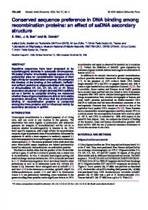
present in the 3⬘-untranslated region and the 3⬘-flanking region of the IL-13 gene, which are highly conserved between mouse and human (Fig. 9). However, homology with replication origins found in other multicellular organisms was not identified. There are three notable features associated with oriIL-13. The first is independency of its origin activity from transcriptional regulation. oriIL-13 was detected in both Th1 and Th2 effector cells. Nascent DNA abundance assays revealed that the abundance and location of nascent DNA in Th1 and Th2 cells were very similar, indicating that the efficiency of origin firing and its location were not affected by Th differentiation. The IL-13 gene is induced specifically in Th2 cells upon Ag stimulation but not in Th1 cells. Epigenetic modification of the chromatin structures is at least partially responsible for this differential expression of cytokine genes. Although the level of histone acetylation in the vicinity of oriIL-13 is very high in Th2 cells and is low in Th1 cells, this did not influence the origin activity of oriIL-13, suggesting that at least the level of histone acetylation near the origin does not affect the activity of oriIL-13. In cells carrying two X chromosomes, one of the X chromosomes is active, in which many genes are expressed, while the other is inactive due to the heterochromatinization and extensive methylation of CpG islands. However, the replication origins associated with CpG islands are equally active on both chromosomes (37). In the chicken -globin locus, little correlation was observed between origin activities and the state of histone acetylation and methylation (36). Thus, origin activation can be regulated independently from histone modification which affects gene expression. Second feature of oriIL-13 is the requirement of a distant element for its origin activity. CNS-1 coincides with a Th2-specific DNaseI HSS and is required for the efficient expression of Th2 cytokines, including IL-13, IL-4, and IL-5. Our data show that CNS-1 is required for replication initiation at oriIL-13 in both Th1 and Th2 cells. Roles of distant elements in origin activation have been reported for other loci. The region upstream of the locus control region of the -globin locus, but not locus control region itself, may be required for initiation at the human -globin replication origin, which is 50 kb distant (35, 49). In the Drosophila chorion gene cluster (24), ACE3 and ori elements are separated by the 1.8-kb chorion gene. Although both elements are recognized by ORC, ori is used as a major replication initiation site while ACE3 is required for the replication initiation as a distant element (52– 54). The c-myc replicator integrated at ectopic loci induced DNA
5452
REPLICATION ORIGIN IN Th2 CYTOKINE LOCUS
FIGURE 8. Replication timing analyses in Th1 and Th2 cells. a, Newly synthesized DNA were pulse-labeled with BrdU and separated into five fractions (G1, S1, S2, S3, G2-M) after staining with PI and sorting by FACS. A representative profile of PI intensity for Th1 and Th2 cells (derived from the wild-type (WT) mice) before sorting (top) and the purity of sorted fractions (bottom) is shown. Abundance of nascent DNA at various loci in Th1 and Th2 cells of the wild-type (b) and in those of CNS-1 deficient (c) mice was determined by real-time PCR. As a control of input sample, primer sets for one unique region of the mitochondrial genome was used for real-time PCR. Relative abundance of the nascent DNA in each cell cycle fraction at the gene loci indicated is shown. Early replicating loci (3⬘ of CNS-1, IL-4 promoter, CD8␣, and CD4) show peaks in the G1 or S1 fraction, while a late replicating locus (-globin) shows peaks in the S3 fraction.
The Journal of Immunology
5453 genes and the neighboring genes such as rad50, KIF3a, septin8, and IRF-1 are expressed in less restricted cell types or even ubiquitously. CNS-1 deletion affects the activity of oriIL-13 but apparently not the neighboring origins, since this locus is still replicated early in CNS-1 mutant cells, presumably by the replication forks derived from the neighboring origins. The delineation of the genomic loci which control the replication timing of the cytokine cluster region would require the analyses of much more extensive genomic DNA segments.
Acknowledgments We thank members of our laboratories for critical reading of this manuscript and valuable discussions. We gratefully acknowledge Noriko Kitamura for excellent technical assistance and Dr. Peter Todd for expert assistance on manuscript editing. FIGURE 9. Sequences highly conserved between human and mouse found within oriIL-13. a, Four AT-rich sequence elements near oriIL-13 that are highly conserved between human and mouse are shown. The sequence elements 1, 2, and 3 are within the 3⬘-untranslated region of IL-13 gene exon 4, while element 4 is located in the 3⬘-flanking region. b, The positions of these elements are shown as black bars below the schematic drawing of the IL-13 exon-intron structure.
synthesis at multiple sites, some of which were as far as 10 kb distant (55). The roles of these distant elements in origin activation are still elusive, but it may induce chromatin modification at distant locations favorable for initiation. Therefore, we examined the histone modification in CNS-1-deficient Th1 and Th2 cells. The extent of histone acetylation and methylation in the segment encompassing the IL-13-coding region and most of the IL-13/IL-4 intergenic regions were similar to those of the wild type in both Th1 and Th2 cells (Fig. 7). Histone modification was reduced in the mutant cells only at the promoter regions for the Th2 cytokines (56). These results strongly suggest that decreased origin activity of oriIL-13 in CNS-1 mutant cells is not due to the change in histone modification. CNS-1 is known to contain binding sites for various transcription factors (56, 57). It has been well documented that transcription factors can stimulate initiation of DNA replication in viral and chromosomal systems, and they can do so by direct interactions with replication factors (34, 58 – 61). Thus, CNS-1 may enhance the oriIL-13 activity by facilitating the prereplicative complex formation or origin activation process through distal proteinprotein interactions. Alternatively, a replication complex, assembled at CNS-1, may somehow lead to origin unwinding at oriIL-13. The third feature of oriIL-13 is that this locus is replicated during early S phase in both Th1 and Th2 cells (Fig. 8b) and that the loss of CNS-1 does not affect the early replication timing (Fig. 8c). This may be consistent with the notion that hundreds of the replication origins in clusters are coregulated (replicon clusters; Refs. 62 and 63). In fact, the 1500-kb DNA segment encompassing the human 5q cytokine cluster locus is replicated early during S phase in both T cells and non-T cells (M. Oda and H. Masai, unpublished data). In Drosophila, it was reported that transcriptional status generally correlates with replication timing when the large chromosomal domains (⬎100 kb) are analyzed (64 – 66). However, at the level of individual loci, the transcriptional status does not always correlate with the replication timing. Thus, the replication timing may be regulated by a regulatory mechanism which exerts its effect over a segment much longer than the clustered cytokine genes. In contrast, gene expression and activation of a replication origin can be governed in a more local manner. Th2-specific expression that is under the regulation of CNS-1 is confined to IL-4, IL-13, and IL-5
Disclosures The authors have no financial conflict of interest.
References 1. Loots, G. G., R. M. Locksley, C. M. Blankespoor, Z. E. Wang, W. Miller, E. M. Rubin, and K. A. Frazer. 2000. Identification of a coordinate regulator of interleukins 4, 13, and 5 by cross-species sequence comparisons. Science 288: 136 –140. 2. Ansel, K. M., D. U. Lee, and A. Rao. 2003. An epigenetic view of helper T cell differentiation. Nat. Immunol. 4: 616 – 623. 3. Murphy, K. M., and S. L. Reiner. 2002. The lineage decisions of helper T cells. Nat. Rev. Immunol. 2: 933–944. 4. Neurath, M. F., S. Finotto, and L. H. Glimcher. 2002. The role of Th1/Th2 polarization in mucosal immunity. Nat. Med. 8: 567–573. 5. Takemoto, N., N. Koyano-Nakagawa, T. Yokota, N. Arai, S. Miyatake, and K. Arai. 1998. Th2-specific DNase I-hypersensitive sites in the murine IL-13 and IL-4 intergenic region. Int. Immunol. 10: 1981–1985. 6. Agarwal, S., and A. Rao. 1998. Modulation of chromatin structure regulates cytokine gene expression during T cell differentiation. Immunity 9: 765–775. 7. Lee, D. U., S. Agarwal, and A. Rao. 2002. Th2 lineage commitment and efficient IL-4 production involves extended demethylation of the IL-4 gene. Immunity 16: 649 – 660. 8. Avni, O., D. Lee, F. Macian, S. J. Szabo, L. H. Glimcher, and A. Rao. 2002. TH cell differentiation is accompanied by dynamic changes in histone acetylation of cytokine genes. Nat. Immunol. 3: 643– 651. 9. Fields, P. E., S. T. Kim, and R. A. Flavell. 2002. Cutting edge: changes in histone acetylation at the IL-4 and IFN-␥ loci accompany Th1/Th2 differentiation. J. Immunol. 169: 647– 650. 10. Yamashita, M., M. Ukai-Tadenuma, M. Kimura, M. Omori, M. Inami, M. Taniguchi, and T. Nakayama. 2002. Identification of a conserved GATA3 response element upstream proximal from the interleukin-13 gene locus. J. Biol. Chem. 277: 42399 – 42408. 11. Baguet, A., and M. Bix. 2004. Chromatin landscape dynamics of the Il4-Il13 locus during T helper 1 and 2 development. Proc. Natl. Acad. Sci. USA 101: 11410 –11415. 12. Mohrs, M., C. M. Blankespoor, Z. E. Wang, G. G. Loots, V. Afzal, H. Hadeiba, K. Shinkai, E. M. Rubin, and R. M. Locksley. 2001. Deletion of a coordinate regulator of type 2 cytokine expression in mice. Nat. Immunol. 2: 842– 847. 13. Solymar, D. C., S. Agarwal, C. H. Bassing, F. W. Alt, and A. Rao. 2002. A 3⬘ enhancer in the IL-4 gene regulates cytokine production by Th2 cells and mast cells. Immunity 17: 41–50. 14. Ansel, K. M., R. J. Greenwald, S. Agarwal, C. H. Bassing, S. Monticelli, J. Interlandi, I. M. Djuretic, D. U. Lee, A. H. Sharpe, F. W. Alt, and A. Rao. 2004. Deletion of a conserved Il4 silencer impairs T helper type 1-mediated immunity. Nat. Immunol. 5: 1251–1259. 15. Lee, G. R., C. G. Spilianakis, and R. A. Flavell. 2005. Hypersensitive site 7 of the TH2 locus control region is essential for expressing TH2 cytokine genes and for long-range intrachromosomal interactions. Nat. Immunol. 6: 42– 48. 16. Van Houten, J. V., and C. S. Newlon. 1990. Mutational analysis of the consensus sequence of a replication origin from yeast chromosome III. Mol. Cell. Biol. 10: 3917–3925. 17. Segurado, M., A. de Luis, and F. Antequera. 2003. Genome-wide distribution of DNA replication origins at A⫹T-rich islands in Schizosaccharomyces pombe. EMBO Rep. 4: 1048 –1053. 18. Dai, J., R. Y. Chuang, and T. J. Kelly. 2005. DNA replication origins in the Schizosaccharomyces pombe genome. Proc. Natl. Acad. Sci. USA 102: 337–342. 19. Abdurashidova, G., M. Deganuto, R. Klima, S. Riva, G. Biamonti, M. Giacca, and A. Falaschi. 2000. Start sites of bidirectional DNA synthesis at the human lamin B2 origin. Science 287: 2023–2026. 20. Paixao, S., I. N. Colaluca, M. Cubells, F. A. Peverali, A. Destro, S. Giadrossi, M. Giacca, A. Falaschi, S. Riva, and G. Biamonti. 2004. Modular structure of the human lamin B2 replicator. Mol. Cell. Biol. 24: 2958 –2967. 21. Aladjem, M. I., L. W. Rodewald, C. M. Lin, S. Bowman, D. M. Cimbora, L. L. Brody, E. M. Epner, M. Groudine, and G. M. Wahl. 2002. Replication
5454
22.
23.
24.
25. 26.
27.
28.
29.
30. 31.
32. 33. 34.
35.
36.
37.
38.
39. 40.
41.
42.
43.
initiation patterns in the -globin loci of totipotent and differentiated murine cells: evidence for multiple initiation regions. Mol. Cell. Biol. 22: 442– 452. Dijkwel, P. A., S. Wang, and J. L. Hamlin. 2002. Initiation sites are distributed at frequent intervals in the Chinese hamster dihydrofolate reductase origin of replication but are used with very different efficiencies. Mol. Cell. Biol. 22: 3053–3065. Mesner, L. D., X. Li, P. A. Dijkwel, and J. L. Hamlin. 2003. The dihydrofolate reductase origin of replication does not contain any nonredundant genetic elements required for origin activity. Mol. Cell. Biol. 23: 804 – 814. Lu, L., H. Zhang, and J. Tower. 2001. Functionally distinct, sequence-specific replicator and origin elements are required for Drosophila chorion gene amplification. Genes Dev. 15: 134 –146. Lee, D. G., and S. P. Bell. 1997. Architecture of the yeast origin recognition complex bound to origins of DNA replication. Mol. Cell. Biol. 17: 7159 –7168. Lee, J. K., K. Y. Moon, Y. Jiang, and J. Hurwitz. 2001. The Schizosaccharomyces pombe origin recognition complex interacts with multiple AT-rich regions of the replication origin DNA by means of the AT-hook domains of the spOrc4 protein. Proc. Natl. Acad. Sci. USA 98: 13589 –13594. Kong, D., and M. L. DePamphilis. 2002. Site-specific ORC binding, pre-replication complex assembly and DNA synthesis at Schizosaccharomyces pombe replication origins. EMBO J. 21: 5567–5576. Vashee, S., C. Cvetic, W. Lu, P. Simancek, T. J. Kelly, and J. C. Walter. 2003. Sequence-independent DNA binding and replication initiation by the human origin recognition complex. Genes Dev. 17: 1894 –1908. Remus, D., E. L. Beall, and M. R. Botchan. 2004. DNA topology, not DNA sequence, is a critical determinant for Drosophila ORC-DNA binding. EMBO J. 23: 897–907. Hyrien, O., C. Maric, and M. Mechali. 1995. Transition in specification of embryonic metazoan DNA replication origins. Science 270: 994 –997. Sasaki, T., T. Sawado, M. Yamaguchi, and T. Shinomiya. 1999. Specification of regions of DNA replication initiation during embryogenesis in the 65-kilobase DNApol␣-dE2F locus of Drosophila melanogaster. Mol. Cell. Biol. 19: 547–555. Delidakis, C., C. Swimmer, and F. C. Kafatos. 1989. Gene amplification: an example of genome rearrangement. Curr. Opin. Cell Biol. 1: 488 – 496. Agarwal, B. D., and B. R. Calvi. 2004. Chromatin regulates origin activity in Drosophila follicle cells. Nature 430: 372–376. Danis, E., K. Brodolin, S. Menut, D. Maiorano, C. Girard-Reydet, and M. Mechali. 2004. Specification of a DNA replication origin by a transcription complex. Nat. Cell Biol. 6: 721–730. Cimbora, D. M., D. Schubeler, A. Reik, J. Hamilton, C. Francastel, E. M. Epner, and M. Groudine. 2000. Long-distance control of origin choice and replication timing in the human -globin locus are independent of the locus control region. Mol. Cell. Biol. 20: 5581–5591. Prioleau, M. N., M. C. Gendron, and O. Hyrien. 2003. Replication of the chicken -globin locus: early-firing origins at the 5⬘ HS4 insulator and the - and Aglobin genes show opposite epigenetic modifications. Mol. Cell. Biol. 23: 3536 –3549. Gomez, M., and N. Brockdorff. 2004. Heterochromatin on the inactive X chromosome delays replication timing without affecting origin usage. Proc. Natl. Acad. Sci. USA 101: 6923– 6928. Giacca, M., C. Pelizon, and A. Falaschi. 1997. Mapping replication origins by quantifying relative abundance of nascent DNA strands using competitive polymerase chain reaction. Methods 13: 301–312. Gilbert, D. M. 1986. Temporal order of replication of Xenopus laevis 5S ribosomal RNA genes in somatic cells. Proc. Natl. Acad. Sci. USA 83: 2924 –2928. Hansen, R. S., T.K. Canfield, M. M. Lamb, S. M. Gartler, and C. D. Laird. 1993. Association of fragile X syndrome with delayed replication of the FMR1 gene. Cell 73: 1403–1409. Azuara, V., K. E. Brown, R. R. E. Williams, N. Webb, N. Dillon, R. Festenstein, V. Buckle, M. Merkenschlager, and A. G. Fisher. 2003. Heritable gene silencing in lymphocytes delays chromatid resolution without affecting the timing of DNA replication. Nat. Cell Biol. 5: 668 – 674. Zhao, Y., R. Tsutsumi, M. Yamaki, Y. Nagatsuka, S. Ejiri, and K. Tsutsumi. 1994. Initiation zone of DNA replication at the aldolase B locus encompasses transcription promoter region. Nucleic Acids Res. 22: 5385–5390. Vassilev, L., and E. M. Johnson. 1990. An initiation zone of chromosomal DNA replication located upstream of the c-myc gene in proliferating HeLa cells. Mol. Cell. Biol. 10: 4899 – 4904.
REPLICATION ORIGIN IN Th2 CYTOKINE LOCUS 44. Giacca, M., L. Zentilin, P. Norio, S. Diviacco, D. Dimitrova, G. Contreas, G. Biamonti, G. Perini, F. Weighardt, S. Riva, et al. 1994. Fine mapping of a replication origin of human DNA. Proc. Natl. Acad. Sci. USA 91: 7119 –7123. 45. Taira, T., S. M. Iguchi-Ariga, and H. Ariga. 1994. A novel DNA replication origin identified in the human heat shock protein 70 gene promoter. Mol. Cell. Biol. 14: 6386 – 6397. 46. Delgado, S., M. Gomez, A. Bird, and F. Antequera. 1998. Initiation of DNA replication at CpG islands in mammalian chromosomes. EMBO J. 17: 2426 –2435. 47. Keller, C., E. M. Ladenburger, M. Kremer, and R. Knippers. 2002. The origin recognition complex marks a replication origin in the human TOP1 gene promoter. J. Biol. Chem. 277: 31430 –31440. 48. Kitsberg, D., S. Selig, I. Keshet, and H. Cedar. 1993. Replication structure of the human -globin gene domain. Nature 366: 588 –590. 49. Aladjem, M. I., M. Groudine, L. L. Brody, E. S. Dieken, R. E. Fournier, G. M. Wahl, and E. M. Epner. 1995. Participation of the human -globin locus control region in initiation of DNA replication. Science 270: 815– 819. 50. Gale, J. M., R. A. Tobey, and J. A. D’Anna. 1992. Localization and DNA sequence of a replication origin in the rhodopsin gene locus of Chinese hamster cells. J. Mol. Biol. 224: 343–358. 51. Dijkwel, P. A., and J. L. Hamlin. 1995. The Chinese hamster dihydrofolate reductase origin consists of multiple potential nascent-strand start sites. Mol. Cell. Biol. 15: 3023–3031. 52. Heck, M. M., and A. C. Spradling. 1990. Multiple replication origins are used during Drosophila chorion gene amplification. J. Cell Biol. 110: 903–914. 53. Delidakis, C., and F. C. Kafatos. 1989. Amplification enhancers and replication origins in the autosomal chorion gene cluster of Drosophila. EMBO J. 8: 891–901. 54. Austin, R. J., T. L. Orr-Weaver, and S. P. Bell. 1999. Drosophila ORC specifically binds to ACE3, an origin of DNA replication control element. Genes Dev. 13: 2639 –2649. 55. Malott, M., and M. Leffak. 1999. Activity of the c-myc replicator at an ectopic chromosomal location. Mol. Cell. Biol. 19: 5685–5695. 56. Grogan, J. L., Z. E. Wang, S. Stanley, B. Harmon, G. G. Loots, E. M. Rubin, and R. M. Locksley. 2003. Basal chromatin modification at the IL-4 gene in helper T cells. J. Immunol. 171: 6672– 6679. 57. Takemoto, N., Y. Kamogawa, H. J. Lee, H. Kurata, K. Arai, A. O’Garra and S. Miyatake. 2000. Cutting edge: chromatin remodeling at the IL-4/IL-13 intergenic regulatory region for Th2-specific cytokine gene cluster. J. Immunol. 165: 6687– 6691. 58. , Kohzaki, H., and Y. Murakami. 2005. Transcription factors and DNA replication origin selection. BioEssays 27: 1107–1116. 59. Beall, E. L., J. R. Manak, S. Zhou, M. Bell, J. S. Lipsick and M. R. Botchan. 2002. Role for a Drosophila Myb-containing protein complex in site-specific DNA replication. Nature 420: 833– 837. 60. Bosco, G., W. Du and T. L. Orr-Weaver. 2001. DNA replication control through interaction of E2F-RB and the origin recognition complex. Nat. Cell Biol. 3: 289 –295. 61. Ghosh, M., G. Liu, G. Randall and J. Bevington and M. Leffak. 2004. Transcription factor binding and induced transcription alter chromosomal c-myc replicator activity. Mol. Cell. Biol. 24: 10193–10207. 62. Nakamura, H., T. Morita, and C. Sato. 1986. Structural organization of replicon domains during DNA synthetic phase in the mammalian nucleus. Exp. Cell Res. 165: 291–297. 63. Jackson, D. A., and A. Pombo. 1998. Replicon clusters are stable units of chromosome structure: evidence that nuclear organization contributes to the efficient activation and propagation of S phase in human cells. J. Cell Biol. 140: 1285–1295. 64. MacAlpine, D. M., H. K. Rodriguez, and S. P. Bell. 2004. Coordination of replication and transcription along a Drosophila chromosome. Genes Dev. 18: 3094 –3105. 65. Woodfine, K., H. Fiegler, D. M. Beare, J. E. Collins, O. T. McCann, B. D. Young, S. Debernardi, R. Mott, I. Dunham, and N. P. Carter. 2004. Replication timing of the human genome. Hum. Mol. Genet. 13: 191–202. 66. White, E. J., O. Emanuelsson, D. Scalzo, T. Royce, S. Kosak, E. J. Oakeley, S. Weissman, M. Gerstein, M. Groudine, M. Snyder, and D. Schubeler. 2004. DNA replication-timing analysis of human chromosome 22 at high resolution and different developmental states. Proc. Natl. Acad. Sci. USA 101: 17771–17776.