APPLIED AND ENVIRONMENTAL MICROBIOLOGY, Apr. 2000, p. 1532–1537 0099-2240/00/$04.00⫹0 Copyright © 2000, American Society for Microbiology. All Rights Reserved.
Vol. 66, No. 4
Sequencing and Expression of Additional Xylanase Genes from the Hyperthermophile Thermotoga maritima FjSS3B.1 ROSALIND A. REEVES,1 MORELAND D. GIBBS,1 DANIEL D. MORRIS,1 KATHERINE R. GRIFFITHS,1 DAVID J. SAUL,2 AND PETER L. BERGQUIST1,3* Department of Biological Sciences, Macquarie University, Sydney New South Wales, Australia,1 and Centre for Gene Technology, University of Auckland,2 and Department of Molecular Medicine, University of Auckland Medical School,3 Auckland, New Zealand Received 19 October 1999/Accepted 10 January 2000
Two genes, xynB and xynC, coding for xylanases were isolated from Thermotoga maritima FjSS3B.1 by a genomic-walking–PCR technique. Sequencing of the genes showed that they encode multidomain family 10 xylanases. Only XynB exhibited activity against xylan substrates. The temperature optimum (87°C) and pH optimum (pH 6.5) of XynB are different from the previously reported xylanase, XynA (also a family 10 enzyme), from this organism. The catalytic domain expressed without other domains has a lower temperature optimum, is less thermostable, and has optimal activity at pH 6.5. Despite having a high level of sequence similarity to xynB, xynC appears to be nonfunctional since its encoded protein did not show significant activity on xylan substrates.
Kraft pulping, a process widely used in paper manufacture, removes about 95% of the lignin by alkaline sulfate cooking. The remaining lignin gives the pulp a brown color that is removed in a multistage bleaching process using a variety of agents. Currently there is concern about the environmental impact of some of the compounds used in the process, particularly chlorine and chlorine dioxide. Enzymes, including xylanases (endo-1,4--xylanase; EC 3.2.1.8), have been shown to reduce the amount of chlorine required to achieve comparable levels of paper brightness (22). However, the mesophilic enzymes currently in use have limitations because high temperatures are used in bleaching, and this fact has encouraged a number of groups to identify new enzymes from hyperthermophilic, xylanolytic microorganisms. Thermotoga maritima FjSS3B.1, which was isolated from an intertidal hot spring on Savu-Savu beach in Fiji (10) by enrichment, has been shown to produce a highly thermostable xylanase. The gene for this enzyme (xynA) has been cloned from a genomic library (18). In this communication, we report the isolation from this organism of xynB and xynC, two more glycosyl hydrolase family 10 xylanase genes, which were not revealed in the analysis of the expression library. We used a genomic-walking–PCR technique which relied on amplification of a short fragment, using consensus primers, followed by directional walking to find the 5⬘ and 3⬘ termini of the genes (14). The full-length genes and the individual catalytic domains were then amplified from the genome and expressed in Escherichia coli.
for genomic walking as described by Morris et al. (13, 14). The two-step PCR method used for the identification and sequencing of the T. maritima FjSS3B.1 xynB and xynC genes has been described previously (13, 14). A combination of double-stranded plasmid sequencing and single-stranded M13 sequencing was performed by using both dye primer and dye terminator chemistries. DNA fragments were sequenced fully on both strands. The full-length genes xynB and xynC and the DNA coding for the catalytic domains of XynB and XynC (XynBcat and XynCcat, respectively) were inserted into the heat-inducible vector pJLA602 (19) as described previously (13). E. coli DH5␣ (7) was transformed by the recombinant pJLA602/xynB and pJLA602/xynC plasmids with selection for ampicillin-resistant transformants. Primer design and PCR. Genomic-walking PCR was carried out as previously described (13). The primers used for the amplification of full-length genes encoding XynB and XynC, as well as the catalytic domains of these genes, for directional insertion into the expression vector pJLA602 are listed in Table 1. Their relative binding positions with respect to the two genes are indicated in Fig. 1. Enzyme assays. Xylanase activity was determined by the p-hydroxybenzoic acid hydrazide method of Lever (12), using birchwood xylan (Sigma) as a substrate. The standard assay reaction mixture consisted of 0.5% (wt/vol) xylan supplemented with 120 mM universal buffer (3), pH 6.5, and the enzyme to give a final volume of 0.1 ml. The reaction mixture was incubated at 70°C for 10 min. One unit of xylanase activity is defined as the amount of enzyme required to liberate 1 mol of xylose reducing-sugar equivalent per min at the assay temperature. The effects of temperature and pH on the stability and activity of the recombinant enzymes were determined as described by Gibbs et al. (6). Purification of recombinant XynB and XynC. Crude cell extracts were prepared by the method of Gibbs et al. (6) except that the heat-induced precipitation of host proteins was performed at 85°C. The T. maritima FjSS3B.1 XynB, XynBcat, XynC, and XynCcat xylanases (Fig. 1) were purified by anion-exchange chromatography. Crude cell extracts were prepared from E. coli DH5␣ strains harboring recombinant pJLA602 plasmids as described previously (6). These extracts were concentrated 10-fold by centrifugation through a 10,000-molecularweight-cutoff size exclusion membrane (Vivascience, Binbrook, Lincoln, United Kingdom) to a final volume of 2.5 ml. Low-molecular-mass materials were removed by passing concentrated extracts through a Pharmacia PD10 Superfine G-25 (AMRAD Pharmacia Biotech, Melbourne, Australia) column equilibrated with buffer A (12.5 mM bis Tris-propane buffer, pH 7.3). The 3.5 ml of PD10 eluate was applied to a Pharmacia 5-ml HiTrap-Q column (equilibrated with buffer A), which was then washed with 50 ml of buffer A. The xylanases were eluted from the column by applying a stepwise NaCl gradient consisting of 0, 25, 50, 75, 100, 125, and 150 mM concentrations of NaCl in buffer A. Three 5-ml fractions were collected at each step. The standard assay was used to detect XynB and XynBcat in HiTrap-Q column fractions, while sodium dodecyl sulfate (SDS)-PAGE was used for XynC and XynCcat because of the absence of detectable xylanase activity in these samples. XynB and XynC eluted from the column in 125 mM NaCl, while XynBcat and XynCcat eluted in 100 mM NaCl. SDS-PAGE. Electrophoresis through 12.5% polyacrylamide was performed by the standard methods described by Laemmli (11). Proteins were visualized by incubating gels with gentle shaking for 30 min in 10% trichloroacetic acid, 4 h in
MATERIALS AND METHODS Bacterial strains and recombinant plasmids. Thermotoga strains were provided as cell pellets by Hugh Morgan of Waikato University, Hamilton, New Zealand. This organism is a strain of Thermotoga maritima, as judged from ribosomal small subunit (16S) gene sequence analysis, since it exhibits 99.6% nucleotide sequence identity to the type strain (J. Thomas, University of Auckland, unpublished data, 1997). Chromosomal DNA of T. maritima FjSS3B.1 was isolated and digested with several restriction enzymes to generate linker libraries
* Corresponding author. Mailing address: Research Office, Macquarie University, Sydney, NSW 2109, Australia. Phone: 61-2-9850-8614. Fax: 61-2-9850-8799. E-mail:
[email protected]. 1532
THERMOTOGA XYLANASES
VOL. 66, 2000
1533
TABLE 1. Oligonucleotides used for genomic walking-PCR and amplification of DNA coding for various domain combinations of T. maritima FjSS3B.1 xynB and xynC Oligonucleotide
Gene
Sequence
Site
XYNBFULLF XYNBFULLR XYNBCATF XYNBCATR XYNCFULLF XYNCFULLR XYNCCATF XYNCCATR TXYNB3 TXYNB4 TXYNB5 TXYNB6 TXYNC1 TXYNC2
xynB xynB xynB xynB xynC xynC xynC xynC xynB xynB xynB xynB xynC xynC
5⬘-GTCTCTCCATATGAAACAGTTCTTGTCCTG 5⬘-GGAAATCTGGATCCTCACTTGATGAGCCTG 5⬘-AAAGCTCGCCATGGGTCCGGAAGAAGAGATACCCG 5⬘-TACCACAGGAGCTCATTCGGAGATCCTGCTTTCT 5⬘-TGTTTCCATGGGGTCTCTTCTGAGACTGTG 5⬘-CGGGGAATTCTTTCATTTCAGGAGT 5⬘-CAAACCGCCCATGGCGTTCGAAGAGGATGTACCTT 5⬘-CACCACGGGAGCTCACTCTGAAATGGTAGAGGTC 5⬘-CCAGAGGCACCGAGGACGGCACTCA 5⬘-CTTGAAAATCTCAAAGAGCTGCATC 5⬘-ATCTCGGCAGAAGTGGTGTCCACTACT 5⬘-AGACGGAGGTCGACAGAGAAGACGTAC 5⬘-GAGGGGCTTCCAAGAAACGTGGAGG 5⬘-TCTGAAGACGCTGAACAGTTGGGCG
NdeI BamHI NcoI SacI NcoI EcoRI NcoI SacI
Coomassie blue staining solution (45% water, 45% ethanol, 10% acetic acid, 0.25% [wt/vol] Coomassie brilliant blue R250), and overnight in destaining solution (67% water, 25% ethanol, 8% acetic acid). Modified gels for the detection of in situ xylanase activity (zymograms) were prepared by substituting a boiled solution of birchwood xylan (0.5%, wt/vol) for water during the preparation of the separating layer of the gel. Following electrophoresis, these gels were incubated for 1 h with gentle agitation in 2.5% (vol/vol) Triton X-100 and then for 30 min at 80°C in preheated buffer (12.5 mM bis-Tris propane, pH 6.0, at 80°C). In situ xylanase activity was detected by staining the gels for 30 min in 0.1% Congo red (20) and then destaining them in 1 M NaCl. The activity gels were rinsed in a dilute acid solution (10 mM HCl) to increase the contrast between the hydrolyzed (clear) and nonhydrolyzed (black) xylan prior to photography.
Hybridization of specific probes to genomic DNAs of Thermotoga species. Genomic DNA was prepared from T. maritima FjSS3B.1, T. maritima MSB8, and Thermotoga neapolitana as previously described (13, 14). Genomic DNAs digested with EcoRI, BamHI, and NcoI were transferred onto a positively charged membrane by using a vacuum manifold (Bio-Rad, Sydney, Australia) and processed by standard techniques (17). The sequences of the xynB and xynC genes (and their equivalents in T. maritima and T. neapolitana) were aligned and compared in order to identify regions suitable for use as specific probes for the xylanase genes. We designed six oligonucleotide primers to amplify an approximately 500-bp PCR fragment containing DNA coding for the first thermostabilizing domain (TSD) of each gene. This region was selected because it showed the maximum variability among the different xylanase genes, both at the primer
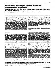
FIG. 1. The structures of the T. maritima FjSS3B.1 xynB and xynC genes and encoded products. The EcoRI and BamHI sites that distinguish xynB and xynC are indicated along with the genomic-walking–PCR products (hatched bars) used for the determination of the sequences of the genes. PCR products for cloning various domain combinations of xynB and xynC into pJLA602 are shown as black bars along with the relative binding positions of the corresponding primers used in the PCR. See Table 1 for a full description of the PCR primers. Abbreviations: GH10, glycosyl hydrolase family 10 (xylanase) domain; CBD IX, family IX CBD.
1534
REEVES ET AL.
APPL. ENVIRON. MICROBIOL.
TABLE 2. PCR amplification of the first TSD domains of xylanase genes from T. maritima MSB8, T. neapolitana, and T. maritima FjSS3B.1 PCR template DNA
T. maritima FjSS3B.1 T. neapolitana T. maritima MSB8 No-DNA control
Result with primer combinationd: APF-APRa
BPF-BPRb
CPF-CPRc
⫺ ⫺ ⫹ ⫺
⫹ ⫹ ⫺ ⫺
⫹ ⫹ ⫺ ⫺
a XYNAPF (5⬘-GGAACAACAGAAGGTGTGGTC) and XYNAPR (5⬘-AC TTCAGATGAAGCTTCGATG), forward and reverse primers for T. maritima MSB8 xynA, respectively. b XYNBPF (5⬘-GGGGACCACAGACGGTGCTTC) and XYNBPR (5⬘-ACT TTTGGAGAAATGGAAATC), forward and reverse primers for T. maritima FjSS3B.1 xynB, respectively. c XYNCPF (5⬘-AAGGAAGAAGATGGGGTTTCA) and XYNCPR (5⬘-AC CCTGGAAGTGACCTGAAGT), forward and reverse primers for T. maritima FjSS3B.1 xynC, respectively. d ⫹, PCR product of expected size observed; ⫺, no PCR product observed.
binding positions and across the region to be amplified. A separate and unique set of primers was designed for each of the T. maritima MSB8 xynA and T. maritima FjSS3B.1 xynB and xynC genes to avoid any possible nonspecific amplification problems. The 500-bp fragments were amplified from the respective genomic DNA preparations. Each primer set was used on the T. maritima MSB8 and T. maritima FjSS3B.1 genomic DNAs to confirm that each gene is unique to each organism. The sequences of the primers for gene-specific probes for T. maritima FjSS3B.1 xynB and xynC and T. maritima MSB8 xynA are listed in footnotes a to c of Table 2. PCR conditions for amplification of the specific probe fragments were as follows: 35 cycles of 95°C for 30 s, 50°C for 30 s, and 72°C for 3 min followed by one cycle of 72°C for 10 min. Each gene fragment was labeled by use of a digoxigenin chemiluminescence system (Roche Diagnostics, Sydney, Australia). The probes were hybridized to the membrane in accordance with the manufacturer’s instructions, following the high-stringency wash protocol (two 5-min room-temperature incubations in 2⫻ SSC (1⫻ SSC is 0.15 M NaCl plus 0.015 M sodium citrate)–0.1% SDS followed by two 15-min washes in 0.5⫻ SSC–0.1% SDS). Computer analysis. Computer analysis of sequence data was carried out with the Genetics Computer Group software package (4). Nucleotide sequence accession numbers. The sequences of the full-length xynB and xynC genes have been deposited in the GenBank database under accession no. AF126689 and AF126690, respectively.
RESULTS Comparison of the genes and their encoded proteins. Family 10 (8) xylanase consensus fragments were amplified from T. maritima FjSS3B.1 genomic DNA by using degenerate PCR primers (15). Sequencing of these fragments revealed three sequence species that corresponded to the previously identified xylanase gene (xynA) plus two new xylanase genes (xynB and xynC). Genomic walking–PCR was employed to obtain the complete nucleotide sequences of xynB (3,168 bp) and xynC (3,063 bp) (Fig. 1). T. maritima FjSS3B.1 xynB and xynC are genes with substantial sequence identity (75%). The putative XynB and XynC peptides exhibit 72% sequence identity and are both multidomain proteins composed of five discrete domains: two repeated N-terminal domains, a central xylanase catalytic domain (glycosyl hydrolase family 10), and two repeated C-terminal domains. A similar domain arrangement has been reported for T. maritima MSB8 XynA (25) and T. neapolitana XynA (26), to which T. maritima FjSS3B.1 XynB (and XynC) exhibit 91.8% (71.0%) and 94.8% (74.1%)sequence identity, respectively. Duplicated N-terminal domains of the type found in T. maritima FjSS3B.1 XynB and XynC are present in several thermophilic family 10 xylanases and have been termed TSDs because of their proposed roles in protein stabilization (2). Winterhalter et al. (25) demonstrated that the duplicated C-terminal
domains of T. maritima XynA represent a novel family of cellulose-binding domains (CBDs) which has since been assigned to CBD family IX (21). Accordingly, by homology, the domain architecture of T. maritima FjSS3B.1 XynB and XynC can be described as follows: TSD-TSD-family 10 xylanaseCBD IX-CBD IX. There are various degrees of sequence identity between the different domains of XynB and XynC. Specifically, the TSDTSD regions of XynB and XynC are 49.5% identical, the family 10 xylanase domains are 72.6% identical, and the CBD IX-CBD IX regions are 91.1% identical. This latter sequence similarity is intriguing given that catalytic domains are typically more conserved than CBDs. The low level of homology between the XynB and XynC TSDs is consistent with the low degree of sequence similarity that can be observed within this domain family. Southern blot analysis to confirm the presence of xynC on the genome of T. maritima FjSS3B.1. The gene xynC is not present on the sequenced genome of T. maritima MSB8 (16). Gene-specific primer combinations were used to amplify a portion of the first TSD of the T. maritima MSB8 xynA and T. maritima FjSS3B.1 xynB and xynC genes. Each primer combination was also used to amplify the equivalent genes from T. neapolitana DNA. The PCR products obtained are shown in Table 2. As expected, the T. maritima FjSS3B.1 xynB-specific primer combination amplified the equivalent fragment from the closely related T. neapolitana xynA gene. Interestingly, the PCR using the T. maritima FjSS3B.1-specific primers amplified a third, previously unidentified T. neapolitana gene, xynC (Table 2). DNA sequencing of the PCR products confirmed that each region was correctly amplified from the appropriate target gene. Over the 495 bp of DNA sequenced, the T. maritima FjSS3B.1 xynC and T. neapolitana xynC gene fragments had 18 sequence differences (95.1% identity), which resulted in eight translated differences (96.4% identity). The PCR products labeled with digoxigenin were used as probes in hybridizations to genomic DNA of each of the three bacteria cut with the restriction enzymes EcoRI, BamHI, and NcoI. These enzymes were chosen via inspection of the respective gene being examined (when known) to allow identification of restriction fragments of known sizes. Figure 2 shows an agarose gel separation of the cut DNAs and three Southern blot filters that had been hybridized with each of the three probes. All hybridizing fragments match the expected sizes (when known). For example, the expected fragment sizes (in base pairs) for T. maritima MSB8 xynA were 8,297 (EcoRI), 8,156 (BamHI), and 2,608 plus 2,704 (NcoI), whereas the observed fragment sizes were ⬎8,000, ⬎8,000, and 2,600 plus 2,700, respectively. The most significant feature of the gel shown in Figure 2 was the complete absence of hybridization of the xynC probe to DNA from T. maritima MSB8, whereas this probe hybridized to both T. neapolitana and T. maritima FjSS3B.1 genomic DNA. There was a small amount of cross-hybridization of the T. maritima MSB8 xynA probe and the T. maritima FjSS3B.1 xynB probe to each other’s genomic DNA, and there is almost 100% sequence identity between T. neapolitana xynA and T. maritima FjSS3B.1 xynB, which resulted in cross-hybridization between the T. maritima FjSS3B.1 xynB probe and T. neapolitana xynA. Cloning of xynB and xynC. PCR primers were designed to amplify the xynB and xynC genes from genomic DNA of T. maritima FjSS3B.1, plus the catalytic domains of these genes, to allow the biochemical characterization of XynB and XynC. Specific primers were designed with consideration for the sub-
VOL. 66, 2000
THERMOTOGA XYLANASES
1535
FIG. 2. Agarose gel separation of the three Thermotoga genomic DNAs cut with restriction enzymes, and Southern hybridization using the three specific probes. Symbols: E, weak cross-hybridization of T. maritima MSB8 xynA probe to FjSS3B.1 xynB gene; , weak cross-hybridization of T. maritima MSB8 xynA probe to T. neapolitana xynA gene; 䊐, weak cross-hybridization of FjSS3B.1 xynB probe to T. maritima MSB8 xynA gene. Abbreviations: FjSS, T. maritima FjSS3B.1; T. mar, T. maritima MSB8; T. nea, T. neapolitana; E, EcoRI; B, BamHI; N, NcoI; L, 1-kb ladder (Life Technologies Inc., Melbourne, Australia).
stantial homology between xynB and xynC. T. maritima FjSS3B.1 genomic-DNA PCRs using these primers yielded xynB and xynC products of the expected sizes that could be discriminated by unique EcoRI sites in the xynB fragments. The PCR products encoding the full-length XynB and XynC xylanases were respectively named XynB and XynC, while the products encoding the isolated XynB and XynC family 10 xylanase catalytic domains were named XynBcat and XynCcat, respectively (Fig. 1). These four fragments were ligated into the pJLA602 expression plasmid and resequenced to confirm the absence of PCR-induced sequence errors. Expression of XynB and XynC. Crude cell extracts prepared from E. coli DH5␣ strains harboring the genes encoding XynB, XynC, XynBcat, and XynCcat fragments were analyzed by SDSPAGE. Heterologous proteins of the appropriate molecular mass could be observed readily following Coomassie blue staining (data not shown). However, unexpectedly, enzymatic activity could be detected only from the XynB and XynBcat samples under standard assay conditions, although a trace level of activity was observed from the XynC sample following extended incubations with excess enzyme (60-min assays using a 10-fold excess of enzyme compared to XynB). PAGE analysis of XynB and XynC. The XynB, XynC, XynBcat, and XynCcat proteins were purified on Q-Sepharose to more than 90% purity (as judged from examination of Coomassie blue-stained SDS-PAGE gels [see Fig. 3]). Following SDS-PAGE, the XynBcat and XynCcat samples migrated, re-
spectively, at higher- and lower-than-expected molecular masses, which had the effect of making XynCcat appear to be slightly truncated. N-terminal sequence data obtained for XynCcat confirmed that this peptide did not contain an Nterminal deletion. However, it is possible that the XynCcat peptide was being proteolytically truncated at the C terminus by E. coli proteinases. Zymogram analysis of the four xylanase samples revealed strong activity for the XynB and XynBcat bands, weak activity for the XynC band, and no visible activity for the XynCcat band. The higher XynC/XynB activity ratio on the zymogram compared to the colorimetric assays for reducing-sugar release could be attributed to the very high sensitivity of the zymogram assay and to the fact that the zymogram was incubated for an extended period to reveal the activity of XynC. XynB in Fig. 3 is probably substrate limited as a result. Additional active xylanase bands from the XynB sample were also apparent in the zymogram. These extra bands were probably partially folded XynB peptides that migrated over a range of apparent molecular mass values or truncated enzymes generated by E. coli proteinases. In our experience, such bands are common when thermophilic glycosyl hydrolases are examined by zymogram techniques because thermostable enzymes are difficult to fully denature prior to being loaded on SDS-PAGE gels. Characterization of XynB, XynBcat, and XynC. Under the assay conditions used, the pH for optimal activity of XynB at 85°C was determined to be 6.5, with 50% of maximum activity
1536
REEVES ET AL.
APPL. ENVIRON. MICROBIOL.
for ⬎50% activity and is significantly more thermostable (Table 3) even though it does not possess TSDs. The complete sequence of the T. maritima MSB8 genome became available (16) after we had completed the cloning and characterization of the xynB and xynC genes and their products from T. maritima FjSS3B.1. When we failed to find a sequence equivalent to xynC on the T. maritima MSB8 genome, it was of concern to establish that this gene was an authentic component of the T. maritima FjSS3B.1 genome and had not been amplified from a trace contaminant, since PCR amplification was central to the identification and isolation of the gene. As has been demonstrated above, not only is a gene equivalent to xynC absent from T. maritima MSB8, but it is present on the genome of T. neapolitana. The substantial similarities between T. maritima FjSS3B.1 xynB and xynC suggest either that these genes arose by duplication or that T. maritima FjSS3B.1 acquired xynC by lateral gene transfer. Alternatively, the xynC gene may have been lost from T. maritima (or its immediate ancestor). However, we do not know at this point whether xynC of T. neapolitana expresses a functional product or is nonfunctional like its counterpart in T. maritima FjSS3B.1. XynC xylanase activity was observed at only a very low level in the purified sample, and there was no detectable xylanase activity from the single-domain XynCcat enzyme. A possible explanation for this observation is that XynC has been adapted to degrade a polysaccharide other than xylan. However, neither XynC nor XynCcat showed any detectable hydrolytic activity against azocarboxymethylcellulose, azo-Avicel, or azogalactomannan (Megazyme, Bray, Ireland) (data not shown). Alternatively, the XynC xylanase domain may incorporate one or more destabilizing missense mutations which are partially overcome through interactions between the xylanase domain and the N-terminal and/or C-terminal flanking domain. Indeed, there have been several previous reports proposing a stabilizing interaction between the TSDs and catalytic domains of other multidomain family 10 xylanases (5, 25). These observations suggest that T. maritima FjSS3B.1 xynC may be an example of a persistent mutant glycosyl hydrolase gene. However, unlike the mutant celC and the orf3 and orf4 genes from Caldicellulosiruptor saccharolyticus (1, 13), the T. maritima FjSS3B.1 xynC gene does not contain any frameshift mutations. Hence, one or more missense mutations are presumed to be responsible for rendering the xynC expression product largely inactive. T. maritima FjSS3B.1 xynC is the third example of a mutant glycosyl hydrolase gene that we have encountered and the second that we have discovered by using consensus PCR and genomic walking. We have argued previously that such defec-
FIG. 3. SDS-PAGE of extracts from induced recombinant plasmids in E. coli. (A) Proteins stained with Coomassie blue; (B) activity gel stained with Congo red. Lanes: L, size markers; 1, full-length XynB; 2, XynBcat (XynB catalytic domain only); 3, full-length XynC; 4, XynCcat (XynC catalytic domain only). The positions of molecular size markers (in kilodaltons) are shown on the left.
being retained between pH 5.5 and pH 7.4 (data not shown). The catalytic domain of XynB was found to have a much narrower pH range, with optimal activity at pH 7.0, and the truncated enzyme was much less thermostable, with maximal enzyme activity being observed at 70°C, and it was determined to have a significantly reduced half-life. The full-length XynC protein had low activity, but it was possible to measure a residual enzyme activity, but it was possible to measure a residual enzyme activity; it exhibited a pH profile similar to that of XynB (Table 3). DISCUSSION Winterhalter et al. (25) have reported the isolation of two xylanases from T. maritima MSB8 which are equivalent to XynA and XynB of T. maritima FjSS3B.1. In subsequent experiments, they showed that the removal of the N-terminal domain of their XynA (our XynB) resulted in a decrease in the thermostability of the catalytic domain, whereas the removal of the C-terminal CBDs did not lower the thermostability. Zerlov et al. (26) also described the sequence of an extracellular xylanase, from T. neapolitana, that also appears to be equivalent to our XynB. Although there are only minor sequence differences among the three genes, the complete T. neapolitana enzyme is significantly more thermostable than the other two enzymes. Our results for full-length XynB are largely in accord with those reported previously (25). The pH optima and halflives of XynB differ from those reported previously for XynA from T. maritima FjSSB.1 (18), which has a broader pH range
TABLE 3. Thermostability and pH and temperature option of T. maritima xylanases XynA and XynB Half-life at temp. (°C) of a: Enzyme
XynAd XynB XynBcat XynC a
pH characteristics
95
90
85
pH optimumb
50% of maximum activityc retained at pH:
Temp. optimuma (°C)
50% of maximum activityc retained at temp. (°C):
12 h ⬍10 min 1.5 min ND
22 h 8h 4 min ND
NLe NL 3h ND
6.3 6.5 6.5 6.5
5.1–8.1 5.5–7.4 6.1–7.4 5.8–7.4
⬎98 87 70 90
NDf 64–98 52–80 80–95
Enzymes were incubated in 25 mM bis-Tris propane, pH 6.5, in the absence of substrate. Determined at 85°C in 25 mM bis-Tris propane adjusted to specified pH. Enzyme activity measured as the release of reducing sugars from birchwood xylan, as previously described (6). d Data for XynA are from reference 18 and D. J. Saul (unpublished). e NL, no detectable loss after 16 h. f ND, not done. b c
Temp. characteristics
THERMOTOGA XYLANASES
VOL. 66, 2000
tive glycosyl hydrolase genes are likely to be widespread in view of the genetic rearrangement mechanisms central to the evolution of this gene family (2, 24). Pseudogenes are common in higher eukaryotes, but the occurrence of nonfunctional genes in prokaryotes is unexpected. Presumably, a mutant bacterial gene could be maintained only because of the presence of multiple gene copies. We have previously reviewed our results for thermophilic cellulolytic bacteria that exhibit very similar sequence structure for individual genes but glycosyl hydrolases whose have totally dissimilar gene arrangements (2). These observations imply that there is a very high degree of fluidity in the organization of these genes and that these regions of the genomes are very volatile. A consequence of such an evolutionary mechanism may be the accumulation of nonfunctional genes by virtue of nonproductive gene rearrangements, but in prokaryotes it would be expected that such mutant genes should be quickly lost because their rapid life cycles favor small, efficient genomes (9, 23). It is reasonable to expect that some of these mutant genes might have persisted in the genomes of organisms if they were closely linked to metabolically important genes, as would be the case in multigene clusters. However, their identification may require systematic examination by sequencing of large portions of genomes, since pseudogenes would be overlooked by standard techniques used for genomic expression libraries (2, 14, 15). ACKNOWLEDGMENTS This work was funded by the Foundation for Research, Science and Technology, Wellington, New Zealand; an ARC small grant; and a Macquarie University research grant. REFERENCES 1. Bergquist, P. L., M. D. Gibbs, D. J. Saul, R. A. Reeves, and V. S. J. Te’o. 1996. Families and functions of novel thermophilic hemicellulases in the facilitated bleaching of pulp. ACS Symp. Ser. 655:85–100. 2. Bergquist, P. L., M. D. Gibbs, D. D. Morris, V. S. Te’o, D. J. Saul, and H. W. Morgan. 1999. Molecular diversity of thermophilic cellulolytic and hemicellulolytic bacteria. FEMS Microbiol. Ecol. 28:99–110. 3. Britton, H. T. S., and R. A. Robinson. 1931. Universal buffer solutions and the dissociation constant of veronal. J. Chem. Soc. 1931:1456–1462. 4. Devereux, J., P. Haeberli, and O. Smithies. 1984. A comprehensive set of sequence analysis programs for the VAX. Nucleic Acids Res. 12:387–395. 5. Fontes, C. M., G. P. Hazlewood, E. Morag, J. Hall, B. H. Hirst, and H. J. Gilbert. 1995. Evidence for a general role for non-catalytic thermostabilizing domains in xylanases from thermophilic bacteria. Biochem. J. 307:151–158. 6. Gibbs, M. D., R. A. Reeves, and P. L. Bergquist. 1995. Cloning, sequencing, and expression of a xylanase gene from the extreme thermophile Dictyoglomus thermophilum Rt46B.1 and activity of the enzyme on fiber-bound substrate. Appl. Environ. Microbiol. 61:4403–4408. 7. Hanahan, D. 1983. Studies on the transformation of Escherichia coli with plasmids. J. Mol. Biol. 179:185–194. 8. Henrissat, B. 1991. A classification of glycosyl hydrolases based on amino acid sequence similarities. Biochem. J. 280:309–316. 9. Herdman, M. 1985. The evolution of bacterial genomes, p. 37–68. In T.
10. 11. 12. 13.
14.
15. 16.
17. 18.
19. 20. 21.
22. 23. 24. 25.
26.
1537
Cavalier-Smith (ed.), The evolution of genome size. John Wiley & Sons, London, United Kingdom. Huser, B. A., B. K. C. Patel, R. M. Daniel, and H. W. Morgan. 1986. Isolation and characterization of a novel extremely thermophilic anaerobic chemoorganotrophic eubacterium. FEMS Microbiol. Lett. 37:121–127. Laemmli, U. K. 1970. Cleavage of structural proteins during the assembly of the head of bacteriophage T4. Nature 227:680–685. Lever, M. 1973. Colorimetric and fluorometric carbohydrate determination with p-hydroxybenzoic acid hydrazide. Biochem. Med. 7:274–281. Morris, D. D., R. A. Reeves, M. D. Gibbs, D. J. Saul, and P. L. Bergquist. 1995. Correction of the -mannanase domain of the celC pseudogene from Caldicellulosiruptor saccharolyticus and activity of the gene product on kraft pulp. Appl. Environ. Microbiol. 61:2262–2269. Morris, D. D., M. D. Gibbs, C. W. J. Chin, M.-H. Koh, K. K. Y. Wong, R. W. Allison, P. J. Nelson, and P. L. Bergquist. 1998. Cloning of the xynB gene from Dictyoglomus thermophilum Rt46B.1 and action of the gene product on kraft pulp. Appl. Environ. Microbiol. 64:1759–1765. Morris, D. D., M. D. Gibbs, M. Ford, J. Thomas, and P. L. Bergquist. 1999. Sequence analysis of the multidomain family 10 and 11 xylanase genes from Caldicellulosiruptor sp. isolate Rt69B.1. Extremophiles 3:103–112. Nelson, K. E., R. A. Clayton, S. R. Gill, M. L. Gwinn, R. J. Dodson, D. H. Haft, E. K. Hickey, J. D. Peterson, W. C. Nelson, K. A. Ketchum, L. McDonald, T. R. Utterback, J. A. Malek, K. D. Linher, M. M. Garrett, A. M. Stewart, M. D. Cotton, M. S. Pratt, C. A. Phillips, D. Richardson, J. Heidelberg, G. G. Sutton, R. D. Fleischmann, J. A. Eisem, O. White, S. L. Salzberg, H. O. Smith, J. C. Venter, and C. M. Fraser. 1999. Evidence for lateral gene transfer between archaea and bacteria from genome sequence of Thermotoga maritima. Nature 399:323–329. Sambrook, J., E. F. Fritsch, and T. Maniatis. 1989. Molecular cloning: a laboratory manual, 2nd ed. Cold Spring Harbor Laboratory Press, Cold Spring Harbor, N.Y. Saul, D. J., L. C. Williams, R. A. Reeves, M. D. Gibbs, and P. L. Bergquist. 1995. Sequence and expression of a xylanase gene from the hyperthermophilic Thermotoga sp. strain FjSS3-B.1 and Characterization of the recombinant enzyme and its activity on kraft pulp. Appl. Environ. Microbiol. 61:4110–4113. Schauder, B., H. Blo ¨cker, R. Frank, and J. E. G. McCarthy. 1987. Inducible expression vectors incorporating the Escherichia coli atpE transcriptional initiation region. Gene 52:279–283. Teather, R. M., and P. J. Wood. 1982. Use of Congo red-polysaccharide interactions in enumeration and characterization of cellulolytic bacteria from the bovine rumen. Appl. Environ. Microbiol. 43:777–780. Tomme, P., R. A. J. Warren, R. C. J. Miller, D. G. Kilburn, and N. R. Gilkes. 1995. Cellulose binding domains: classification and properties, p. 142–161. In J. N. Saddler and M. H. Penner (ed.), Enzymatic degradation of insoluble polysaccharides, vol. 618. American Chemical Society, Washington, D.C. Viikari, L., A. Kantelinen, J. Sundquist, and M. Linko. 1994. Xylanases in bleaching: from an idea to an industry. FEMS Microbiol. Rev. 13:335–350. Watanabe, H., H. Mori, T. Itoh, and T. Gojobori. 1997. Genome plasticity as a paradigm of eubacteria evolution. J. Mol. Evol. 44:S57–S64. West, C. A., A. Elzanowski, L.-S. Yeh, and W. C. Barker. 1989. Homologues of catalytic domains of Cellulomonas glucanases found in fungal and Bacillus glycosidases. FEMS Microbiol. Lett. 50:167–172. Winterhalter, C., P. Heinrich, A. Candussio, G. Wich, and W. Liebl. 1995. Identification of a novel cellulose-binding domain within the multidomain 120 kDa xylanase XynA of the hyperthermophilic bacterium Thermotoga maritima. Mol. Microbiol. 15:431–444. Zverlov, V., K. Piotukh, O. Dakhova, G. Velikodvorskaya, and R. Borriss. 1996. The multidomain xylanase A of the hyperthermophilic bacterium Thermotoga neapolitana is extremely thermoresistant. Appl. Microbiol. Biotechnol. 45:245–247.