Sequential Binding of Import Ligands to Distinct Nucleopore Regions During Their Nuclear Import Nelly Pante* and Ueli Aebi
Import of proteins into the nucleus occurs through the NPCs, which are -125-megadalton supramolecular assemblies embedded in the double-membraned nuclear envelope (NE) (1). This process is highly selective and energy- and temperature-dependent, and requires at least four cytosolic factors (2, 3). At the molecular level, nuclear import of proteins first involves the recognition of specific sequences of the protein (4) by the cytosolic transport factor importin ot (5, 6) in the cytoplasm. Next, this "targeting complex" docks to the cytoplasmic periphery of an NPC by association of importin ot with importin (6, 7). At least two additional transport factors, the small guanosine triphosphatase (GTPase) Ran (8), which has to be in the guanosine diphosphate (GDP)-bound form (9), and the nuclear transport factor 2 (NTF2) (10) are required in vitro after docking at the NPC. However, the site or sites on the NPC where these factors exert their function and their mechanism of action remain to be established. Moreover, it is unclear how the Ran-GTPase cycle is coupled with the import of nuclear proteins. Repeated interactions between the import ligand complex and distinct NPC components are thought to be the driving force for translocation of nuclear proteins through the NPC (11, 12). Because the NPC is a highly organized macromolecular assembly with a distinct three-dimensional architecture (1, 13), any model for the molecular mechanism underlying nuclear import of proteins must consider interactions between the import ligand complex and all of the components of the NPC in their native conformation. To identify distinct NPC binding regions for the import ligand M. E. Muller Institute for Microscopy, Biozentrum, University of Basel, Klingelbergstrasse 70, CH-4056 Basel, Switzerland.
*To whom correspondence should be addressed. E-mail:
[email protected]
complex along the import pathway of nuclear proteins, we followed by electron microscopy (EM) the spatial and temporal fate of colloidal gold-labeled nucleoplasmin (NP-gold) during its import into Xenopus oocyte nuclei. NP-gold is readily imported into Xenopus oocyte nuclei (Fig. 1) (14, 15), and the kinetics of its import depend on the site of microinjection and the size of the colloidal gold bound to the import ligand. When NP-gold is microinjected close to the nucleus, its nuclear import occurs very rapidly: Two minutes after microinjection most of the gold particles are in the process of being translocated, some have already been imported into the nucleus, and many NPgold particles are associated with the NPC. To dissect the import process into distinct steps, we examined the import of individual import ligands through single NPCs by microinjecting Xenopus oocytes with small amounts of NP-gold far away from the nucleus. In this experimental setup, NP-gold particles reached the NE within 10 min, and after this time they started to be translocated into the nucleus, with three or fewer gold particles being observed per NPC (Fig. 1). Quantitative analysis of the distribution of NP-gold associated with the NPC in cross-sectioned NEs (Fig. 2) revealed the import ligand in two distinct regions on the cytoplasmic face of the NPC (i) at about 50 nm from the central plane of the NPC, representing NP-gold particles associated with the distal part of the cytoplasmic filaments (Fig. 3A); and (ii) at about 13 nm from the central plane of the NPC, representing gold particles residing at the cytoplasmic periphery of the central gated channel (Fig. 3B). Because the amount of NP-gold in the first region was initially high and decreased with time, whereas the number of NP-gold particles in the second region increased with time (Fig. 2), we concluded that NP-gold sequentially bound first to the cytoplasmic filaments and SCIENCE
*
VOL. 273
*
20 SEPTEMBER 1996
30 mir *
£-4
50 minA
::W;
;-E
k.s>g,
i,+> E¢
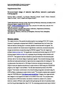
Fig. 1. Visualization of nuclear import of NP-gold through the NPC. Eight-nanometer NP-gold particles (21) were microinjected into the cytoplasm of Xenopus oocytes (22). After incubation at room temperature for 10 min, 20 min, 30 min, 40 min, and 50 min, the injected oocytes were processed for embedding and thin-section EM by conventional procedures as described (16). Shown are views along cross-sectioned Xenopus oocyte NEs. c, cytoplasmic side of the NE; n, nuclear side of the NE. Scale bar, 100 nm.
then at the cytoplasmic entry to the central gated channel. At the nuclear side of the NPC, NP-gold was predominantly found at 10 to 20 nm from the central plane of the NPC (that is, at the nuclear periphery of the gated channel; see Fig. 1, 50 min), rather than being associated with the filamentous assembly called nuclear baskets (16). The two distinct cytoplasmic NPC binding regions for NP-gold (presumably complexed with cytosolic factors) involving the cytoplasmic filaments and the cytoplasmic entry to the gated channel were better resolved when nuclear import was inhibited by the lectin wheat germ agglutinin (WGA) or by low temperature.
For this
purpose, we
microin-
jected WGA conjugated to 8-nm colloidal gold particles 1 hour before microinjection of 14-nm NP-gold. WGA-gold, which binds to the NPC via its specific interaction with nucleoporins that contain 0-linked N-acetylglucosamine residues (17), inhibited the import of NP-gold by "plugging" the cytoplasmic opening of the central pore (Fig. 4A, middle panel). Like 8-nm NP-gold (see Figs. 1 and 3), 14-nm NP-gold sequentially associated with the two distinct cytoplasmic NPC binding regions in the absence of WGA (Fig. 4A, top panel). However, when 8-nm WGA-gold was microin1729
Downloaded from www.sciencemag.org on April 19, 2010
Protein import into nuclei is mediated by the nuclear pore complex (NPC) and by cellular factors. To structurally characterize this process, nuclear import of gold-labeled nucleoplasmin was followed by electron microscopy to identify NPC components interacting with the import ligand complex in vivo. Before translocation into the nucleus, nucleoplasmin sequentially bound to two distinct regions: first to the distal part of the cytoplasmic filaments and then at the cytoplasmic entry to the central gated channel. Evidence that the delivery of the import ligand from the first to the second binding region occurred by bending of the cytoplasmic filaments is presented here.
Fig. 2. Analysis of the position and distribution of NP-gold associated with NPCs reveals two distinct and sequential binding regions for NP-gold before translocation into the nucleus. In the four panels, the dots represent the positions of gold particles, which are defined by horizontal distance (the distance from the central axis of the NPC perpendicular to the NE) and vertical distance (the distance perpendicular to the central plane of the NE). The positions of NP-gold associated with NPCs were measured from cross-sectioned Xenopus oocyte NEs (as in Fig. 1) for samples prepared for EM (A) 10 min, (B) 20 min, (C) 30 min, and (D) 40 min after microinjection of the oocytes with 8-nm NP-gold. Because of the lack of resolution, each dot may represent several gold particles, and the actual distributions of gold particles for horizontal and vertical distances are shown on adjacent panels. Before translocation into the nucleus (that is, for vertical distances >0 nm), the gold particles yielded a binomial distribution for the vertical distances with major peaks at -50 nm and - 1 3 nm, whereas the distribution for the horizontal distances was more or less similar for all time points with a major peak at the center. Cross-sectioned NEs from four different experiments were analyzed, which yielded 67, 102, 106, and 84 particles for (A), (B), (C), and (D), respectively.
j20 min
50
Gold particles 25 (%) 10 20
I
60 *@
30
.1--
0 -30
30 -30 0
40 min
D
30 6
p - 75
-50 particles -25 (% 10 20
30-
O..
01
I:
-30 . __ -60 -30 0 30 60 Horizontal distance (nm)
A
B
E
z
9
co
OC O
3.0
Fig. 3. Gallery of selected examples of NPC cross-sections revealing NP-gold particles associated with the distal part of the cytoplasmic filaments (A) and with the cytoplasmic entry to the gated channel (B). c, cytoplasmic side of the NE; n, nuclear side of the NE. Scale bar, 100 nm.
jected before 14-nm NP-gold ro block import, the N P-gold parricles predominantly associated with the NPC at a distance of about 50 nm from the central plane of the NPC (Fig. 4A, bottom panel). This posi-
tion, which was also observed without WGA inhibition (Fig. 3A), corresponded to the initial binding region for NP-gold at the distal part of the cytoplasmic filaments. Hence, plugging by WGA inhibited the delivery of NP-gold from the initial binding region to the second binding region at the cytoplasmic entry to the gated channel. At low temperature, nuclear proteins acCUMuIlate at the cytoplasmic face of the NE (15, 18). By EM we found that NP-gold accumulated about the central axis at an average distance of 13 nm from its central plane when Xenopus oocytes were kept at 40C after microinjection of NP-gold (Fig. 1730
j!v
.
_
80
-
z~~~~~~~~~~~~~
~60 _
_
=
-
W/// I0
a Fig. 4. Inhibition of nuclear import by WGA or low ' 20 temperature also reveals two distinct NPC binding re0 gions for NP-gold. (A) WGA inhibits translocation of NP-gold through the NPC but not its initial binding to the cytoplasmic filaments. Shown is a gallery of select0 10 20 30 40 50 60 ed cross-sectioned NPCs revealing nuclear import of Vertical distance (nm) 14-nm NP-gold in the absence of WGA (top row), binding of 8-nm WGA-gold (middle row), and accumulation of 14-nm NP-gold near the distal part of the cytoplasmic filaments in the presence of 8-nm WGA-gold (bottom row). (B) Cross-sectioned Xenopus oocyte NEs from oocytes that were microinjected with NP-gold and kept at 40C to inhibit or attenuate nuclear import. (C) Distribution of NP-gold particles for the vertical distance (the distance perpendicular to the central plane of the NE). NP-gold particles were measured in cross-sectioned Xenopus oocytes NEs after inhibition of nuclear import by WGA (light shading) or at 40C (dark shading). Xenopus oocytes were microinjected with 14-nm NP-gold (21) as described in Fig. 1. For inhibition of nuclear import by WGA, 8-nm WGA-gold was microinjected into the cytoplasm of the oocytes 1 hour before microinjection of 14-nm NP-gold. For inhibition of nuclear import at 40C, the oocytes were cooled to 40C 30 min before microinjection and were kept at this temperature for 1 hour after microinjection of 8-nm NP-gold. c, cytoplasmic side of the NE; n, nuclear side of the NE. Scale bars in (A) and (B), 100 nm.
4B). This position corresponded to the second binding region (that is, the cytoplasmic entry to the gated channel) that was also SCIENCE * VOL. 273 * 20 SEPTEMBER 1996
depicted at room temperature (Fig. 3B). Because the delivery of the import ligand complex from the initial docking region
Downloaded from www.sciencemag.org on April 19, 2010
60-
plasmic filaments and with the central pore (Fig. 5A). Moreover, in stereo pairs (Fig. 5B), NP-gold-bearing cytoplasmic filaments could frequently be seen bending inward so as to reach down into the central pore, possibly "handing over" the NP-gold complex to the second binding region at the entry to the gated channel. At this stage, we cannot determine whether such bending of the cytoplasmic filaments was an artifact caused by sample preparation, or whether it is indeed involved in the delivery of the import ligand complex to the gated channel. However, because this bending of the cytoplasmic filaments is also depicted when nuclei are embedded and thinsectioned (Figs. 1 and 3A) (20), we favor the hypothesis that delivery of the import ligand complex from the first to the second binding region involves bending (conceivably driven by Brownian motion) of the cytoplasmic filaments so as to bring the import ligand complex into the vicinity of the gated channel entry, where it is handed
A
over to the second binding site (20). Taken together, our morphological analysis of nuclear protein import suggests that before the actual translocation of the import ligand complex through the gated channel, it sequentially associates with two distinct regions at the cytoplasmic face of the NPC. Hence, we suggest that after formation of the targeting complex by association of the nuclear protein with at least two cytosolic factors [importin a (5) and importin 13 (7)1, this complex first binds to the distal part of a cytoplasmic filament. From this initial NPC docking site, the import ligand complex is then delivered to a second site near or at the cytoplasmic entry to the gated channel, which involves bending of the cytoplasmic filament. Once in the vicinity of the central channel entry, the import ligand complex might increase its affinity for the second binding site through association with Ran-GDP and possibly NTF2. Such a sequential binding model does not require multiple associations and dissociations of the import ligand complex to move from the initial docking site to the central gated channel (10). REFERENCES AND NOTES 1. N. Pantb and U. Aebi, Curr. Opin. Struct. Biol. 4, 187 (1994); Int. Rev. Cytol. 162B, 225 (1995). 2. F. Melchior and L. Gerace, Curr. Opin. Cell Biol. 7, 310 (1995); D. Gorlich and 1. W. Mattaj, Science 271. 1513 (1996). 3. N. Pante and U. Aebi, Curr. Opin. Cell Biol. 8, 397
(1996).
4. Signals mediating import of nuclear proteins (called nuclear localization signals) usually comprise one or two short stretches of basic amino acids [C. Dingwall and R. A. Laskey, Trends Biochem. Scd. 16, 478 (1991); J. Garcia-Bustos, J. Heitman, M. N. Hall, Biochim. Biophys. Acta 1071, 83 (1991); T. Boulikas,
B
Fig. 5. Quick freezing, freeze drying, and rotary metal shadowing also reveal two distinct NPC binding regions for NP-gold before translocation into the nucleus. (A) Cytoplasmic face of quick-frozen, freezedried, rotary metal-shadowed Xenopus oocyte NE prepared for EM 1 0 min after microinjection of 8-nm NP-gold. Shown at right is a gallery of selected examples of NPCs with associated gold particles near the distal part of the cytoplasmic filaments. Scale bar, 1 00 nm. (B) Stereopairs of the cytoplasmic face of quick-frozen, freeze-dried, rotary metal-shadowed Xenopus oocyte NEs revealing NP-gold bound to the distal part of the cytoplasmic filaments of NPCs. The two stereopairs display examples of cytoplasmic filaments with bound NP-gold that are in the process of delivering the import ligand complex to the second NPC binding region at the cytoplasmic entry to the gated channel. Xenopus oocytes were isolated and microinjected with 8-nm NP-gold as described in Fig. 10. After microinjection and subsequent incubation at room temperature for 1s0 min, nuclei were manually isolated and prepared for EM as described (16). The stereopairs were recorded by tilting the goniometer stage by t 100. Scale bar, 50 nm. SCIENCE * VOL. 273 * 20 SEPTEMBER 1996
Crit. Rev. Biochem. 62, 219 (1993)]. 5. D. Gdrlich, S. Prehn, R. A. Laskey, E. Hartmann, Cell 79, 767 (1994). 6. Importin a, and importin X were identified at the same time and independently by different groups in different species with different techniques, and have been named differently. For the different names of the various homologs of these transport factors, see table 1 of (3). 7. E. J. H. Adam and S. A. Adam, J. Cell Biol. 125, 547 (1994); D. Gorlich et al., Curr. Biol. 5, 383 (1995). 8. F. Melchior, B. Paschal, J. Evans, L. Gerace, J. Cell Biol. 123, 1649 (1993); M. S. Moore and G. Blobel, Nature 365, 661 (1993). 9. D. Gbrlich, N. Pant6, U. Kutay, U. Aebi, F. R. Bischoff, EMBO J., in press. 10. M. S. Moore and G. Blobel, Proc. Natl. Acad. Sci. U.S.A. 91, 10212 (1994); B. M. Paschal and L. Gerace, J. Cell Biol. 129, 925 (1995). 11. M. K. lovine, J. L. Watkins, S. R. Wente, J. Cell Biol. 131, 1699 (1995); U. Nehrbass and G. Blobel, Science 272, 120 (1996). 12. A. Radu, M. S. Moore, G. Blobel, Cell 81, 215 (1995); M. Rexach and G. Blobel, ibid. 83, 683 (1995). 13. J. E. Hinshaw, B. 0. Carragher, R. A. Milligan, ibid. 69, 1133 (1992); W. Akey and M. Radermacher, J. Cell Biol. 122, 1 (1993). 14. C. M. Feldherr, E. Kallenbach, N. Schultz, J. Cell Biol. 99,2216 (1984).
15. W. D. Richardson, A. D. Mills, S. M. Dilworth, R. A. Laskey, C. Dingwall, Cell 52, 655 (1988).
1731
Downloaded from www.sciencemag.org on April 19, 2010
involving the distal part of the cytoplasmic filaments to the gating area occurred at 4°C (Fig. 4B), we also investigated the effect of nucleotide depletion. Contrary to earlier findings (15), we did not observe binding of NP-gold to the NPC by pre-injection of saturating amounts of apyrase. This indicates that some initial step of the import pathway before docking of the import ligand complex to the NPC requires nucleotide triphosphate hydrolysis. This finding is in agreement with the recent discovery of a protein kinase that phosphorylates Srpl (19), the yeast homolog of importin oX. The two distinct cytoplasmic NPC binding regions for NP-gold were also depicted when the Xenopus oocyte nuclei were isolated after microinjection, and their NEs were spread on an EM grid, then quickfrozen, freeze-dried, and rotary metal-shadowed. By this EM preparation, which reveals the cytoplasmic filaments of the NPC (16), NP-gold particles were found to be associated with the distal part of the cyto-
(1985)]. Nucleoplasmin was conjugated to colloidal gold particles as described [W. Baschong and N. G. Wrigley, J. Electron Microsc. Tech. 14, 313 (1990)]. 22. Mature (stage six) oocytes were surgically removed from female Xenopus laevis as described (16) and stored in modified Barth's saline [MBS; see (16) for buffer composition]. Oocytes were defolliculated by treatment with collagenase (5 mg/ml) (Sigma) in calcium-free MBS for 3 hours. Oocytes were then washed with MBS and used for microinjection within the next 2 days. NP-gold (50 nl) was microinjected into the cytoplasm of each oocyte far away from the nucleus. The injected oocytes were incu-
(1988).
19. Y. Azuma, M. M. Tabb, L. Vu, M. Nomura, Proc. Natl. Acad. Sci. U.S.A. 92, 5159 (1995). 20. D. Byrd et al., J. Cell Biol. 1 27, 1 515 (1 994); N. Pante and U. Aebi, J. Cell Sci. 108 (suppl. 19), 1 (1995). 21. Colloidal gold particles (-8 nm or 14 nm in diameter) were prepared by the method described by J. W. Slot and H. J. Geuze [Eur. J. Cell Biol. 38, 87
bated in MBS buffer at room temperature for the times indicated. 23. We thank U. Sauder for her help with embedding and thin-sectioning, M. Dobeli for help with the data analysis, M. Hall for providing nucleoplasmin, and H. Frefel and M. Zoller for their expert photographic work. Supported by the Canton BaselStadt, the M. E. Muller Foundation of Switzerland, and by research grants from the Swiss National Science Foundation and the Human Frontier Science Program Organization.
8 May 1996; accepted 1 1 July 1996
New Location. Discover SCIENCE On-line at our new location and take advantage of these great features... *
*
Fully searchable database of abstracts and news summaries in current & past SCIENCE issues
Interactive
projects, special
features and
additional
data found in the Beyond the Printed Page section * Classified Advertising & Electronic Marketplace Tap into the sequence below and see SCIENCE On-line for yourself.
http://www.sciencemag.org SCIENCE 1 732
SCIENCE * VOL. 273 * 20 SEPTEMBER 1996
Downloaded from www.sciencemag.org on April 19, 2010
16. M. Jarnik and U. Aebi, J. Struct. Biol. 107, 291 (1991). 17. C. M. Snow. A. Senior, L. Gerace, J. Cell Biol. 104, 1143 (1987). 18. D. D. Newmeyer and D. J. Forbes, Cell 52, 641