4 Marmont AM, Horowitz MM, Gale RP, Sobocinski K, Ash RC, van-. Bekkum DW .... 46 Lamb LSJ, Robbins NF, Abhyankar S, Joyce M, Stetler SM, Henslee.
Leukemia (2001) 15, 293–302 2001 Nature Publishing Group All rights reserved 0887-6924/01 $15.00 www.nature.com/leu
Sequential monitoring of chimerism and detection of minimal residual disease after allogeneic blood stem cell transplantation (BSCT) using multiplex PCR amplification of short tandem repeat-markers C Thiede1, M Bornha¨user1, U Oelschla¨gel1, C Brendel1, R Leo2, H Daxberger3, B Mohr1, M Florek1, F Kroschinsky1, G Geissler1, R Naumann1, M Ritter1, G Prange-Krex1, T Lion3, A Neubauer1 and G Ehninger1 1
Medizinische Klinik und Poliklinik I, Universita¨tsklinikum Carl Gustav Carus der Technischen Universita¨t, Dresden, Germany; 2Medizinische Klinik und Poliklinik, Innere Medizin A der westfa¨lischen Wilhelms-Universita¨t Mu¨nster, Mu¨nster, Germany; and 3Forschungsinstitut fu¨r Krebskranke Kinder (CCRI), St Anna Kinderspital, Wien, Austria
Sequential analysis of chimerism after allogeneic blood stem cell transplantation (BSCT) has been shown to be predictive for graft failure and relapse. We have explored the impact of a novel approach for the quantitative determination of chimerism using a commercial PCR assay with multiplex amplification of nine STR-loci and fluorescence detection. The feasibility was studied in 121 patients transplanted from related or unrelated donors. Follow-up investigation was performed in 88 patients. Twenty-eight of these patients had received a transplantation after dose-reduced conditioning therapy. Results were compared to data obtained by FISH analysis in a subgroup of patients receiving grafts from sex-mismatched donors. The analysis was possible in all patients, the median number of informative alleles was 4 (range 1–8) compared to 7 (range 1– 9) in the related and unrelated situation, respectively. A good correlation was seen in 84 samples from 14 patients analyzed in parallel with STR-PCR and FISH. Decreasing values of donor chimerism were detected prior to or concomitantly with the occurrence of graft failure and relapse of disease in all patients investigated prospectively. Using FACS-sorted material, eg peripheral blood CD34+ cells, the assay permitted the detection of residual recipient cells with high sensitivity (down to one CD34+ Kasumi cell in 40 000 normal WBC). Evaluation of the inter-laboratory reproducibility revealed that in 20 samples analyzed in three different centers, the median coefficient of variation was 2.1% (range 0.7–9.6%). Taken together, the results support the use of the test as a valuable tool in the follow-up of patients undergoing allogeneic BSCT. In cases lacking PCRdetectable disease-specific gene products, this assay may represent an alternative to recently established real-time PCR methods. Leukemia (2001) 15, 293–302. Keywords: STR-PCR; chimerism; transplantation; minimal residual disease
Introduction Transplantation of allogeneic bone marrow (BM) or peripheral blood stem cells (PBSC) currently represents the only available curative therapeutic approach in several malignant and nonmalignant hematological diseases. After being restricted to related donors first, transplantation of bone marrow or peripheral blood stem cells from HLA-matched unrelated donors has become a widely used strategy in recent years. Since acute graft-versus-host disease (aGVHD) is a major complication, especially in PBSC transplantation, T cell depletion of the graft is frequently performed. To further extend the availability of donors, transplantation from haploidentical related donors is increasingly applied, especially in children, and has been shown to be an effective treatment option.1,2 In addition,
Correspondence: C Thiede, Medizinische Klinik und Poliklinik I, Universita¨tsklinikum Carl Gustav Carus der Technischen Universita¨t, Fetscherstrasse 74, 01307 Dresden, Germany; Fax: 49 351 458 5362 Received 13 September 1999; accepted 18 August 2000
novel strategies using less intensive preparative regimens have been established exploiting the antileukemic effect of the donor’s immune system.3 As a major drawback, however, many of these procedures are associated with a substantially increased risk of graft failure and relapse.4 Thus, careful monitoring of the dynamics of chimerism has become especially important to schedule therapeutic intervention.5–9 In sex-mismatched transplantation, FISH analysis of X and Y chromosomes can easily be performed to quantify donor and recipient hematopoiesis.10–14 Established procedures are commercially available, thus making standardization of the methods possible and ensuring comparable results.15 In sexmatched transplantation, PCR-based procedures using amplification of polymorphic DNA sequences, namely short tandem repeat (STR) or variable number of tandem repeat (VNTR) markers, have become a widely used approach. These assays are attractive, because they need only minimal amounts of material and are not dependent on a sex mismatch. Numerous methods have been published so far,16–26 however, the majority of these assays allow only qualitative or semi-quantitative information on the degree of chimerism. Most of these methods also require extensive pretesting and validation of each individual donor/recipient constellation to find informative peak combinations.22,24 Furthermore, none of the assays published so far is used by several centers, but most laboratories use ‘in-house’ techniques. Taken together, these factors hamper comparison and standardization of the results between different institutions. To overcome these limitations, we have recently established a novel PCR-based quantitative technique for the detection of chimerism.27 This assay takes advantage of a commercial multiplex PCR procedure originally designed and validated for forensic purposes. We performed extensive in vitro validation and showed that by calculation of the mean value obtained by amplification of up to nine STR loci this assay gives quantitative information with low variability and high reproducibility. Here we describe an extended analysis, and present clinical follow-up of the first 88 patients investigated prospectively. The results obtained show that especially in patients with high risk of graft failure or relapse, regular monitoring of donor chimerism provided a basis for early intervention. Furthermore, analysis of cellular subsets was found to be a valuable diagnostic tool, capable of detecting minimal populations of recipient cells with high sensitivity. In some patients we were able to trace cells carrying disease-associated chromosomal aberrations, eg loss of chromosome 7, in the CD34+ population. This possibility renders the assay an attractive alternative for the sensitive detection of minimal residual disease (MRD).
Monitoring of chimerism after BSCT C Thiede et al
294
Patients, material and methods
Definition
Patients
Overall chimerism: Result of the chimerism analysis of total WBC preparations.
One hundred and twenty-one patients undergoing allogeneic transplantation between June 1997 and May 1999 were studied, 52 from HLA matched (n = 49) or haploidentical (n = 3) related donors, and 69 from HLA-matched unrelated donors. In 88 of these patients, sufficient follow-up material was available. Patient details are given in Table 1. The standard preparative regimen in the myeloablative setting consisted either of intravenous busulfan (4 × 3.3 mg/kg body weight), cyclophosphamide 200 mg/kg body weight or fractionated total body irradiation (12 Gy), etoposide (30 mg/kg) and cyclophosphamide (120 mg/kg). Patients treated with the dose-reduced regimen received two doses of intravenous busulfan (3.3 mg/kg body weight) and fludarabine (150 mg/m2). All patients transplanted from unrelated donors received ATG (Merieux, 4 × 2.5 mg/kg body weight, total dose 10 mg/kg; Pasteur Me´rieux, Lyon, France) on days −5 to −2. GVHD prophylaxis consisted of cyclosporine A (CsA; 3 mg/kg), recipients of unmodified BM grafts received 10 mg/m2 methotrexate on days +1, +3 and +6. After dose-reduced conditioning, 5 mg MTX/m2 were given to recipients of unmodified BM grafts. In patients conditioned with the dose reduced concept, CsA was used in combination with oral mycophenolate mofetil (MMF; 4 × 500 mg/day). For overall chimerism analysis, 10 ml heparinized peripheral blood or 2 ml of bone marrow were collected. To obtain the pre-transplantation STR profiles, blood samples were collected from the recipient and the donor prior to transplantation. In four cases, DNA extracted from buccal swabs taken after transplantation was used to obtain the pre-transplantation STR profile of the recipient.28
Table 1
Clinical data of the 88 patients investigated prospectively
Disease Acute myelocytic leukemia Chronic myelogenous leukemia Acute lymphoblastic leukemia Non-Hodgkin’s lymphoma Myelodysplastic syndrome Other malignant Non-malignant Sex (F/M) Age (years; median/range) Donor Sex matched/mismatched HLA matched related haploidentical HLA matched unrelated Conditioning therapy Myeloablative Bu/Cy TBI/Eto/Cy Dose reduced Graft source Bone marrow PBSC PBSC (CD34-enriched)
Leukemia
24 35 13 6 5 3 (CMMoL, M Hodgkin, Multiple myeloma) 2 (aplastic anemia, severe aplastic anemia) 38/50 39 (16–62) 50/38 22 3 63 44 16 28 19 43 26
Fluorescent in situ hybridization (FISH) FISH analysis of interphase nuclei was performed by standard techniques as described recently.29 Briefly, MNC were prepared from peripheral blood or bone marrow using hypotonic KCl lysis and fixation in acetic acid/methanol (1:3). Dual-color FISH was done using the CEP X SpectrumOrange/Y SpectrumGreen DNA probe kit (Vysis, Stuttgart, Germany). Every attempt was made to analyze at least 500 nuclei.
Fluorescence activated cell sorting (FACS) Fluorescence activated cell sorting was performed on MNC enriched by density gradient centrifugation (Lymphoprep; Pharmacia, Freiburg, Germany) from 30 to 60 ml of peripheral blood, depending on the actual cell counts. One milliliter of the pellet obtained after density centrifugation (containing the erythrocyte and granulocyte fraction) was recovered and erythrocytes were removed by hypotonic lysis. CD34+ cells were enriched from the MNC fraction using one step of an immunomagnetic procedure as recommended by the manufacturer (MiniMacs; Miltenyi Biotec, Bergisch-Gladbach, Germany). Aliquots of the CD34-negative fraction were then incubated with the following monoclonal fluorophore-conjugated antibodies: anti-CD3 PECy5 and anti-CD3 PE (Coulter Immunotech Diagnostics, Hamburg, Germany), anti-CD4 PE (Pharmingen, Hamburg, Germany), anti-CD8 FITC (DAKO Diagnostika, Hamburg, Germany) and anti-CD56 FITC, antiCD19 PE and anti-CD14 FITC (Becton-Dickinson, Heidelberg, Germany) after washing in phosphate-buffered saline solution and sorted to obtain the following populations: CD3+/CD4+ and CD3+/CD8+, CD3−/CD56+, CD14+ and CD19+. Likewise, the CD34 pre-enriched fraction, usually showing a CD34+ content between 5 and 30%, was incubated with anti-CD34 PE (Pharmingen) and sorted to achieve a purity above 95%. The sample containing the granulocytes was incubated with anti-CD15 PE (Coulter Immunotech) and sorted as indicated above. When sufficient cell numbers were available, between 1500 and 10 000 cells were sorted for each population. Samples were also collected to assess the purity of the sort for at least three populations, whenever possible. The median purity, as measured by repeated FACS analysis, was 98%.
Isolation of DNA, STR-multiplex PCR and quantification DNA extraction, multiplex STR-PCR and quantification was performed as described recently27 with minor modifications. For a detailed description of the procedure for quantitative chimerism analysis see appendix, ‘Method in Focus’.
Dilution experiment A blood sample (150 ml) was drawn from a volunteer donor. The cell count was 6.5 × 10⁹ WBC/l. The concentration of CD34+ cells in this sample was 0.05%. Two aliquots were
Monitoring of chimerism after BSCT C Thiede et al
prepared, each containing 74 ml of whole blood. To the first aliquot, 1.2 × 10⁵ cells of a CD34+ cell line (Kasumi, DSMZ, Braunschweig, Germany) carrying the translocation t(8;21) were added (dilution 1: 1.2 × 10⁵ Kasumi cells in 4.81 × 10⁸ normal WBC, 1/4000), whereas to the second aliquot 1.2 × 10⁴ cells of this cell line were added (dilution 2: 1.2 × 10⁴ Kasumi in 4.81 × 10⁸ normal WBC, 1/40 000). Thirty-seven ml of each aliquot were then processed for the enrichment of CD34+ cells (see above). The remaining 37 ml were used for extraction of total RNA. RNA extraction and reverse transcription were performed as described recently.30 First round PCR for the AML1-ETO rearrangement associated with the translocation t(8;21) was performed in PE 9600 thermocyclers using primers AML2 (5′CCTCAGGTTTGTCGGTCGAAGTGGA-3′) and ETO4 (5′GCTGTAGGAGAATGGCTCGTGCCA-3′). Total volume of the PCR reaction was 25 l, containing 200 mol/l deoxynucleoside-triphosphates, 0.001% gelatine, 50 mmol/l KCl, 1.5 mmol/l MgCl2, 10 mmol/l Tris-HCl, 500 nmol/l of the primers and 1 U of Taq-polymerase (AmpliTaq, PE Biosystems, Weiterstadt, Germany). PCR-conditions were: pre-cycle 94°C/2.5 min, followed by 34 cycles with 94°C/0.5 min, 65°C/0.5 min and 72°C/0.5 min followed by a final step with 94°C/0.5 min, 72°C/10 min. The nested PCR was performed with 5 l of the first round PCR-product (diluted 1:100) in a total volume of 25 l. Primer sequences were AML3 (5′AAGTCGCCACCTACCACAGAGCCA-3′) and ETO5 (5′GTGGCATTGTTGGAGGAGTCAGCCT). The same PCR conditions were used, but only 30 cycles were performed. Dilution experiments revealed a limit of detection of 1 Kasumi cell in 1 × 10⁵ normal WBC for the nested RT-PCR assay. Results
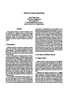
Analysis of informative alleles in related and unrelated donor/recipient constellations We have recently shown that due to the combination of nine STR markers, an informative peak constellation can be identified in all patients. However, extended analysis revealed that one of the possible combinations, namely a homozygous peak of one individual displaying the same size as one of the heterozygous alleles of the other individual (see Appendix, section 4 for details), is inferior in accuracy compared to the other constellations. Although there is a good correlation between cell mixtures and the peak ratio, the variability of results is increased, and in case of nearly complete chimerism, calculated values can reach results above 100%, thus making the interpretation difficult. For this reason, we now perform the analysis only for those STRs with at least two clearly discernible alleles. Figure 1 shows a comparison of the number of STRs fulfilling these criteria in patients transplanted from related and unrelated donors. The median number of informative STRs was four in the 52 related patients (range 1 to 8) and seven (range 1 to 9) in the 69 unrelated donor/recipient pairs. As shown in patients transplanted from unrelated donors, there was only one in 69 patients (1.4%) with a single informative STR. In contrast, five of 52 patients (9.6%) transplanted from related donors had only one allele that could used for calculation, thus clearly reducing the accuracy of quantitative results in some of these patients. However, even under these more restricted criteria for selection of STR markers for quantitative analysis in this extended group of patients, we did not find cases lacking informative signals. These obser-
295
Figure 1 Comparison of the number informative STRs in patients transplanted from related donors (n = 52) or unrelated donors (n = 69).
vations confirm our previous results demonstrating the ability of the assay to adequately discriminate between donor and recipient cells. Thus far, identical profiles in donor/recipient pairs were found only in identical twins.
Correlation of results obtained with FISH and STRPCR We also asked whether the values obtained are comparable to data obtained with standard two-color FISH in patients transplanted from sex-mismatched donors. As illustrated in Figure 2, in 84 randomly chosen samples from 14 of the 38 patients with sex-mismatched donor analyzed in parallel on the same day, a good correlation was found between the assays. Using a linear regression analysis, the value for the coefficient of determination r2 was 0.972.
Figure 2 Linear regression of results obtained with standard XYFISH and the multiplex STR-PCR during routine investigation in 14 patients analyzed with both methods on the same day. The coefficient of determination r2 for these 84 analyses was 0.972. Leukemia
Monitoring of chimerism after BSCT C Thiede et al
296
Application in the clinical setting
Subset analysis and detection of minimal residual disease
In our institution, the procedure described has been used for monitoring of the post-transplant chimerism in 88 patients. In each patient, a median number of 18 samples (range 5–55) covering a median time period of 299 days (range 26–754 days) were analyzed in a prospective manner. Table 1 summarizes the clinical characteristics of this cohort. Several of these patients were at high risk of graft failure or relapse, ie they were transplanted from mismatched donors or received a T cell-depleted graft. Twenty-eight patients were transplanted after conditioning with a dose reduced regimen. In this strategy, the recipient’s immune system is not completely ablated, and the risk of graft failure may therefore be increased.31,32 The diagnostic value of the novel technique in this subset of patients was our major focus of interest. In one patient (UPN 238), primary graft failure was observed which could be documented by the dynamics of donor chimerism even at time points when WBC counts were below 0.1 × 10⁹/l (Figure 3). Monitoring of chimerism also allowed early detection of secondary graft failure, which was seen in seven patients between days 35 and 60. Two patients who were transplanted from unrelated donors after non-myeloablative conditioning are presented in Figure 3 (UPN 169 and 188). In these seven cases, donor chimerism decreased 2–10 days before the diagnosis of graft rejection was made. Early relapse or refractory disease were seen in three patients, and 12 additional patients relapsed during follow-up. In all cases, decreasing values of overall donor chimerism were detected prior to or concomitantly with the clinical diagnosis of relapse.
The onset of relapse can display different kinetics. Especially in cases of acute leukemia or blast crisis of CML, relapse can occur very rapidly. We thus asked, whether quantification of chimerism in peripheral blood CD34+ cells might provide a more sensitive means of predicting relapse. To evaluate the sensitivity of this approach, we performed a spiking experiment, where we added cells of a CD34-positive cell line (Kasumi) to WBC of a healthy volunteer. Two dilutions were prepared (1 Kasumi cell/4000 and 40 000 WBC), and the samples were divided into two aliquots. From one aliquot, CD34+ cells and T cell were isolated as outlined in the Methods section. From the second aliquot, total RNA was prepared, subjected to cDNA synthesis and nested RT-PCR for the AML1-ETO fusion mRNA was performed. As shown in Figure 4, STR-PCR analysis permitted detection of Kasumi cells in both dilutions. Similar results were obtained in the RTPCR assay, however a nested step was necessary to detect the 1/40 000 dilution. We also addressed the utility of this assay in the clinical setting. Figure 5a shows the development of overall chimerism in a patient with AML after transplantation with dose-reduced conditioning. As depicted, the patient had a prolonged increase in overall donor chimerism, which is frequently seen in this kind of transplantation. On day +54, the patient reached a mixed donor chimerism of 92%, and remained stable at this level. However, starting on day +69, gradually decreasing donor chimerism values were observed. To get more information on the source of increasing host signals, we performed an analysis of individual cell subsets on day +88, when the donor chimerism had declined to 86%. The values of donor chimerism were between 96 and 98% in lymphoid (CD4, CD8, CD19, CD56) and myeloid (CD14, CD15) lineage (Figure 5b). However, only 20% of the cells were derived from the donor in the CD34+ fraction. Careful investigation of the signal intensity obtained in CD34+ cells revealed an interesting observation. The intensity of one allele of the STR marker D7S820 on chromosome 7 was much lower than expected (Figure 5c), indicating loss of heterozygosity (LOH) at this locus. Since the pretransplantation karyotype was 44, X, −Y, −7, del(11)(q23) it appears reasonable to assume that the CD34+ cells of recipient origin were in part of the reappearing malignant clone. In addition, loss of the Y-chromosome could be also demonstrated in the CD34+ population (data not shown). We have subsequently observed similar changes in eight additional patients carrying aberrations of chromosome 7 (monosomy 7 or 7q−), chromosome 5 or chromosome 13, in whom LOH of the corresponding STR marker could be detected using the STR procedure during follow-up after transplantation. In this situation, the use of multiplex PCR with simultaneous information on several other alleles allowed us to rule out a sampling error, because the remaining STR markers always showed a pattern identical to the original sample. The use of STRs located in chromosomal regions frequently lost in malignancy, eg chromosomes 5, 7 and 17p, should be avoided for chimerism analysis in PCR assays targeting a single locus because of the high risk of false negative detection of recipient alleles. In multiplex PCR assays, each locus showing a strong deviation of the quantitative result from the mean value of the other STRs should be checked for the presence of LOH. Several patients were also tested serially to investigate whether sequential chimerism analysis of CD34+ cells allows
Figure 3 Analysis of chimerism in peripheral blood leukocytes in three patients with early graft failure. UPN 169 is a male patient with CML in accelerated phase. He was transplanted with 7.2 × 10⁶ CD34+ cells/kg body weight from an HLA-matched unrelated donor. After primary engraftment, this patient experienced secondary graft failure at day +38. UPN 188 is a female patient with c-ALL (Ph1 pos). This patient was transplanted with 16 × 106 CD34+ cells/kg body weight. After primary engraftment at day +14, this patient experienced secondary graft failure at day +32 and received an autologous back-up. UPN 238 is a male patient with acute myeloid leukemia in first CR, who was transplanted with 12.1 × 10⁶/kg body weight CD34+ cells from his two-antigen mismatched niece. He developed fever at day +10 and persistent pancytopenia. Primary graft failure was diagnosed at day +17 and an autologous back-up was given. Leukemia
Monitoring of chimerism after BSCT C Thiede et al
297
Figure 4 Dilution experiment comparing the sensitivity of the CD34-chimerism analysis with a nested RT-PCR assay. (a) Results of the STR analysis: donor profile; CD34+ fraction of dilution 1: 1 Kasumi cell/4000 normal WBC, 7000 cells were sorted, the purity was 98%; CD34+ fraction of dilution 2: 1 Kasumi cell/40 000 normal WBC, 6000 cells were sorted. (B) Ethidium bromide stained 3% agarose gel showing the results of the first and the second (nested) PCR for the AML1-ETO rearrangement. MW, molecular weight marker 123 bp (Gibco BRL, Karlsruhe, Germany); pos. co., patient sample carrying a t(8;21); NTC, no template control.
Figure 5 (a) Development of overall chimerism in a patient with AML, FAB type M1/2 in second complete remission, who was transplanted with 7.6 × 10⁶ CD34+ selected PBSC/kg body weight from an HLA-matched unrelated donor after dose reduced conditioning. For details see text. (b) Chimerism analysis in subsets at day +88. (c) Detailed analysis of the STR profile obtained in the CD34+ fraction compared to the initial recipient and donor profiles for the D7S820 STR. The recipient pretransplantation allele profile is 9, 12 the donor constellation is 9, 11. The CD34+ fraction at day +88 contains the alleles 9, 11, and 12 however, the intensity of allele 9 is much lower than expected, since it has only the height of the residual second donor allele.
the detection of relapse. Figure 6 shows the comparison of overall chimerism in peripheral blood with analysis of CD34+ and CD15+ cells in four patients with high-risk disease, two with CML (accelerated phase) and two with MDS. One patient was transplanted with CD34+ haploidentical cells from his mother, the three others received unrelated HLA-matched transplants after reduced intensity conditioning. Detailed clinical courses are indicated in the figure legend. All patients developed donor chimerism ⬎95% between days +10 and +51. Decreasing overall donor chimerism was found between days +28 and +175 post transplantation. In contrast, donor chimerism in CD34+ cells already decreased 26 to 50 days earlier in three patients who relapsed, whereas in the fourth patient with secondary graft failure, no dominant donor chim-
erism in CD34+ cells was achieved at any time. Despite this finding, the CD15+ cells showed high levels of donor chimerism for a long time, indicating that even small numbers of residual donor CD34+ cells can proliferate and differentiate very efficiently.
Monitoring of therapy Donor lymphocyte infusion (DLI) has been established as an effective treatment of relapsing disease after allogeneic bone marrow transplantation, especially in CML. Figure 7 shows the follow-up of a patient with CML who had relapsed 19 months after unrelated bone marrow transplantation. The patient Leukemia
Monitoring of chimerism after BSCT C Thiede et al
298
Figure 6 Analysis of CD34+ cells in patients after allogeneic transplantation. (a) Case 1: a patient with CML in second accelerated phase was transplanted with 13.4 × 10⁶/kg CD34+ selected G-CSF mobilized PBSC from his haploidentical mother. The patient showed rapid engraftment with ⬎500 granulocytes/l at day +12 and ⬎50 000 platelets at day +17, complete donor chimerism was found at day +10. This level of chimerism remained stable with minute changes until day +175, when the donor chimerism level dropped instantly to 29% and a hematological relapse was diagnosed. The patient was retransplanted, but died from severe complications. Subset analysis indicated almost complete chimerism in CD34+ cells which dropped to 65% at day +124, almost 50 days before the relapse was seen in the peripheral blood. (b) Case 2: a patient with MDS (RAEB-T) received 9.7 × 10⁶/kg CD34+ selected G-CSF mobilized PBSC of a matched unrelated donor after dose reduced conditioning. This patient showed rapid engraftment (⬎500 granulocytes/l day +11, ⬎50 000 platelets day +25) and 97% donor chimerism at day +17. Between days +111 and +122 the chimerism in the peripheral blood started to decrease. Relapse of disease was diagnosed at day +124. Chimerism analysis of CD34+ cells had revealed decreasing values of donor cells at day +84. (c) Case 3: this patient with MDS was transplanted with 3.8 × 10⁶/kg CD34+ selected G-CSF mobilized PBSC from a matched unrelated donor after dose reduced conditioning. Neutrophil engraftment (⬎500/l) was seen at day +12, platelet engraftment at day +16, and the donor chimerism reached 96% at day +21. Chimerism in the peripheral blood slowly decreased again, and secondary graft failure was diagnosed at day +58. Donor chimerism in the CD34+ subset reached only 51% and already dropped at day +28. (d) Case 4: a patient with accelerated phase CML grafted with BM from a matched unrelated donor after dose reduced conditioning. Neutrophil and platelet engraftment was observed at days +15 and +32, respectively. The donor chimerism in the PBL reached 98% at day +51. At day +110, the value declined to 94%, and bone marrow aspiration showed 4% Ph1 cells. The immunosuppression was rapidly tapered and the donor chimerism increased again, however the patient died at day +138 due to GVHD of the liver and pulmonary Aspergillus infection. Analysis of CD34+ cells had shown this decrease of donor hematopoiesis at day +84.
received three increasing doses of donor CD3+ cells (5 × 10⁶/kg, 1 × 10⁷/kg and 5 × 10⁷/kg body weight). As shown, an increase of donor chimerism was documented 12 weeks after the third DLI, and was accompanied by limited chronic GVHD.
Discussion
Figure 7 Monitoring of chimerism after donor lymphocyte infusion (DLI). The arrows indicate the time points of infusion of increasing doses of donor CD3+ T cells (first dose: 5 × 10⁶ CD3+ cells/kg body weight, second dose: 1 × 10⁷/kg, third dose: 5 × 10⁷/kg). Leukemia
Sequential monitoring of chimerism after allogeneic BSCT has been shown to be predictive for graft failure and relapse.6,8,9,33–35 Although persistent recipient cells are not necessarily associated with an increased relapse rate,36 several reports have shown, that the incidence of recurrent disease is significantly increased in the group of patients with unstable mixed chimerism showing increasing host signals compared to patients presenting with stable complete chimerism or patients with mixed chimerism turning into complete chimeras.24 In this situation it is of critical importance to have a quantitative method for the evaluation of chimerism to provide useful information which could serve as a basis for clinical decision making. Knowledge on the variability of the results is important to discriminate between technical artifacts and real changes in the percentage of donor chimerism. Thus, a validated procedure with low variability and high reproduci-
Monitoring of chimerism after BSCT C Thiede et al
bility would undoubtedly contribute to an improvement in the management of patients after allogeneic BSCT. We have recently established a novel method for quantitative assessment of chimerism based on a commercial multiplex PCR kit for forensic purposes.27 In vitro data showed that the assay has a high reproducibility and precision and is therefore suitable for quantitative analysis of chimerism after allogeneic BSCT. In the current study, we were interested in the applicability and the predictive value of this assay in the clinical setting. PCR analysis performed in 121 recipient/donor pairs showed that the assay was informative in all cases with the exception of identical twins. Since we have shown that for reproducible quantitative results it is important to calculate the mean value for several STRs to compensate for random variability in amplification, we have assessed the number of STR markers eligible for analysis in patients transplanted from related donors. In the 52 patients analyzed, a median value of four STR markers were adequate for quantification. As expected, the number is lower than in the unrelated setting, where a median of seven STR markers were informative. These data show that in 80% of patients with a related donor, three or more data points were available for the calculation of the mean value. In those patients with only a single informative allele, we would strongly recommend using FISH analysis whenever possible, because the quantitative reliability of the STR-PCR assay could be reduced. Alternatively, another multiplex STR-kit (AmpFlSTR SGM plus; Perkin Elmer, Weiterstadt, Germany) could be used. This novel assay is based on co-amplification of 10 STRs and contains six STR loci (D16S539; D2S1338; D8S1179; D2S11; D18S51; D19S433) not included in the Profiler kit. Since both assays are run under identical PCR conditions, they can be readily combined in the diagnostic setting. Preliminary data indicate that the SGM multiplex assay has similar reproducibility and sensitivity in the context of chimerism analysis (data not shown). Re-evaluation of the six samples showing only a single informative allele in the Profiler assay revealed that 5/6 samples had two or more (1; 2; 2; 2; 4; 5) informative STRs in the SGM multiplex assay. Prospective chimerism analyses were performed in 88 patients. Especially in the group of patients transplanted from unrelated donors after reduced intensity conditioning, a high proportion of graft failures and relapses were observed37 and (Bornha¨user et al, manuscript in preparation). In these instances, detection of decreasing donor signals provided a basis for therapeutic intervention, ie reduction of immunosuppression or DLI. However, in three cases, decreasing levels of chimerism were observed at the time of hematologic diagnosis of relapse. In this context, a major drawback of the technique described is the relatively low level of sensitivity, with an upper limit of detection of 5%. Several PCR techniques described have much higher sensitivity.16,24 Using such highly sensitive techniques, long-term persistence of recipient cells has been demonstrated.38 However, because DNA-based assays cannot provide information on the cell type involved, it is difficult to assess the clinical relevance of mixed chimerism at low levels. In agreement with this notion, several studies have shown that stable mixed chimerism at a very low level is compatible with disease-free survival.36,39 Quantitative monitoring of disease specific markers, eg the bcr-abl fusion mRNA in CML or Ph1-positive ALL is also frequently performed to monitor patients after BSCT. These methods have been shown to be predictive for relapse in several reports.40–44 However, similar genetic markers are avail-
able only in the minority of patients. Several groups have demonstrated the usefulness of analyzing lineage-specific chimerism by applying either FISH or PCR-based methods to overcome this limitation.36,45–47 Other investigators have analyzed chimerism in CD34+ cells obtained after FACS sorting of bone marrow samples.48 Bone marrow aspiration is distressful for the patient and cannot be performed at short intervals. We therefore asked whether CD34+ cells isolated from peripheral blood can be used to obtain early information on imminent relapse. We assumed that given a median frequency of 0.1% of CD34+ cells in peripheral blood and a sensitivity of 5% for the STR-PCR assay, residual recipient hematopoiesis should be detectable with a sensitivity of about 0.005%, which means that five in 100 000 cells should be detectable. Dilution experiments revealed a sensitivity of 1/40 000 (Figure 4). Thus, the combination of FACS sorting and quantitative STR-PCR is able to achieve a level of sensitivity comparable to nested PCR assays detecting disease-specific translocations. As demonstrated in Figures 5 and 6, this method might be useful for early detection of relapse. In some patients it was also possible to determine the malignant origin of these cells, because disease-specific markers like monosomy 7 were detected using the STR-PCR in the CD34+ fraction (Figure 5c). It is obvious that analyses of leukocyte subsets are timeconsuming and cannot be performed routinely. However, in high-risk patients or in situations where mixed chimerism or slowly decreasing values are observed in overall chimerism, this method can provide important additional information on T cell chimerism and the stem cell fraction and may therefore herald disease relapse several weeks before it becomes detectable in overall chimerism analyses performed on peripheral blood leukocytes (Figure 6). In addition, information of the amount of residual donor CD34+ cells might be important for planning of therapeutic interventions. In a recent study, patients with less than 5% CD34+ donor cells in the BM were found to be at a high risk for severe aplasia after DLI.49 Larger patient numbers with longer follow-up are needed to assess the long-term predictive value of these analyses. As shown in Figures 3, 5 and 6, the kinetics of graft failure and relapse can show high variability. There are reports indicating that in certain high-risk diseases like acute leukemia or blast crisis of chronic myelogenous leukemia, hematological relapse can occur within a short period of time.50 Thus, at least in these situations, it seems appropriate to analyze chimerism at short intervals. Patients in our institution are currently investigated twice a week in the first month, and at 1-week intervals during the first 3 months post-transplant. This implies that the total number of analyses is increasing rapidly. For the current study more than 1700 samples were analyzed. As a major advantage of the assay described, the same reagents and primers are used under identical conditions in all patients. This substantially simplifies the procedure. Furthermore, the whole concept will eventually allow automatic control of most of the steps involved (DNA extraction, PCR set-up, automated sample loading and fragment analysis on capillary electrophoresis systems), which might be useful for large scale applications. Currently, great effort is made to develop standards for laboratory techniques.51 Although this is already practice in the laboratories for clinical chemistry, it is only beginning in the field of molecular diagnostics in hematology, oncology and transplantation medicine. Approval of laboratory tests by institutions like the Food and Drug Administration (FDA) is also strongly recommended. Apparently, this cannot be done for all ‘in-house’ tests currently used in different institutions. The
299
Leukemia
Monitoring of chimerism after BSCT C Thiede et al
300
Figure 8 Comparison of quantitative results obtained in three different centers using the multiplex STR-PCR assay described. Twenty samples obtained from six patients were analyzed independently in three laboratories (Vienna, Munster and Dresden). All samples were derived from states of mixed chimerism. For each sample, the measured value is given. The patients had 3–7 informative STRs. Two centers performed the analyses with a capillary electrophoresis instrument (ABI 310, PE Biosystems), the other center used a gel-based system (ABI 377, PE Biosystems).
availability of an assay which is already validated and approved for forensic purposes may be helpful as a first step in the direction of standardization in molecular detection of chimerism after BSCT. It will permit relatively easy comparison of results between different transplantation centers. This could be important for multi-center studies, or when the follow-up of a patient is not performed at the same center as the transplantation. In order to check the variability of results obtained at different centers, 20 DNA samples derived from six individual patients were analyzed independently in three different laboratories with the AmpFlSTR Profiler assay. Two of these centers used capillary electrophoresis (ABI 310) for the detection. As displayed in Figure 8, the measured donor values show a very low degree of variability, with a median coefficient of variation of 2.4% (range: 0.7–9.6%). This is comparable with the inter-assay variability of analyses performed in a single laboratory.27 Thus, this method yields highly reproducible results, even if performed at different locations. In conclusion, our preliminary experience derived from prospective clinical studies indicates that the multiplex STR-PCR described is a valuable tool for early diagnosis of graft failure and relapse in patients after allogeneic BSCT. It appears useful in the follow-up of patients with high risk of treatment failure, especially in patients treated with dose-reduced conditioning. The ability to quantify chimerism in cell populations with very low abundance, such as peripheral blood CD34+ cells, and the possibility to detect disease-specific chromosomal aberrations has great potential for sensitive detection of minimal residual disease. In patients lacking specific PCR-detectable markers, this method may be an alternative to recently introduced real-time quantitative PCR assays.52 This possibility has to be investigated in larger prospective trials.
Acknowledgements The authors would like to thank Mrs Ulrike Lo¨wel, Marita Hartwig and Heidrun Klose for excellent technical assistance. Furthermore, the help of the clinical and laboratory staff of the Dr Mildred-Scheel bone marrow transplantation center at the university clinic in Dresden is highly acknowledged. This study was supported in part by grants from the Deutsche Krebshilfe (Project No. 70–2210-Eh5), the German Jose´ Carreras foundation (AN) and funding from the Technical University Dresden Research Program (CT/MB). Leukemia
References 1 Henslee DP, Abhyankar SH, Parrish RS, Pati AR, Godder KT, Neglia WJ, Goon JK, Geier SS, Lee CG, Gee AP. Use of partially mismatched related donors extends access to allogeneic marrow transplant. Blood 1997; 89: 3864–3872. 2 Aversa F, Tabilio A, Velardi A, Cunningham I, Terenzi A, Falzetti F, Ruggeri L, Barbabietola G, Aristei C, Latini P, Reisner Y, Martelli MF. Treatment of high-risk acute leukemia with T-cell-depleted stem cells from related donors with one fully mismatched HLA haplotype. N Engl J Med 1998; 339: 1186–1193. 3 Giralt S, Estey E, Albitar M, van Besien K, Rondon G, Anderlini P, O’Brien S, Khouri I, Gajewski J, Mehra R, Claxton D, Andersson B, Beran M, Przepiorka D, Koller C, Kornblau S, Korbling M, Keating M, Kantarjian H, Champlin R. Engraftment of allogeneic hematopoietic progenitor cells with purine analog-containing chemotherapy: harnessing graft-versus-leukemia without myeloablative therapy. Blood 1997; 89: 4531–4536. 4 Marmont AM, Horowitz MM, Gale RP, Sobocinski K, Ash RC, vanBekkum DW, Champlin RE, Dicke KA, Goldman JM, Good RA et al. T-cell depletion of HLA-identical transplants in leukemia. Blood 1991; 78: 2120–2130. 5 Gardiner N, Lawler M, O’Riordan JM, Duggan C, De-Arce M, McCann SR. Monitoring of lineage-specific chimaerism allows early prediction of response following donor lymphocyte infusions for relapsed chronic myeloid leukaemia. Bone Marrow Transplant 1998; 21: 711–719. 6 Bader P, Beck J, Schlegel PG, Handgretinger R, Niethammer D, Klingebiel T. Additional immunotherapy on the basis of increasing mixed hematopoietic chimerism after allogeneic BMT in children with acute leukemia: is there an option to prevent relapse? Bone Marrow Transplant 1997; 20: 79–81. 7 Rapanotti MC, Arcese W, Buffolino S, Iori AP, Mengarelli A, DeCuia MR, Cardillo A, Cimino G. Sequential molecular monitoring of chimerism in chronic myeloid leukemia patients receiving donor lymphocyte transfusion for relapse after bone marrow transplantation. Bone Marrow Transplant 1997; 19: 703–707. 8 Dubovsky J, Daxberger H, Fritsch G, Printz D, Peters C, Matthes S, Gadner H, Lion T, Muller-Berat N. Kinetics of chimerism during the early post-transplant period in pediatric patients with malignant and non-malignant hematologic disorders: implications for timely detection of engraftment, graft failure and rejection. Leukemia 1999; 13: 2060–2069. 9 Bader P, Klingebiel T, Schaudt A, Theurer-Mainka U, Handgretinger R, Lang P, Niethammer D, Beck JF. Prevention of relapse in pediatric patients with acute leukemias and MDS after allogeneic SCT by early immunotherapy initiated on the basis of increasing mixed chimerism: a single center experience of 12 children. Leukemia 1999; 13: 2079–2086. 10 Wessman M, Ruutu T, Volin L, Knuutila S. In situ hybridization using a Y-specific probe – a sensitive method for distinguishing residual male recipient cells from female donor cells in bone marrow transplantion. Bone Marrow Transplant 1989; 4: 283–286.
Monitoring of chimerism after BSCT C Thiede et al
11 Oberkircher AR, Strout MP, Herzig GP, Fritz PD, Caligiuri MA. Description of an efficient and highly informative method for the evaluation of hematopoietic chimerism following allogeneic bone marrow transplantation. Bone Marrow Transplant 1995; 16: 695–702. 12 Palka G, Stuppia L, Di-Bartolomeo P, Morizio E, Peila R, Franchi PG, Calabrese G. FISH detection of mixed chimerism in 33 patients submitted to bone marrow transplantation. Bone Marrow Transplant 1996; 17: 231–236. 13 Najfeld V, Burnett W, Vlachos A, Scigliano E, Isola L, Fruchtman S. Interphase FISH analysis of sex-mismatched BMT utilizing dual color XY probes. Bone Marrow Transplant 1997; 19: 829–834. 14 Lapointe C, Forest L, Lussier P, Busque L, Lagace F, Perreault C, Roy DC, Gyger M. Sequential analysis of early hematopoietic reconstitution following allogeneic bone marrow transplantation with fluorescence in situ hybridization (FISH). Bone Marrow Transplant 1996; 17: 1143–1148. 15 Dewald G, Stallard R, Al Saadi A, Arnold S, Bader PI, Blough R, Chen K, Elejalde BR, Harris CJ, Higgins RR, Hoeltge GA, Hsu WT, Kubic V, McCorquodale DJ, Micale MA, Moore JW, Phillips RM, Scheib-Wixted S, Schwartz S, Siembieda S, Strole K, VanTuinen P, Vance GH, Wiktor A, Zinsmeister A. A multicenter investigation with interphase fluorescence in situ hybridization using X- and Ychromosome probes. Am J Med Genet 1998; 76: 318–326. 16 Lawler M, Humphries P, McCann SR. Evaluation of mixed chimerism by in vitro amplification of dinucleotide repeat sequences using the polymerase chain reaction. Blood 1991; 77: 2504–2514. 17 Ugozzoli L, Yam P, Petz LD, Ferrara GB, Champlin RE, Forman SJ, Koyal D, Wallace RB. Amplification by the polymerase chain reaction of hypervariable regions of the human genome for evaluation of chimerism after bone marrow transplantation. Blood 1991; 77: 1607–1615. 18 Martinelli G, Trabetti E, Zaccaria A, Farabegoli P, Buzzi M, Testoni N, Calori E, Bandini G, Rosti G, Belardinelli A et al. In vitro amplification of hypervariable DNA regions for the evaluation of chimerism after allogeneic BMT. Bone Marrow Transplant 1993; 12: 115–120. 19 Muniz ES, Plassa F, Amselem S, Goossens M, Vernant JP. Molecular analysis of polymorphic loci to study chimerism after allogeneic bone marrow transplantation. Heteroduplex analysis in denaturing gradient gel electrophoresis: a new approach to detecting residual host cells. Transplantation 1994; 57: 451–456. 20 Suttorp M, Schmitz N, Dreger P, Schaub J, Loffler H. Monitoring of chimerism after allogeneic bone marrow transplantation with unmanipulated marrow by use of DNA polymorphisms. Leukemia 1993; 7: 679–687. 21 Socie G, Lawler M, Gluckman E, McCann SR, Brison O. Studies on hemopoietic chimerism following allogeneic bone marrow transplantation in the molecular biology era. Leukemia Res 1995; 19: 497–504. 22 Scharf SJ, Smith AG, Hansen JA, McFarland C, Erlich HA. Quantitative determination of bone marrow transplant engraftment using fluorescent polymerase chain reaction primers for human identity markers. Blood 1995; 85: 1954–1963. 23 Stuppia L, Calabrese G, Di-Bartolomeo P, Peila R, Franchi PG, Morizio E, Palka G. Retrospective investigation of hematopoietic chimerism after BMT by PCR amplification of hypervariable DNA regions. Cancer Genet Cytogenet 1995; 85: 124–128. 24 Bader P, Holle W, Klingebiel T, Handgretinger R, Niethammer D, Beck J. Quantitative assessment of mixed hematopoietic chimerism by polymerase chain reaction after allogeneic BMT. Anticancer Res 1996; 16: 1759–1763. 25 Briones J, Urbano IA, Rozman C, Marin P, Carreras E, Rovira M, Sierra J, Colomer D, Martinez C, Montserrat E. Study of hematopoietic chimerism following allogeneic peripheral blood stem cell transplantation using PCR amplification of short tandem repeats. Ann Hematol 1996; 72: 265–268. 26 Rothberg PG, Gamis AS, Baker D. Use of DNA polymorphisms to monitor engraftment after allogeneic bone marrow transplantation. Clin Lab Med 1997; 17: 109–118. 27 Thiede C, Florek M, Bornha¨user M, Ritter M, Mohr B, Brendel C, Ehninger G, Neubauer A. Rapid quantification of mixed chimerism using multiplex amplification of short tandem repeat markers and fluorescence detection. Bone Marrow Transplant 1999; 23: 1055–1060.
28 Thiede C, Prange-Krex G, Freiberg-Richter J, Bornhauser M, Ehninger G. Buccal swabs but not mouthwash samples can be used to obtain pretransplant DNA fingerprints from recipients of allogeneic bone marrow transplants. Bone Marrow Transplant 2000; 25: 575–577. 29 Bornha¨user M, Mohr B, Ehrenlechner U, Neubauer A, Ehninger G. Fluorescence in situ hybridization for the BCR/ABL rearrangement is dependent on the percentage of nonlymphocytic cells in peripheral blood stem cell harvests. J Hematother 1998; 7: 425– 430. 30 Ritter M, Thiede C, Schakel U, Schmidt M, Alpen B, Pascheberg U, Mohr B, Ehninger G, Neubauer A. Underestimation of inversion(16) in acute myeloid leukaemia using standard cytogenetics as compared with polymerase chain reaction: results of a prospective investigation. Br J Haematol 1997; 98: 969–972. 31 Khouri IF, Keating M, Korbling M, Przepiorka D, Anderlini P, O’Brien S, Giralt S, Ippoliti C, von-Wolff B, Gajewski J, Donato M, Claxton D, Ueno N, Andersson B, Gee A, Champlin R. Transplant-lite: induction of graft-versus-malignancy using fludarabinebased nonablative chemotherapy and allogeneic blood progenitor-cell transplantation as treatment for lymphoid malignancies. J Clin Oncol 1998; 16: 2817–2824. 32 Childs R, Bahceci E, Clave E, van Rhee F, Jayasekera D, Mayo G. Non-myeloablative allogeneic peripheral blood stem cell transplants (PBSCT) for malignant diseases reduces transplant-related mortality (TRM). Blood 1998; 92: A552. (Abstr.). 33 Bader P, Holle W, Klingebiel T, Handgretinger R, Benda N, Schlegel PG, Niethammer D, Beck J. Mixed hematopoietic chimerism after allogeneic bone marrow transplantation: the impact of quantitative PCR analysis for prediction of relapse and graft rejection in children. Bone Marrow Transplant 1997; 19: 697–702. 34 Gardiner N, Lawler M, O’Riordan J, De’Arce M, McCann SR. Donor chimaerism is a strong indicator of disease free survival following bone marrow transplantation for chronic myeloid leukaemia. Leukemia 1997; 11 (Suppl. 3): 512–515. 35 Roman J, Martin C, Torres A, Garcia A, Andres P, Garcia MJ, Baiget M. Importance of mixed chimerism to predict relapse in persistently BCR/ABL positive long survivors after allogeneic bone marrow transplantation for chronic myeloid leukemia. Leuk Lymphoma 1998; 28: 541–550. 36 van Leeuwen JE, van Tol MJ, Joosten AM, Wijnen JT, Verweij PJ, Khan PM, Vossen JM. Persistence of host-type hematopoiesis after allogeneic bone marrow transplantation for leukemia is significantly related to the recipient’s age and/or the conditioning regimen, but it is not associated with an increased risk of relapse. Blood 1994; 83: 3059–3067. 37 Bornha¨user M, Neubauer A, Thiede C, Naumann R, Ritter M, Geissler G, Mohr B, Schuler U, Ehninger G. Allogeneic blood stem cell transplantants from unrelated donors after nonablative conditioning therapy. Blood 1998; 92: 355b (Abstr.). 38 Mangioni S, Balduzzi A, Rivolta A, Rovelli A, Nesi F, Rossi V, Busca A, Uderzo C, Miniero R, Biondi A. Long-term persistence of hemopoietic chimerism following sex-mismatched bone marrow transplantation. Bone Marrow Transplant 1997; 20: 969–973. 39 Gardiner N, Lawler M, O’Riordan J, DeArce M, Humphries P, McCann SR. Persistent donor chimaerism is consistent with disease-free survival following BMT for chronic myeloid leukaemia. Bone Marrow Transplant 1997; 20: 235–241. 40 Lion T, Henn T, Gaiger A, Kalhs P, Gadner H. Early detection of relapse after bone marrow transplantation in patients with chronic myelogenous leukaemia. Lancet 1993; 341: 275–276. 41 Cross NC, Feng L, Chase A, Bungey J, Hughes TP, Goldman JM. Competitive polymerase chain reaction to estimate the number of BCR-ABL transcripts in chronic myeloid leukemia patients after bone marrow transplantation. Blood 1993; 82: 1929–1936. 42 Drobyski WR, Endean DJ, Klein JP, Hessner MJ. Detection of BCR/ABL RNA transcripts using the polymerase chain reaction is highly predictive for relapse in patients transplanted with unrelated marrow grafts for chronic myelogenous leukaemia. Br J Haematol 1997; 98: 458–466. 43 Lin F, van-Rhee F, Goldman JM, Cross NC. Kinetics of increasing BCR-ABL transcript numbers in chronic myeloid leukemia patients who relapse after bone marrow transplantation. Blood 1996; 87: 4473–4478.
301
Leukemia
Monitoring of chimerism after BSCT C Thiede et al
302
Leukemia
44 Radich JP, Gehly G, Gooley T, Bryant E, Clift RA, Collins S, Edmands S, Kirk J, Lee A, Kessler P, Schoch G, Buckner CD, Sullivan KM, Appelbaum FR, Thomas ED. Polymerase chain reaction detection of the BCR-ABL fusion transcript after allogeneic marrow transplantation for chronic myeloid leukemia: results and implications in 346 patients. Blood 1995; 85: 2632–2638. 45 Kogler G, Wolf HH, Heyll A, Arkesteijn G, Wernet P. Detection of mixed chimerism and leukemic relapse after allogeneic bone marrow transplantation in subpopulations of leucocytes by fluorescent in situ hybridization in combination with the simultaneous immunophenotypic analysis of interphase cells. Bone Marrow Transplant 1995; 15: 41–48. 46 Lamb LSJ, Robbins NF, Abhyankar S, Joyce M, Stetler SM, Henslee DP, Gee AP. Flow cytometric cell sorting combined with molecular chimerism analysis to detect minimal recurrent leukemia: good news and bad news. Bone Marrow Transplant 1997; 19: 1157– 1161. 47 Gyger M, Baron C, Forest L, Lussier P, Lagace F, Bissonnette I, Belanger R, Bonny Y, Busque L, Roy DC, Perreault C. Quantitative assessment of hematopoietic chimerism after allogeneic bone marrow transplantation has predictive value for the occurrence of irreversible graft failure and graft-versus-host disease. Exp Hematol 1998; 26: 426–434. 48 Briones J, Urbano IA, Orfao A, Marin P, Sierra J, Rovira M, Carr-
49
50
51
52
eras E, Rozman C, Montserrat E. Demonstration of donor origin of CD34+ HLA-DR− bone marrow cells after allogeneic peripheral blood transplantation with a long follow-up. Bone Marrow Transplant 1998; 21: 189–194. Keil F, Haas OA, Fritsch G, Kalhs P, Lechner K, Mannhalter C, Reiter E, Niederwieser D, Hoecker P, Greinix HT. Donor leukocyte infusion for leukemic relapse after allogeneic marrow transplantation: lack of residual donor hematopoiesis predicts aplasia. Blood 1997; 89: 3113–3117. Bader P, Beck J, Frey A, Schlegel PG, Hebarth H, Handgretinger R, Einsele H, Niemeyer C, Benda N, Faul C, Kanz L, Niethammer D, Klingebiel T. Serial and quantitative analysis of mixed hematopoietic chimerism by PCR in patients with acute leukemias allows the prediction of relapse after allogeneic BMT. Bone Marrow Transplant 1998; 21: 487–495. Lion T, Muller-Berat N. Chimerism testing after allogeneic stem cell transplantation: importance of timing and optimal technique for chimerism testing in different clinical-biological situations. Leukemia 1999; 13: 2059. Mensink E, van-de-Locht A, Schattenberg A, Linders E, Schaap N, Geurts-van KA, De-Witte T. Quantitation of minimal residual disease in Philadelphia chromosome positive chronic myeloid leukaemia patients using real-time quantitative RT-PCR. Br J Haematol 1998; 102: 768–774.