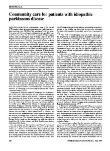
Journal of Neuroinflammation
BioMed Central
Open Access
Research
Serum antibodies from Parkinson's disease patients react with neuronal membrane proteins from a mouse dopaminergic cell line and affect its dopamine expression Victor C Huber1, Tapan Mondal1, Stewart A Factor2, Richard F Seegal1 and David A Lawrence*1 Address: 1Wadsworth Center, New York State Department of Health, Albany, NY 12201, USA and 2Parkinson's Disease & Movement Disorders Center, Albany Medical College, Albany, NY 12208, USA Email: Victor C Huber -
[email protected]; Tapan Mondal -
[email protected]; Stewart A Factor -
[email protected]; Richard F Seegal -
[email protected]; David A Lawrence* -
[email protected] * Corresponding author
Published: 20 January 2006 Journal of Neuroinflammation 2006, 3:1
doi:10.1186/1742-2094-3-1
Received: 16 November 2005 Accepted: 20 January 2006
This article is available from: http://www.jneuroinflammation.com/content/3/1/1 © 2006 Huber et al; licensee BioMed Central Ltd. This is an Open Access article distributed under the terms of the Creative Commons Attribution License (http://creativecommons.org/licenses/by/2.0), which permits unrestricted use, distribution, and reproduction in any medium, provided the original work is properly cited.
Abstract Evidence exists suggesting that the immune system may contribute to the severity of idiopathic Parkinson's disease (IPD). The data presented here demonstrates that antibodies in the sera of patients with IPD have increased binding affinity to dopaminergic (DA) neuronal (MN9D cell line) membrane antigens in comparison to antibodies in sera from healthy controls. In general, the degree of antibody reactivity to these antigens of the mouse MN9D cell line appears to correlate well with the disease severity of the IPD patients contributing sera, based on the total UPDRS scores. Surprisingly, the sera from IPD patients enhanced the DA content of MN9D cells differentiated with n-butyrate; the n-butyrate-differentiated MN9D cells had a greater concentration of DA (DA/mg total protein) than undifferentiated MN9D cells, especially early in culture. Although the IPD sera did not directly harm MN9D cellular viability or DA production, in the presence of the N9 microglial cell line, the amount of DA present in cultures of untreated or n-butyrate-treated MN9D cells was lowered by the IPD sera. The results suggest the involvement of antibodies in the decline of dopamine production and, thus, the potential of immune system participation in IPD.
Introduction Idiopathic Parkinson's disease (IPD) is a progressive neurological disorder that affects approximately 1 million people in North America [1,2]. It is characterized clinically by a loss of motor control as evidenced by muscular rigidity, resting tremor, bradykinesia, and gait dysfunction with postural instability [1,2]. Pathological features include, predominantly, the degeneration of dopaminergic (DA) neurons within the substantia nigra (SN) and intracytoplasmic inclusions (Lewy bodies) within surviv-
ing neurons [3]. To date, the cause of this disease remains unknown [4]; however, certain gene mutations, e.g., alpha-synuclein, parkin, DJ1, LRKK2, PINK1, and ND5 have been implicated [5]. Expression of any of these mutated genes may enhance the likelihood of IPD by itself or after an environmental insult. Although potentially only a consequence of IPD pathology, abnormal immune activity has been considered a possible cause of IPD based on post-mortem analysis of Page 1 of 9 (page number not for citation purposes)
Journal of Neuroinflammation 2006, 3:1
http://www.jneuroinflammation.com/content/3/1/1
Table 1: Clinical Data of IPD patients with High (H), Intermediate (I), or Low (L) Relative Western Analysis Values
Lane 1 2 3 4 5 6 7 8 9 10 11 12 13 14 15 16 17
Western Value*
Age
Age at onset
H&Y Stage
UPDRS (total)
UPDRS(motor)
L L L-I L L H L-I H L H H H L H L H H
59 59 51 69 67 77 80 79 72 57 71 69 72 48 57 72 41
59 58 50 65 55 57 71 78 65 51 64 65 70 43 53 54 36
1 2 2 2 2 2 3 4 2 2 2 2 1 2 1 4 3
11 8 14 28 30 -33.5 64 11 82.5 33 20 19 17.5 16 84 56
7 6 10 18 9 -22.5 25 9 26.5 12 10 10 40.5 7 49 34
* Relative western values are based on the summation of each band intensity above the background level); H, high; I, intermediate; L, low values (see Fig. 2). H&Y is the Hoehn and Yahr scale of Parkinson's disease (Hoehn and Yahr, 1967). UPDRS is the unified Parkinson's disease rating scale.
IPD patients' brains [6-8] and utilization of mouse models of parkinsonism [9-12]. Specifically, roles for both the innate immune system, as evidenced by increased expression of pro-inflammatory cytokines [10,13-16], and the adaptive immune system, in the form of increased levels of neuron-specific antibodies in the sera of IPD patients [17-24], have been posited. To date, the strongest evidence for specific immune involvement in the development of IPD was published by Chen et al. when they reported a selective loss of DA neurons within the SN region of rat brains upon administration of immunoglobulin (Ig) G from sera of patients with IPD [25]. Furthermore, in later studies by the same group [26,27], in vivo and in vitro models demonstrated an important contribution of Fc receptor-bearing cells in the induction of TNF-α, which, in turn, resulted in a reduction of DA neurons as evidenced by decreased tyrosine hydroxylase (TH) activity [26]. However, there have been no reports detailing the specific reactivities of IPD sera with neuronal cell membrane antigens. In this study, we set out to examine the interaction between antibodies in sera from IPD patients and DA neurons. We determined that serum IgG from IPD patients react with membrane proteins from mouse MN9D neuronal cells to a greater extent than serum IgG from healthy control individuals. Additionally, we found that IPD sera have differential modulatory effects on DA expression by MN9D cells cultured in the presence and absence of N9
microglia. The observed interactions and their possible implications are discussed.
Methods Sera and IPD patients During a routine office visit, IPD patients were asked if they would consider participation in a research project to evaluate their sera for antibodies to DA neurons in vitro. The consent form was approved by the Institutional Review Boards for Human Research of two institutions of the investigators. Most control sera were from the spouses of the IPD patients. Venous bloods were collected in EDTA vacutainers, centrifuged to remove cells, and the sera stored at -20°C until utilized as described. Clinical information regarding the IPD patients is provided (Table 1). Cell lines The MN9D cell line (provided by Dr. Alfred Heller, Department of Department of Pharmacological and Physiological Sciences, University of Chicago) was derived from rostral mesencephalic tegmentum (RMT) of the 14day-old embryonic mouse employing somatic cell fusion techniques [28]. This clonal hybrid cell line expresses a high amount of DA, which is efficiently depleted by Nmethyl-4-phenylpyridinium ion (MPP+), the active metabolite of the neurotoxin N-methyl-4-phenyl-1,2,3,6tetrahydropyridine (MPTP). The N9 microglial cell line (provided by Dr. P. Ricciardi-Castagnoli, Department of Biotechnology and Bioscience, University of Milano-Bic-
Page 2 of 9 (page number not for citation purposes)
Journal of Neuroinflammation 2006, 3:1
OD (405 nm)
1.0 0.8 0.6 0.4
*
0.2 0.0
Control Sera
IPD Sera
Figure ELISA lated from reactivity 1 MN9D of neuronal human sera cells with membrane proteins isoELISA reactivity of human sera with membrane proteins isolated from MN9D neuronal cells. Black bars represent reactivity of sera from 8 control individuals, and gray bars represent sera from 19 IPD patients. Bars represent the mean and standard error of the mean of five individual experiments. *P < 0.05 compared to control sera.
occa) was derived by retroviral immortalization of day 13 embryonic mouse brain cultures; they are similar to primary microglia in that, upon activation, they produce proinflammatory cytokines [29] and nitric oxide [30]. Cell viability Cell viability was assessed in the separate culture and cocultures of the MN9D and N9 cells in the absence and presence of the human sera. Viability was determined by a MTT assay as described [31] or by exclusion of propidium iodide assayed by flow cytometry [32]. Membrane protein isolation Membrane proteins from MN9D cells were obtained using lysis buffer containing 1.5% Triton X-114 (Sigma, St. Louis, MO), 1 mM MgCl2 (Fisher Scientific Co., Fair Lawn, NJ), 5 µg mL-1 each of RNase (Sigma) and DNase (Invitrogen Corporation, San Diego, CA) in cold phosphate-buffered saline (PBS) as previously described [33]. Briefly, cells were treated with this mixture for 15 min on ice with vortexing, and then centrifuged at 27,000 × g for 10 min at 4°C to remove nuclei. The supernatants were collected and placed at 37°C for 4 min and centrifuged at 400 × g in a swinging bucket rotor for 10 min at 25°C. The pellet containing membrane proteins were resuspended in 200 µL of 10 mM Tris•HCl, pH 7.5, and protein was quantified using the BCA assay (Pierce, Rockford, IL) using bovine serum albumin (BSA) as a protein standard.
http://www.jneuroinflammation.com/content/3/1/1
ELISA ELISA 96-well plates (Corning, Inc., Corning, NY) were coated with 10 µg mL-1 MN9D membrane protein in PBS. Plates were washed with PBS containing 0.1% (v/v) Tween-20 (PBS-T), blocked with 10% fetal bovine serum (FBS) in PBS, washed again, and then sera was added at a 1:100 dilution in 20% normal goat serum in PBS. Plates were again washed, and alkaline-phosphatase-conjugated goat anti-human IgG (H + L) (Jackson Immunoresearch Laboratories, Inc.) (1:10,000 in 10% FBS-PBS) was added. After washing, 1 mg mL-1 p-nitrophenyl phosphate substrate (Sigma) in buffer (0.1 M glycine (Sigma), 1 mM MgCl2 (Fisher), 1 mM ZnCl2 (Fisher), pH 10.4) was used to measure reactivity. Plates were read at 405 nm on a CERES UV900C microplate reader (Bio-Tek Instruments, Winooski, VT). Sera from 27 individuals, including 19 IPD patients and 8 controls, have been analyzed. ELISA for IL-1β, TNF-a and IL-6 were run as previously described [34] with DuoSets of capture and detection antibodies purchased from R & D (Minneapolis, MN). Western blot analysis MN9D membrane proteins (100 µg) were resolved by SDS-PAGE electrophoresis on a 12 % polyacrylamide gel (single 67 mm loading well) for 2.5 hr at 100 v. The proteins were transferred to a PVDF (Millipore, Bedford, MA) membrane (30 min at 20 v) and blocked with 5% (v/v) fish gelatin (Sigma) in PBS containing 0.05% Tween20 (PBS-T). The blot was washed with PBS-T, and affixed to a slot-blotting apparatus (Bio-Rad) that allows multiple sera to be screened simultaneously. Sera from 17 IPD patients and 2 controls (1:50 dilution in 5% normal goat serum in PBS-T) were applied in separate slots and incubated overnight at 4°C while rocking. The blot was then washed with PBS-T, and incubated with biotin-conjugated goat anti-human IgG (gamma chain specific) (Tago, Inc., Burlingame, CA) (1:5,000 dilution in 5% normal goat serum in PBS-T). The blot was again washed with PBS-T followed by addition of HRP-conjugated streptavidin (1:20,000 dilution; Pierce, Rockford, IL). After a final wash with PBS-T, Super Signal West Pico (Pierce), a chemiluminescent substrate was added. Reactivity was observed with a Fuji LAS 1000 system (FujiFilm Medical Systems USA, Inc., Stamford, CT), and analyzed using ImageGauge software (FujiFilm Medical Systems USA, Inc.). Cell culture conditions MN9D and N9 cell lines were cultured in Dulbecco's modified Eagle's medium with L-glutamine and 4500 mg L-1 glucose, without sodium bicarbonate (Sigma). Importantly, this medium contains pyridoxal•HCl, which is required for the survival of the MN9D mesencephalic cell line [28]. This medium was supplemented with 10% FBS (Hyclone, Logan, UT), 50 U mL-1 penicillin and 50 µg mL-
Page 3 of 9 (page number not for citation purposes)
Journal of Neuroinflammation 2006, 3:1
http://www.jneuroinflammation.com/content/3/1/1
Total Peak Areas
70000 60000 50000 40000 30000 20000 10000 0 0
20
40
60
80
100
UPDRS (total) Figure Western teins isolated 2 blot from reactivity MN9D of human neuronal sera cells with membrane proWestern blot reactivity of human sera with membrane proteins isolated from MN9D neuronal cells. In this figure, 17 individual IPD sera (lane 1–17) and 2 control sera (lane 18 & 19) were used to probe the PVDF membrane. Numbers designated to the left of the blot reveal the migration of molecular weight standards in kDa. Data are representative of two individual experiments.
1
streptomycin (Invitrogen Corporation), 3.7 g L-1 NaHCO3 (J.T. Baker Chemical Co., Phillipsburg, NJ), and 50 µM 2-mercaptoethanol (Sigma). Cell culture conditions were set up and analyzed as previously described (Le et al., 2001), with minor modifications. In 24-well tissue culture plates (Corning Inc.), 2 × 10⁴ N9 cells were seeded for 24 hr at 37°C under 5% CO2. After 24 hr, medium was removed, and MN9D cells (4 × 10⁴) in fresh medium were added in the presence of individual human sera (1%) samples. The N9:MN9D ratio was maintained at 1:2, as previously described (Le et al., 2001). Cells were co-cultured for three days. Differentiation of MN9D cells was performed as described [28] by culturing 4.6 × 10⁴ cells per well in 4 mL medium in 6-well tissue culture plates (Corning). Cells were exposed to 1 mM n-butyrate (Sigma) throughout the seven day culture and designated wells were harvested daily, beginning with day 2. Medium was replaced every 48 hr with fresh medium containing 1 mM n-butyrate. Co-cultures of differentiated MN9D cells were established by culturing 9.2 × 10³ MN9D cells in 24-well tissue culture plates (Corning Inc.) in the presence of 1 mM n-butyrate (Sigma) and 1% human sera. After 48 hr, the medium was removed, and fresh medium supplemented with 1 mM nbutyrate and 1% human sera was added. Twenty-four hr later, medium was again removed, and cells were washed twice with 1 mL PBS. Indicated wells received 4.6 × 10³ N9
Figure Correlation the sum 3of the analysis peak of areas clinical fromscore the Western (UPDRS analysis – total) (Fig. with 2) Correlation analysis of clinical score (UPDRS – total) with the sum of the peak areas from the Western analysis (Fig. 2). The r2 value was assessed by linear regression analysis (SigmaPlot 2000) and the significance (p) was calculated by Pearson correlation analysis with SigmaStat (Jandel Corp). microglia and 1% human sera in n-butyrate-free medium. Cells cultured in the absence of N9 microglia received 1% human sera in n-butyrate-free medium. Cells were co-cultured for three days. Quantification of DA expression At the end of the specified culturing period, plates were centrifuged at 200 × g for 10 min at 4°C, medium was removed, and cells were washed with 1 mL PBS. Cells were exposed to 1 mL 0.2 M HClO4 and sonicated as described. This mixture was then centrifuged to remove proteins from the samples, and DA expression was analyzed using high performance liquid chromatography with electrochemical sensors, and quantified using Waters Millenia software (Waters, Milford, MA), as described [35]. The protein pellet was resuspended in 50 µL 0.5 M NaOH, and protein was quantified using the BCA method with BSA as a standard.
Results When compared with sera from healthy controls, IgG in the sera of IPD patients had significantly increased (P < 0.05) binding to ELISA wells coated with MN9D neuronal membrane proteins (Fig. 1). Western blot analysis also demonstrated greater IgG reactivity to MN9D cell membrane proteins (Fig. 2); only two sera from healthy individuals, which had the greatest activity for the control sera, are shown. The Western analysis of the IPD sera revealed antibody reactivity to a number of proteins present in the MN9D neuronal membrane isolates; proteins of 30 to 65 kDa molecular weights were especially
Page 4 of 9 (page number not for citation purposes)
Journal of Neuroinflammation 2006, 3:1
http://www.jneuroinflammation.com/content/3/1/1
100 12
A
80
ng DA/well
ng DA/well
14
10 8 6 4
B
150
*
100 50
Control Sera
IPD Sera
ng DA/mg protein
ng DA/mg protein
40 20
2 200
60
1000 800 600 400 200
1
2
3
4
5
6
Day 4 cells/ml) 4 Figure After 724hr assessed cells/ml) and in co-cultures exposure N9 microglia toofhuman MN9D (2 × 10 sera neuronal at 37°C, cells DA(4levels × 10were After 72 hr exposure to human sera at 37°C, DA levels were assessed in co-cultures of MN9D neuronal cells (4 × 10⁴ cells/ml) and N9 microglia (2 × 10⁴ cells/ml). Black bars represent DA values upon exposure to 8 control sera and gray bars represent values upon exposure to 19 sera from IPD patients. Results are reported as both (A) ng/well and (B) ng/ mg protein. Bars represent the mean and standard error of the mean for four individual experiments. *P < 0.05 compared to control sera.
predominant. While this reactivity revealed no consistent differences between IPD and control sera and no major common protein band amongst the IPD sera, in general, there was a noticeable increase in the intensity of bands with sera from IPD patients with greater unified Parkinson's disease rating scale (UPDRS) scores (Table 1). The UPDRS is a combined score from the physician's evaluation of motor activity including temors, rigidity, posture, gait, and bradykinesia. For example, the sera from patients 7, 8, 10, 16 and 17 had the most severe IPD (UPDRS-total, 33.5–82.5; UPDRS-motor, 22.5–49), whereas sera from the least severe (UPDRS-total, 8–16; UPDRS-motor, 6– 10) IPD patients (1, 2, 9, 15) generally had binding as low as most normal sera.
Figure Time exposure course 5 to n-butyrate of DA expression by MN9D neuronal cells after Time course of DA expression by MN9D neuronal cells after exposure to n-butyrate. Black bars represent untreated cells and gray bars represent cells differentiated in the presence of 1 mM n-butyrate. Results are reported as both (A) ng/well and (B) ng/mg protein. Results represent the data from three individual wells per group. *P < 0.05 compared to untreated cells. Pearson correlational analysis does suggest that the antibodies in the IPD sera significantly correlate (r2 = 0.21; p = 0.05) with the total UPDRS score (Fig. 3). However, there was no correlation of the antibody binding to MN9D antigens (Fig. 2) with regard to UPDRS motor values or the duration of IPD. To assess if the observed interactions between IPD sera and neuronal antigens correlated with any adverse effects on neuronal cells, in vitro assays were performed. This analysis revealed that, compared to control sera, IPD sera had no significant effect on the levels of DA regardless of whether the quantification was calculated as ng/well or ng/mg protein (approximately 10 ng/well and 160 ng/mg protein). Alternatively, if the N9 microglial cells were co-cultured with the MN9D neuronal cells and sera (Fig. 4), there was a noticeable loss of DA. Although there was not a signifi-
Page 5 of 9 (page number not for citation purposes)
Journal of Neuroinflammation 2006, 3:1
5
6
A
*
ng DA/well
ng DA/well
6
http://www.jneuroinflammation.com/content/3/1/1
4 3 2
A
4
*
3 2 1
B
*
60
40
20
Control
IPD Sera
ng DA/mg protein
1
ng DA/mg protein
5
B 60
*
40
20
Control
IPD Sera
ronal After assessed Figure 72 cells 6hr in(4 cultures exposure × 104 cells/ml) ofton-butyrate-differentiated human sera at 37°C, DAMN9D levels were neuAfter 72 hr exposure to human sera at 37°C, DA levels were assessed in cultures of n-butyrate-differentiated MN9D neuronal cells (4 × 10⁴ cells/ml). Black bars represent DA values upon exposure to a pool of 8 control sera; gray bars represent values upon exposure to a pool of 19 sera from IPD patients. Results are reported as both (A) ng/well and (B) ng/ mg protein. Bars represent the mean and standard error of the mean for three individual wells. *P < 0.05 compared to control sera.
4 cells/ml) 4 Figure cells/ml) and N9 microglia (2MN9D × 10 neuronal After assessed 727hr in cells co-cultures exposure (4 × 10to ofhuman n-butyrate-differentiated sera at 37°C, DA levels were After 72 hr exposure to human sera at 37°C, DA levels were assessed in co-cultures of n-butyrate-differentiated MN9D neuronal cells (4 × 10⁴ cells/ml) and N9 microglia (2 × 10⁴ cells/ml). Black bars represent DA values upon exposure to a pool of 8 control sera; gray bars represent values upon exposure to a pool of 19 sera from IPD patients. Results are reported as both (A) ng/well and (B) ng/mg protein. Bars represent the mean and standard error of the mean for three individual wells. *P < 0.05 compared to control sera.
cant reduction of DA within these co-cultures on a ng/well basis (Fig. 4A), the difference between IPD and control sera became significant (P < 0.05) when corrected for protein content (Fig. 4B). Additionally, analysis of the viability of these cells revealed that the observed reductions in DA levels within these co-cultures were not due to the viability of these cells (data not shown). Analysis of the expression of the pro-inflammatory cytokines IL-1β, IL-6, TNF-α, and IFN-γ revealed no difference between IPD and control sera with regard to the expression levels of these cytokines (data not shown). Recent studies have suggested that LPS-activated microglia cause DA neuronal cell death via molecules