This article was downloaded by : Amit Agrawal Created On : Friday, January 06, 2012 Publisher Information :Pediatric Oncall 1/B,Saguna,271/B, St.Francis Road, Vile Parle(W), Mumbai-400056, India.
SEVER DISEASE: A COMMON BUT COMMONLY UNDIAGNOSED CAUSE OF HEEL PAIN IN GROWING CHILDREN Amit Agrawal, Rashmi Agrawal Department of Pediatrics, Chirayu Medical College and Hospital, Bhopal
The online version of this article, along with updated information and services, is located on the World Wide Web at: http://www.pediatriconcall.com/fordoctor/viewersChoice/heel_pain.asp
PLEASE SCROLL DOWN FOR ARTICLE This article may be used for research, teaching and private study purposes. Any substantial or systematic reproduction, re-distribution, re-selling, loan or sub-licensing, systematic supply or distribution in any form to anyone is expressly forbidden. The publisher does not give any warranty express or implied or make any representation that the contents will be complete or accurate or up to date. The accuracy of any instructions, formulae and drug doses should be independently verified with primary sources. The publisher shall not be liable for any loss, actions, claims, proceedings, demand or costs or damages whatsoever or howsoever caused arising directly or indirectly in connection with or arising out of the use of this material.
http://www.pediatriconcall.com
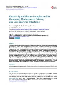
LETTER TO EDITOR (VIEWERS CHOICE) SEVER’S DISEASE: A COMMON BUT COMMONLY UNDIAGNOSED CAUSE OF HEEL PAIN IN GROWING CHILDREN Amit Agrawal, Rashmi Agrawal Keywords: Sever’s disease, heel pain, children A 10 years old girl presented with history of bilateral heel pain, which started about one year back. It was gradually increasing in severity, and had worsened over one month period. Parents or child were not able to correlate the pain with any trauma and it was spontaneous in onset. It was aggravated by physical activity especially running or jumping and relieved by rest. On physical examination, child was wellnourished, with normal vitals and normal systemic examination findings. She had a limping gait with toewalking in between. Local examination of feet revealed no swelling, warmth or redness; however on palpation posterior part of heel was markedly tender, more on left side. Results of her blood investigations including complete blood count, erythrocyte sedimentation rate, C-reactive proteins, serum calcium, liver and renal function tests, anti-streptolysin O (ASO) titre and rheumatoid factor were normal. Radiograph of feet showed increased sclerosis and fragmentation within the calcaneal apophysis (Figure 1). A diagnosis of Sever’s disease was made and patient was advised to take rest and to avoid activities that cause heel pain e.g. sports, jumping or running. Medication in the form of diclofenac gel for local application and oral ibuprofen for two weeks was prescribed. On follow-up after one and half months, pain was decreased significantly and now she was able to walk comfortably. Fig 1 - X-ray ankle showing increased sclerosis and fragmentation of calcaneal apophysis
Sever’s disease or calcaneal apophysitis is a painful inflammation of the calcaneal apophysis, which is thought to be caused by overuse or repetitive microtrauma due to increased traction by achilles tendon on its insertion site i.e. secondary growth plate of the calcaneus. (1-3) The calcaneal growth centre appears at approximately seven years of age and fuses by around 13 years in girls and 15 years
in boys, (4,5) hence it is more commonly seen in growing children. Sever’s disease is characterised by heel pain near lower posterior aspect of the calcaneus. Incidence or prevalence of Sever’s disease is not very well documented and according to western literatures, posterior heel pain associated with Sever’s disease comprise 2-16% of musculoskeletal injuries in children. (6) James Warren Sever first described this condition in 1912 as an inflammation of the calcaneal apophysis associated with muscle strain in immature skeleton, resulting in posterior heel pains, swelling and difficulty in walking. (7) Several different theories are proposed to explain the etio-pathogenesis of posterior heel pain in Sever’s disease: 1) Rapid growth spurt during pubertal age increases relative tension in the tendoachilles/ triceps surae complex with resultant increased traction on apophysis and microfractures. (2,3,8,9) 2) Children with cavus or planus foot are more prone to calcaneal apophysitis, probably due to increased strain on the affected area secondary to harder heel strike placing.(10,11) 3) Repetitive microtrauma causing mechanical disruption is an important causative factor.(12,13) 4) Obesity and weight bearing has also been proposed as an influential factor in calcaneal apophysitis.(5,7) 5) Infection of the affected area as a causative agent was also proposed by some authors but it has been included in differential diagnosis rather than cause by most of the authors.(2,14) However, of these no single theory could gain widespread acceptance due to limitation of clinical data to support them. Recently conducted case control studies reported biomechanical malalignment and gastrocnemius equinus as possible predisposing factors. (11,15) Usual age of presentation of sever’s disease is typically adolescent period, usually between 8 and 15 years. (2,3) Diagnosis can be made on clinical examination alone. Radiographic changes seen in Sever’s disease include increased sclerosis and fragmentation within the calcaneal apophysis, which are non-specific and can be a normal feature of developing calcaneus. (16) Other causes of heel pain in children include achilles tendonitis, calcaneal stress fracture, retrocalcaneal bursitis, calcaneal cysts, osteomyelitis, and plantar fasciitis, which can usually be differentiated by thorough clinical examination and good radiological evaluation. Treatment aims at reducing the present pain and preventing recurrences. Parents must also be reassured about its self-limiting nature and that surgery is usually not required in majority of cases. Just rest and cessation of sports activity resolves symptoms with resumption of sports in 1-2 months in majority. (3) In patients with mild to moderate symptoms other options include wearing inner-shoe heel lift, padding for shock absorption, strapping of heel, monitored stretching or strengthening, pre and post-sport icing, and judicious use of anti-inflammatory agents (NSAIDs). In severe cases, immobilisation using cast in mild equinus for 2-3 wk relieves pain and surgery in the form of total or partial removal of apophysis is rarely required. (17)
Pediatric Oncall October - December 2011. Volume 8 Issue 4
109
http://www.pediatriconcall.com
Contributors list: RA: Acquisition of data, drafting the article, and literature review; AA: Concept, manuscript review, manuscript editing, revising the article critically for important intellectual content; AA will act as guarantor. Both the authors approved the final manuscript. REFERENCES 1. Cassas KJ, Cassettari-Wayhs A. Childhood and adolescent sports-related overuse injuries. Am Fam Physician. 2006; 73: 1014?1022 2. Scharfbillig RW, Jones S, Scutter SD. Sever’s disease: what does the literature really tell us? J Am Podiatr Med Assoc. 2008; 98: 212-223 3. Micheli LJ, Ireland ML. Prevention and management of calcaneal apophysitis in children: an overuse syndrome. J Pediatr Orthop. 1987; 7: 34-38. 4. Volpon J, De Carvalho Filho G. Calcaneal apophysitis: a quantitative radiographic evaluation of the secondary ossification centre. Arch Orthop Trauma Surg. 2002; 122: 338. 5. Harding VV. Time schedule for the appearance and fusion of a second accessory center of ossification of the calcaneus. Child Dev. 1952; 23: 180-184. 6. de Inocencio J. Musculoskeletal pain in primary pediatric care: analysis of 1000 consecutive general pediatric clinic visits. Pediatrics. 1998; 102: E63. 7. Sever JW. Apophysitis of the os calcis. NY Med J. 1912; 95: 1025-1029. 8. Ogden JA, Ganey TM, Hill JD, Jaakkola JI. Sever’s injury: a stress fracture of the immature calcaneal metaphysis. J Pediatr Orthop. 2004; 24: 488-492. 9. Price RJ, Hawkins RD, Hulse MA, Hodson A. The football association medical research programme: an audit of injuries in academy youth football. Br J Sports Med. 2004; 38: 466-471.
10. Szames SE, Forman WM, Oster J, Eleff JC, Woodward P. Sever’s disease and its relationship to equines: a statistical analysis. Clin Podiatr Med Surg. 1990; 7: 377-384. 11. Becerro de Bengoa Vallejo R, Losa Iglesias ME, Rodrihuez Sanz D, Prados Frutos JC, Salvadores Fuentes P, Chicharro JL. Plantar pressures in children with and without sever’s disease. J Am Podiatr Med Assoc. 2011; 101: 17-24. 12. Brenner JS. Overuse injuries, overtraining, and burnout in child and adolescent athletes. Pediatrics. 2007; 119: 1242-1245. 13. Hendrix CL. Calcaneal apophysitis (Sever’s disease). Clin Podiatr Med Surg. 2005; 22: 55-62. 14. Ozgocmen S, Kocakoc E, Kiris A, Sen Y, Ardicoglu O. Calcaneal apophysitis due to brucellosis. J Trop Pediatrics. 2003; 49: 55-58. 15. Scharfbillig RW, Jones S, Scutter S. Sever’s disease: a prospective study of risk factors. J Am Podiatr Med Assoc. 2011; 101: 133-145. 16. Kose O, Celiktas M, Yigit S, Kisin B. Can we make a diagnosis with radiographic examination alone in calcaneal apophysitis (sever’s disease)? J Pediatr Orthop B. 2010; 19: 396-398. 17. Caspi I, Ezra E, Horoszowski H. Partial apophysectomy in sever’s disease. J Orthop Sports Phys Ther. 1989; 10: 370-373.
From:Department of Pediatrics, Chirayu Medical College & Hospital, Bhopal, India. Address for Correspondence: Dr. Amit Agrawal, 28, Ravidas Nagar, Near Nizamuddin Colony, Indrapuri, Bhopal, MP - 462023. India. E-mail :
[email protected] E-published: 1st November 2011. Art#73
LETTER TO EDITOR (VIEWERS CHOICE) IMIDACLOPRID POISONING RESULTING IN DEATH J Harish, N Girish, R Nisarga A 15 years old girl was referred to our hospital with history of consumption of some insecticide poison. She had consumed the poison about 16 hours prior to admission to our hospital. She was first taken to a nearby hospital where she was suspected to have consumed organophosphorus poison and was given stomach wash and treated with atropine before referring to our hospital. On admission she was irritable with dystonic movements. Her vitals were stable with very minimal oral secretions. Her pupils were bilaterally mid-dilated sluggishly reactive to light. She was maintaining saturation at room air. Examination of cardiovascular and respiratory system was within normal limits. She was shifted to pediatric intensive care unit (PICU) and given stomach wash and her clothes were changed. She received symptomatic treatment. The poison she had consumed
was Confidor® which contains imidacloprid. Within few hours her condition deteriorated. She became stuporous with shallow breathing and her oxygen saturation was dropping even with supplemental oxygen. She was intubated and connected to ventilator. Blood gases, renal functions, blood glucose, coagulation profile, pseudocholinestarase were normal. In the next few hours, she went into shock and was put on ionotropic support. However she succumbed inspite of all resuscitative efforts. Imidacloprid, is a synthetic nicotinoid insecticide which has become an important pest control agent on many crops. The neonicotinoid insecticides are related to nicotine in their structure and action at the nicotinic acetylcholine receptor. (1) It is commonly used on rice, cereal, maize, potatoes and vegetable. Imidacloprid is most active against suckling insects because of their
Pediatric Oncall October - December 2011. Volume 8 Issue 4
110