Jul 14, 2011 - science can make in discerning the inner work- ings of the behavioral ... Glutamate. Glutamate receptor.
The
n e w e ng l a n d j o u r na l
of
m e dic i n e
clinical implications of basic research
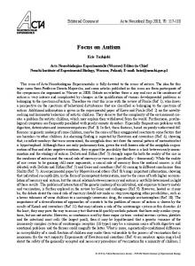
SHANK3, the Synapse, and Autism Martha R. Herbert, M.D., Ph.D. Autism spectrum disorders present a paradox of great heterogeneity and great specificity. Well over 100 genetic disorders yield an autism phenotype,1 most through specific but distinct mechanisms, and many of which affect the synapse. SHANK3 (SH3 and multiple ankyrin repeat domains 3) is known to be disrupted in the 22q13 deletion syndrome (Phelan–McDermid syndrome), and variants of SHANK3 have been linked to autism in some patients without the 22q13 deletion and to schizophrenia2 or intellectual disability3 in those without autism. Using two mouse models, Peça et al.4 have advanced our knowledge of both SHANK3 and autism by probing the functions of the orthologous gene (Shank3) in the mouse. In so doing, they have provided an exemplar of the deep contributions molecular neuroscience can make in discerning the inner workings of the behavioral manifestations of the autism spectrum disorders and in clarifying the path toward the translational links that address these disorders, which have devastating consequences for individual and public health and costly economic implications. The Shank family of proteins provides scaffolding for signaling molecules in the postsynaptic density (a multiprotein complex attached to the postsynaptic membrane) of glutamatergic synap ses, making these molecules vulnerable to Shank3 mutations. By targeting separate portions of Shank3, the authors engineered two mouse models, each a harboring different mutant allele of the gene. Each mutant allele has distinct effects on the three isoforms of Shank3 — α, β, and γ. Mice engineered to lack expression of the α isoform but in which the β and γ isoforms were unaffected showed little behavioral change as compared with controls. But Shank3B−/− mice, in which both the Shank3α and Shank3β isoforms were absent and the level of Shank3γ was markedly reduced, had an autism-like combination of behavioral features that could not be explained
by rearing. These features included anxiety, selfinjurious behavior, avoidance of social interaction, and poor perception of social novelty (Fig. 1). Shank3 is highly expressed in the striata of humans and mice. The striata of the Shank3B−/− mice showed several microanatomical features consistent with reduced intensity of neuronal signaling. The postsynaptic densities were thinner and shorter than those of control mice, and the levels of several scaffolding proteins and glutamate-receptor subunits were reduced. Consistent with this observation was the comparatively low signal intensity in the corticostriatal synaptic circuitry, and on stimulation, the lower amplitude and frequency of miniature excitatory postsynaptic currents (mEPSCs). This reduction in postsynaptic activity seemed to be restricted to the striatum. Cellular changes pertinent to signal intensity were also found: the arborization of dendrites in medium spiny neurons was more complex, with longer dendrites and increased surface area. This finding is consistent with the larger brain size in many children with autism spectrum disorders (although there may be other explanations for brain enlargement). Even though overall brain size was not increased in the Shank3B−/− mice, there was a small but significant increase in caudate volume; a larger caudate has been found in persons with autism spectrum disorders and may be linked to the repetitive behaviors that are part of the core deficiency features of autism spectrum disorders. Could the development of longer and more complex dendrites be the neuron’s compensatory response to weaker synaptic function? If the cell is programmed to expect a certain level of stimulation that does not arrive, might it not grow out its dendrites to seek more? Yet at the same time these longer dendrites had fewer spines, even though the width of the spine necks was slightly larger than that in controls. The study does not
n engl j med 365;2 nejm.org july 14, 2011
The New England Journal of Medicine Downloaded from nejm.org at HARVARD UNIVERSITY on March 25, 2012. For personal use only. No other uses without permission. Copyright © 2011 Massachusetts Medical Society. All rights reserved.
173
The
A
n e w e ng l a n d j o u r na l
of
m e dic i n e
Synapse in Shank3B –/– knockout mouse
Synapse in wild-type mouse Presynaptic
Presynaptic
Glutamate
Glutamate
β-Neurexin
Glutamate receptor PSD-95 GKAP α β γ
Neuroligin-1,3
PSD-95
Shank
GKAP γ
Postsynaptic
B
β-Neurexin
Glutamate receptor
Neuroligin-1,3
Shank
Postsynaptic
WT
KO
Normal postsynaptic density
Smaller postsynaptic density
Normal dendrites
Dendrites with more complex arborization
C
D
10 pA 1s
0.2 s
E
Normal-sized striatum Brain of mouse
Enlarged striatum Brain of mouse
Normal behavior
Anxiety, self-injurious behavior, avoidance of social interaction
explore the functional consequences of these morphologic features, but one wonders whether they stifle the neuron’s quest — if it exists — for input. At what levels can the authors’ multileveled elucidation of the specific phenotype of Shank3B−/− mice be generalized across many different mu174
n engl j med 365;2
tations or mechanisms? The elegant methods they used to identify this array of abnormalities can also be used to flesh out the molecular, anatomical, and functional components of other varieties of synaptic dysfunction that may also contribute to the daunting heterogeneity of autism spec-
nejm.org
july 14, 2011
The New England Journal of Medicine Downloaded from nejm.org at HARVARD UNIVERSITY on March 25, 2012. For personal use only. No other uses without permission. Copyright © 2011 Massachusetts Medical Society. All rights reserved.
clinical implications of basic research
Figure 1 (facing page). An Angle on Autism Spectrum Disorder. Knocking out Shank3 in the mouse results in the elimination of the Shank3α and Shank3β isoforms and a reduction of the putative Shank3γ isoform at the postsynaptic density. Levels of the scaffolding proteins, including guanylate kinase-associated protein (GKAP, also known as SAPAP), and several subunits of the glutamate receptor are reduced (Panel A). In addition to forming a key scaffold at glutamatergic synapses, PSD95–GKAP–Shank complex proteins interact with other synaptic proteins in the postsynaptic density (Panel B, arrows); also shown are synaptic contacts with presynaptic vesicles (arrowheads) and the dendritic spine (stars). Dendrites in the mutant mice show more arborization and spines with a lower density than those in wild-type mice (Panel C), and the amplitude and frequency of miniature excitatory postsynaptic currents (mEPSCs) are compromised (Panel D). Although the overall size of the brain in mutant mice is no different from that in wild-type mice, the striatum is slightly larger (Panel E). The behavioral manifestations associated with these neurobiologic changes are autistic-like, including reduced social interaction, less seeking of social novelty, excessive grooming, and self-injurious behavior.
trum disorders. Given the molecular complexity of the postsynaptic density alone, one could imagine many different genetic alterations (or for that matter, pathophysiologic or xenobiotic influences) that could compromise the amplitude and frequency of miniature excitatory postsynaptic currents (mEPSCs). Shank3 is also expressed in the gut, the kidney, and other body tissues and may play a role in epithelial turnover and mucosal immune development; in addition, host Shank3 is recruited by gut pathogens in their induction of actin rearrangement.5 These observations suggest that the systemic effects of alterations in Shank3 might contribute to the systemic features of autism spectrum disorders found in many patients. A broad view is important, since at present the vast majority of cases are idiopathic, and both heterogeneous environmental influences and genetic variation may be contributing to synaptic perturbations and phenotypic variability.
n engl j med 365;2
One of the most intriguing developments in the study of autism spectrum disorders is the emerging evidence of transient or persistent remission. Molecular interventions have reversed symptoms in animal models of three genetic syndromes associated with autism spectrum disorder: the fragile X syndrome, the Rett syndrome, and tuberous sclerosis. Symptoms of autism spectrum disorder can transiently remit with fever, treatment with steroids, or a diet restricted to clear fluids in preparation for a medical procedure. In addition, in up to 20% of children with autism spectrum disorders, symptoms seem to abate after intensive intervention. Which among the dysfunctional synaptic routes to autism spectrum disorder might be most amenable to modulation or even reversal? It is noteworthy that the array of autistic-like behaviors in this model occurred without a complete knock-out of Shank3 production: significant expression of the Shank3γ isoform remained. Could these effects just as easily have been caused by perturbations in the splicing of the Shank3 primary transcript? Such mechanisms may contain elements of reversibility. Disclosure forms provided by the author are available with the full text of this article at NEJM.org. From the Massachusetts General Hospital and Harvard Medical School — both in Boston. This article (10.1056/NEJMcibr1104261) was updated on November 9, 2011, at NEJM.org. 1. Betancur C. Etiological heterogeneity in autism spectrum
disorders: more than 100 genetic and genomic disorders and still counting. Brain Res 2011;1380:42-77. 2. Gauthier J, Champagne N, Lafrenière RG, et al. De novo mutations in the gene encoding the synaptic scaffolding protein SHANK3 in patients ascertained for schizophrenia. Proc Natl Acad Sci U S A 2010;107:7863-8. 3. Hamdan FF, Gauthier J, Araki Y, et al. Excess of de novo deleterious mutations in genes associated with glutamatergic systems in nonsyndromic intellectual disability. Am J Hum Genet 2011;88:306-16. 4. Peça J, Feliciano C, Ting JT, et al. Shank3 mutant mice display autistic-like behaviours and striatal dysfunction. Nature 2011;472:437-42. 5. Huett A, Leong JM, Podolsky DK, Xavier RJ. The cytoskeletal scaffold Shank3 is recruited to pathogen-induced actin rearrangements. Exp Cell Res 2009;315:2001-11. Copyright © 2011 Massachusetts Medical Society.
nejm.org
july 14, 2011
The New England Journal of Medicine Downloaded from nejm.org at HARVARD UNIVERSITY on March 25, 2012. For personal use only. No other uses without permission. Copyright © 2011 Massachusetts Medical Society. All rights reserved.
175