Short Technical Reports cular Biology, Georgetown University Medical Center, Washington, D.C. 20057, USA. e-mail:
[email protected] Received 15 May 2002; accepted 16 August 2002.
Negative Purification Method for the Selection of Specific Antibodies from Polyclonal Antisera BioTechniques 33:1050-1054 (November 2002)
Peter D. Burbelo, Adam E. Kisailus, and Jeremy W. Peck Georgetown University Medical Center Washington, D.C., USA
For reprints of this or any other article, contact
[email protected]
ABSTRACT We developed a protocol to remove nonspecific antibodies from polyclonal antisera by adsorption on non-target antigens immobilized on nitrocellulose membranes. This “negative” purification method is simple and provides better immunoreagents than the blocking of nonspecific antibodies in solution or the enrichment of specific antibodies on nitrocellulose membranes. For routine applications, this method is quicker and cheaper than the purification protocols based on selective precipitations and affinity chromatography.
INTRODUCTION Antibodies are instrumental in protocols for immunolocalization, immunopurification, Western blot analysis, EMSA super-shifts, or the screening of cDNA expression libraries—not to mention their use in detection kits. Each of these applications has their own requirements in terms of the purity of the antibodies. Although monoclonal antibodies are free from interfering host immunoglobulins, their production is expensive and requires technical knowledge and animal cell culture facilities. Alternatives such as the phage display technology and the in vitro production of recombinant antibodies suffer from similar limitations. Thus, the use of polyclonal antisera remains very popular but necessitates the removal of nonspecific antibodies. Purification typically begins with a combination of ammonium sulfate/ caprylic acid precipitation or ammonium sulfate/DEAE chromatography (5), and nearly pure antibodies are recovered, although frequently at low titers because of their limited solubilization or inactivation (2). They are typically submitted to an additional affinity pu-
rification. Protein A, protein G, or antiIgG affinity chromatography of IgG from ascites or polyclonal antisera (1) is a straightforward method but not always suitable for all species and antibody subclasses (4), and nonspecific IgG antibodies still end up in the final eluate. Antigen affinity chromatography is the best technique to obtain highly purified and specific antibodies (3) but often requires harsh conditions (e.g., extreme pH, chaotropic and denaturating agents, etc.) to elute the antigen, causing a loss of activity (2,3). Finally, whichever protocol is used, it has to be optimized for each antigen, which is hardly conceivable for the routine production of sera against a significant number of the proteins present on a 2-D electrophoresis gel. Quicker protocols have been used to obtain partially purified preparations. These consist simply of the centrifugation of the nonspecific antibodies inhibited by blocking mixtures (8) or of the adsorption of specific antibodies on nitrocellulose-bound antigens and their subsequent recovery (7). Here we discuss what we call a negative antibody purification in which we remove nonspecific antibodies by incubating polyclonal antisera with non-target proteins that are adsorbed on a membrane. The efficiency of each method is evaluated using Western blot analysis. MATERIALS AND METHODS Bacterial Strains and Growth Conditions E. coli BL21 (DE3)® (Novagen, Madison, WI, USA) clones were grown in liquid LB medium at 37°C under constant agitation in the presence of 100 µg/mL ampicillin. Two bacterial test clones were used. The pET15b-GMD clone contained the pET15b® plasmid (Novagen) with a 1122-bp NdeI/XhoI cDNA fragment of the Arabidopsis thaliana GDP-D-mannose-4,6-dehydratase gene (GMD) (GenBank® accession no. U81805) and the pET21c-GE clone contained the pET21c® plasmid (Novagen) with a 1056-bp NotI/SalI cDNA fragment corresponding to the A. thaliana UDP-glucose-4-epimerase gene (GE) (accession no. Z54214). Vol. 33, No. 5 (2002)
Short Technical Reports Protein Induction The bacterial clones were inoculated in 100 mL LB medium and incubated overnight at 37°C under constant agitation. LB medium (1 L) was inoculated with the pre-culture and incubated at 37°C until A600 = 0.4. Induction was obtained by incubating with IPTG (1 mM final concentration) for 3 h. The cells were collected by centrifugation at 3800× g for 5 min at 4°C. Protein Purification The overexpressed proteins were recovered from bacterial cells as inclusion bodies, according to the manufacturer’s protocol (Novagen). Insoluble GE proteins were resuspended in 10 mL solubilizing buffer (4.29 mM Na2HPO4, 1.47 mM KH2PO4, 2.7 mM KCl, 0.137 mM NaCl, 0.1 % Tween® 20, and 0.002% sodium azide, pH 7.3) containing 6 M urea, sonicated three times for 1 min using a Bandelin Sonoplus sonicator type GM-2070 (Bandelin electronic, Berlin, Germany), and centrifuged at 39 000× g for 20 min at 4°C. The supernatant was filtered on a 0.45-µm MILLEX-HA® filter (Millipore, Bedford, MA, USA) before further purification. Insoluble GMD proteins were resuspended in 10 mL solubilizing buffer (20 mM Tris-HCl, pH 7.9, 5 mM imidazole, 0.5 mM NaCl) containing 6 M urea and were processed as described for GE. The resulting solubilized protein fraction was diluted by adding two volumes of the appropriate urea-free solubilizing buffer before loading it on the affinity column. GE was further purified on a T7-Tag® affinity resin, and GMD was further purified on a Hisbind® affinity resin, according to the manufacturer’s protocols (Novagen). Immunization Procedure The purified proteins (100 µg in 0.25 mL) were mixed with one volume Quil A adjuvant (330 µg/mL in 137 mM NaCl, 2.7 mM KCl, 10 mM Na2HPO4, 1.8 mM KH2PO4, pH 7.4) (Superfos Biosector, Frederikssund, Denmark) and used to perform the intramuscular immunization of two rabbits (New Zealand Californian hybrid, IFFA Cre1052 BioTechniques
do). Boost injections (100 µg in 0.5 mL) were performed 7, 28, 58, and 65 days later. Polyclonal antisera were collected three days after the last injection. Polyclonal Antisera Purification Procedures Preincubation with E. coli proteins. The removal of unwanted antibodies was performed by incubating polyclonal antisera with an excess of E. coli proteins isolated from a BL21 (DE3) strain containing the appropriate pET® plasmid without insert. Briefly, cells from a 1-L overnight culture were collected by centrifugation at 3800× g for 5 min at room temperature. The cells were then sonicated for 1 min at 4°C using a Bandelin Sonoplus sonicator and centrifuged at 17 500× g for 15 min at 4°C. The supernatant was incubated with a 1:50 dilution of the appropriate polyclonal antiserum for 1 h at room temperature under mild agitation. This mixture was finally centrifuged at 2000× g for 5 min at 4°C before it was used in Western analysis. Positive purification of specific antibodies. Positive purification of the polyclonal antisera was performed by immobilizing the target antigens on a nitrocellulose membrane and then capturing specific antibodies by soaking the membrane in the serum, according to a modification of a previously described method (7). T7-Tag or His-bind affinity-purified proteins from a 100mL IPTG-induced culture of each corresponding BL21 (DE3) test clone were separated on a preparative 10% SDSpolyacrylamide gel (10 cm × 8 cm × 1 mm; 1 well) and transferred to a Hybond®-C extra nitrocellulose membrane (Amersham Biosciences, Piscataway, NJ, USA) (see Western Blot Analysis section). The membrane was stained briefly with Ponceau S (Sigma, St. Louis, MO, USA) to visualize the overexpressed proteins and dried to achieve the protein fixation. The area containing the target protein was cut out of the membrane and blocked in 10 mL modified TBS containing 3% BSA for 1 h at room temperature under slow agitation. The saturated piece of membrane was then incubated overnight in 10 mL of a 1:100 dilution of the appropriate polyclonal antiserum at 4°C and washed
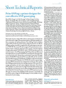
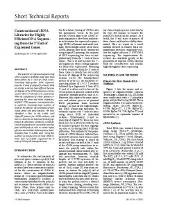
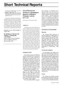
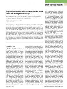
four times in 10 mL modified TBST for 10 min under mild agitation at room temperature. Specific antibodies were eluted by incubating the nitrocellulose sample in 5 mL 0.2 M glycine-HCl, pH 2.5, for 2.5 min. The eluted antibody solution was neutralized by the addition of 0.85 mL 1 M Tris-HCl, pH 8.8, containing 6.9% BSA and used as primary antibody in Western blots at 1:1000 and 1:10 000 final dilutions of anti-GE and anti-GMD polyserum, respectively. Negative purification of specific antibodies. Negative purification of the polyclonal antisera was performed by the absorption of nonspecific antibodies on their antigens immobilized on a nitrocellulose membrane. The total protein fraction of a 1 L IPTG-induced culture of the BL21 (DE3) clone transformed with either empty pET15b or empty pET21c plasmid was isolated and processed as described earlier for the positive purification procedure. The membrane was incubated overnight in the presence of 10 mL 1:100 dilution of the appropriate polyclonal antiserum at 4°C to remove nonspecific antibodies from the solution. The supernatant was finally used as the primary antibody in Western blots. Western Blot Analysis Proteins were separated on a 10% SDS-polyacrylamide gel (10 cm × 8 cm × 0.75 mm) and transferred to a Hybond-C extra nitrocellulose membrane using a Transphor Tank electro-transfer unit (Hoefer Scientific Instruments, San Francisco, CA, USA). The membrane was incubated in anti-GE or anti-GMD polyclonal antisera and processed according to Reference 8. Primary antibodies were detected by a 1:30 000 dilution of anti-rabbit IgG alkaline phosphatase conjugate (Sigma) for 30 min at room temperature under mild agitation. After several washes in modified TBST, complexes were revealed using the NBT/BCIP colorimetric assay, according to the manufacturer’s protocol (Roche Applied Science, Indianapolis, IN, USA). RESULTS AND DISCUSSION Figure 1, A and B, shows the results Vol. 33, No. 5 (2002)
of the Western blots obtained on the recombinant GE (41 kDa) and GMD (44 kDa), respectively. The raw polyclonal antisera detected the antigens along with nonspecific protein bands (lanes 1 and 2), and blocked polyclonal antisera did not yield much better results (lanes 3 and 4). Both polyclonal antisera detected proteins of unexpected molecular weight on the affinity-purified fractions (lanes 1 and 3) as on the crude protein fractions (lanes 2 and 4).
The detection of higher molecular weight bands in the purified protein samples was confirmed with an anti-tag antibody (data not shown), which suggested the multimerization of the overexpressed protein, a feature that has been previously reported (9). This was confirmed by the persistence of these bands when the target protein of expected size was electro-eluted, re-electrophoresed, blotted, and probed (data not shown). The low molecular bands
A
B Figure 1. Effect of three polyclonal antiserum purification methods on the detection of two test proteins overexpressed in E. coli. (A) GE and (B) GMD, whose sequences were obtained from A. thaliana, were detected by Western blot analysis using a 1:1000 and 1:10 000 dilution of anti-GE and anti-GMD polyclonal antiserum, respectively. Lanes 1 and 2, polyclonal antiserum without purification; lanes 3 and 4, purified polyclonal antiserum obtained by preincubation with an excess of E. coli proteins in solution; lanes 5 and 6, positive purification method; and lanes 7 and 8, negative purification method. Each polyclonal antiserum was used to detect GE or GMD in an affinity-purified antigen fraction (lanes 1, 3, 5, and 7; 100 ng) and in a crude IPTG-induced E. coli protein fraction from the corresponding test clone (lanes 2, 4, 6, and 8; 100 ng). Molecular weights were estimated with the pre-stained broad range SDS-PAGE standards (Bio-Rad Laboratories, Hercules, CA, USA). Vol. 33, No. 5 (2002)
Short Technical Reports probably corresponded to truncated expression products that resulted either from proteolytic degradation due to protein instability (10) or secondary site translation initiation (6), as mentioned by the pET plasmid’s manufacturer (Novagen). Aside from these oligomerization and truncation artifacts, the blocked polyclonal antiserum is of limited specificity in Western blotting experiments. In our hands, the second purification method (specific antibodies desorbed from their target immobilized on membrane) (7) gave protein bands of unexpected molecular weight in Western blot analysis (data not shown). The nitrocellulose band was therefore washed in modified TBST, with an ionic strength increased from 150 mM to 500 mM NaCl to maximize the removal of nonspecific antibodies. As shown in Figure 1, the positively purified polyclonal antisera recognized the target antigen with almost the same efficiency as before the purification step. However, they contained fewer antibodies of undetermined specificity because only a limited number of non-target proteins were recognized in the case of the antiGE polyclonal antiserum, and almost no other protein was recognized in the case of the anti-GMD polyclonal antiserum for either the purified fraction (lanes 5) or the crude E. coli protein fractions (lanes 6). However, this positive purification method has three limitations. First, the excision of the target antigen is not very reproducible because of the fuzziness of the Rouge Ponceau coloration. The size of the excised protein band may vary from experiment to experiment, and variable amounts of nonspecific antibodies will still bind the saturated nitrocellulose and end up in the eluted antibody fraction. Second, the positive purification method becomes time consuming when the specific antibody titer is high, in which case it is impossible to recover the antibodies from the polyclonal antiserum in a single round. Third, the strongest limitation of this method is that the most specific antibodies that bind the antigen with the highest affinity cannot be desorbed from the nitrocellulose by a 2.5-min incubation at a pH of 2.5 (standard protocol). Harsher experimental conditions such as in1054 BioTechniques
creased elution time, incubation in the presence of denaturing and reducing agents (50 mM Tris, pH 6.8, 2% SDS, 100 mM β-mercaptoethanol), or heating at 70°C resulted in significant loss of the binding activity of the serum (data not shown). Therefore, we chose a purification method in which antisera were purified by adsorbing contaminating antibodies on their antigens immobilized on nitrocellulose, leaving the specific antibodies in solution. The negative purification protocol consists of the incubation of the polyclonal antiserum with the crude protein fraction of the IPTG-induced BL21 (DE3) clone harboring the corresponding empty plasmid blotted on a nitrocellulose membrane. This third serum specifically recognized its target protein from purified (lanes 7) and crude protein fractions (lanes 8), with virtually no non-target binding. In conclusion, here we described a method for the simple and efficient affinity purification of polyclonal antisera on membranes blotted with contaminating antigens. This simple protocol leaves the specific antibodies in solution throughout the whole procedure and needs no optimization for desorbing them from the nitrocellulose membrane, thus allowing the automation of the procedure.
Baluska, J. Samaj, D. Volkmann, and J. Kendrick-Jones. 1999. Characterization of the unconventional myosin VIII in plant cells and its localization at the post-cytokinetic cell wall. Plant J. 19:555-567. 8.Sambrook, J., E.F. Fritsch, and T. Maniatis. 1989. Molecular Cloning: A Laboratory Manual (2nd ed). CSH Laboratory Press, Cold Spring Harbor, NY. 9.Sprules, T., N. Green, M. Featherstone, and K. Gehring. 1998. Nickel-induced oligomerization of proteins containing 10-histidine tags. BioTechniques 25:20-22. 10.Tobias, J.W., T.E. Shrader, G. Rocap, and A. Varshavsky. 1991. The N-end rule in bacteria. Science 254:1374-1377.
The authors gratefully acknowledge the DGTRE (Région Wallonne, Belgium) for financial support, J. Messiaen and L. Le for helpful discussions, and P. Cambier for excellent technical assistance. Address correspondence to Pierre Van Cutsem, Unité de Recherche en Biologie Cellulaire végétale, Facultés Universitaires Notre-Dame de la Paix, 61 rue de Bruxelles, B-5000 Namur, Belgium. e-mail: pierre.vancutsem@ fundp. ac.be Received 15 November 2002; accepted 16 July 2002.
Cindy Benet and Pierre Van Cutsem Facultés Universitaires Notre-Dame de la Paix Namur, Belgium
REFERENCES 1.Akerstrom, B. and L. Bjorck. 1986. A physicochemical study of protein G, a molecule with unique immunoglobulin G-binding properties. J. Biol. Chem. 5:10240-10247. 2.Bazin, H., F. Cormont, and L. De Clercq. 1986. Purification of rat monoclonal antibodies. Methods Enzymol. 121:638-652. 3.Kristiansen, T. 1978. Matrix-bound antigens and antibodies, p. 191-206. In O. HoffmanOstenhof (Ed.), Affinity Chromatography. Pergamon Press, Oxford. 4.Lindmark, R., K. Thoren-Tolling, and J. Sjoquist. 1983. Binding of immunoglobulins to protein A and immunoglobulin levels in mammalian sera. J. Immunol. Methods 62:1-13. 5.Mamiushichev, V.B., L.B. Potapenko, A.A. Dement’ev, and I.N. Sokolov. 2001. Optimization of a method for purifying antibodies for immunofluorencent diagnosis of influenza. Vopr. Virusol. 46:44-47. 6.Preibisch, G., H. Ishihara, D. Tripier, and M. Leineweber. 1988. Unexpected translation initiation within the coding region of eukaryotic genes expressed in Escherichia coli. Gene 72:179-186. 7.Reichelt, S., A.E. Knight, T.P. Hodge, F.
For reprints of this or any other article, contact
[email protected]
Vol. 33, No. 5 (2002)