www.nature.com/scientificreports
OPEN
received: 28 January 2016 accepted: 04 April 2016 Published: 15 April 2016
Sight restoration after congenital blindness does not reinstate alpha oscillatory activity in humans Davide Bottari1, Nikolaus F. Troje2,3, Pia Ley1, Marlene Hense1, Ramesh Kekunnaya4 & Brigitte Röder1 Functional brain development is characterized by sensitive periods during which experience must be available to allow for the full development of neural circuits and associated behavior. Yet, only few neural markers of sensitive period plasticity in humans are known. Here we employed electroencephalographic recordings in a unique sample of twelve humans who had been blind from birth and regained sight through cataract surgery between four months and 16 years of age. Two additional control groups were tested: a group of visually impaired individuals without a history of total congenital blindness and a group of typically sighted individuals. The EEG was recorded while participants performed a visual discrimination task involving intact and scrambled biological motion stimuli. Posterior alpha and theta oscillations were evaluated. The three groups showed indistinguishable behavioral performance and in all groups evoked theta activity varied with biological motion processing. By contrast, alpha oscillatory activity was significantly reduced only in individuals with a history of congenital cataracts. These data document on the one hand brain mechanisms of functional recovery (related to theta oscillations) and on the other hand, for the first time, a sensitive period for the development of alpha oscillatory activity in humans. The posterior alpha oscillatory activity (8–12 Hz) of the human electroencephalogram (EEG) has been associated with the inhibition of task-irrelevant neural circuits1 and has been considered as a mechanism for coordinating neural activity in a large number of perceptual and cognitive tasks2. Alpha activity increases (alpha synchronization) result in an inhibition of task-irrelevant neural circuits, alpha decreases (alpha desynchronization) are associated within an engagement of task-relevant neural populations1,2. Moreover, the level of pre-stimulus alpha was found to predict visual processing efficiency3,4 and seems to be important for the control of attention5. Alpha oscillations dominate in the granular and infragranular layers of the cortex; they most likely result from the activity of pyramidal cells modulated by pulsed GABA (Gamma-amino-butyric acid) mediated inhibition of fast-spiking inhibitory interneurons1,6. The EEG of a newborn is characterized by lower frequency activity including theta oscillations7. Posterior alpha activity seems to hardly exist in the EEG of newborns7 and shows a protracted developmental time course lasting until late childhood8. Posterior alpha activity during rest9,10 and in somatosensory tasks11,12 has been found to be markedly reduced or even absent in humans who had been totally blind since birth or who had experienced no more than unstructured light sensations. In contrast, oscillatory activity in lower (e.g. delta) and higher frequency bands (e.g. gamma) during rest have been reported to be relatively unimpaired in individuals with permanent congenital blindness13. Interestingly, people who had lost their sight as adults showed a gradual decrease of alpha activity over the following months11. These findings suggest that alpha oscillations crucially depend on structured visual input. However, it is not yet known, whether the emergence of the neural mechanisms generating alpha oscillations is linked to a sensitive period during which visual experience must be available. Individuals who had their sight restored after a congenital total blindness offer the unique possibility to address this question. In fact, individuals who were born with dense bilateral cataracts which were removed later in life provide such 1
Biological Psychology and Neuropsychology, University of Hamburg, Von-Melle-Park 11 20146 Hamburg, Germany. Department of Psychology, Queen’s University, 62 Arch Street, K7L 3N6 Kingston, Ontario, Canada. 3Canadian Institute for Advanced Research, 180 Dundas Street West, Suite 1400, M5G 1Z8 Toronto, Ontario, Canada. 4Jasti V Ramanamma Children’s Eye Care Center, LV Prasad Eye Institute, Kallam Anji Reddy Campus, L V Prasad Marg, Banjara Hills, 500 034 Hyderabad, Andhra Pradesh, India. Correspondence and requests for materials should be addressed to D.B. (email:
[email protected]) 2
Scientific Reports | 6:24683 | DOI: 10.1038/srep24683
1
www.nature.com/scientificreports/
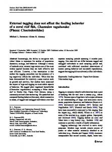
Figure 1. Behavioral and EEG/event-related brain potential (ERP) results. (a) Thresholds of single participants (black diamond = congenital cataract group, cc; gray dots = developmental cataract group, dc; white triangles = matched controls, mc) in the behavioral biological motion (BM) detection task22; higher values indicate higher sensitivity. The three groups did not differ in detecting BM. (b) Hit rates in the EEG biological motion task are separately shown for all groups. The three groups did not differ in their ability to detect the visual targets. (c) Relative power change of the evoked theta response (4–7 Hz; averaged across posterior electrodes, TP8/9, P7/8, and O1/2). Data are shown separately for each group (cc, dc, mc) and condition (BM and scrambled biological motion, SBM). Evoked theta activity was higher for BM than for SBM in all three groups; in addition, the mc group showed higher evoked theta response compared to both dc and cc groups. (d) Mean amplitudes of the N1 wave of the event-related potentials elicited by BM and SBM stimuli, displayed separately for the cc, dc and mc groups. The N1 was enhanced to biological compared to scrambled biological motion stimuli irrespectively of group (data of cc and mc groups from20). *indicates significant condition and/or group effects. a model in humans14–16 and allow uncovering sensitive periods analogous to the extensive work in animals17,18. Strong evidence in favor of a sensitive period for the emergence of the neural mechanisms underlying alpha oscillatory activity would be if it could be shown that alpha oscillations do not or not fully recover after restoring sight following a congenital blindness while alpha oscillations would be recorded in visually impaired individuals who had had some vision at birth. Here we recorded the electroencephalogram (EEG) from twelve individuals who had been born blind due to dense bilateral congenital cataracts (cc) while they were processing intact and scrambled biological motion stimuli. Recent evidence has suggested that the behavioral sensitivity to biological motion is indistinguishable for congenital cataract-reversal individuals and matched controls19,20. Thus, investigating oscillatory activtiy in the context of biological motion processing allows us to identify the neural mechanisms of functional recovery. Moreover, by studying alpha activity we were able to assess neural mechanisms related to the control of the excitatory/inhibitory balance of neural ciricuits. Since the latter crucuially rely on GABA-mediated mechanisms, we consider alpha oscillatory activity as an index for the functionality of inhbitory circuits whose establishment has often been postulated to constitute a hallmark of brain development21. In order to control for the impact of factors related to cataract surgery (e.g. seeing with an interocular lense) or residual visual impairments, we included six individuals with a history of either developmental or incomplete congenital cataracts (refered to as developmental cataract individuals, dc). Moreover an additional group of healthy age matched controls (mc) was tested. It was recently shown that congenital cataract individuals and matched controls display a similar modulation of the N1 of the ERPs while processing biological motion20. Since phase-locked theta oscillatory activity has been related to the N1, we predicted evoked theta oscillatory activity to vary as a function of biological motion processing in all groups. By contrast, alpha activity was hypothesized to depend on visual input from birth.
Results
Behavioral recovery. We ran the subtest “detection” of the Motion Lab Battery22, which is a standardized assessment of the ability to detect biological motion stimuli (point light displays of walking humans) in subgroups of our participants since data was not available for all due to time limitations and technical problems (see Material and Methods). This test did not reveal any group difference (group effect (F(2, 15) = 1.2, p > 0.3) (Fig. 1a): The cc group’s (n = 7, see Table 1) performance did neither differ from the performance of the mc group (n = 7; t(12) = − 1.3, p > 0.2) nor from the performance of the dc group (n = 4, see Table 1; t(9) = 0.1, p > 0.9), and the dc group did not differ from the mc group (t(9) = 1.2, p > 0.2). Biological Motion experiment. Behavioral performance. While recording the EEG, intact and scram-
bled point light displays of walking humans (biological motion, BM, and scrambled biological motion, SBM, respectively) were presented intermixed with rare “target” point light displays of a moving cat which had to be detected. All groups (cc: N = 12, dc: N = 6 and mc: N = 12, see Table 1) performed at a high level and no difference in detection rates between groups was observed (mean hit rates: cc = 98.8%, SE = 0.6, dc = 99.3%, SE = 0.7 and mc = 98.6%, SE = 1.0 and; F(2, 26) = 0.1, p > 0.8, Fig. 1b). The false alarms rate was below 1% in all groups. EEG results. Oscillatory brain activity in the theta (4–7 Hz) and in the alpha band (8–12 Hz) were analyzed. The results for the evoked theta activity replicated previous ERP-N1 findings (see23): Higher theta power was observed
Scientific Reports | 6:24683 | DOI: 10.1038/srep24683
2
www.nature.com/scientificreports/ Last postsurgical visual acuity in best eye
Mean accuracy in BM EEG logMar task, %
BM detection task, noise dots n**
Gender
Cataract onset
Nystagmus
Presurgical visual acuity in best eye*
cc-a
23
M
congenital
48
Unknown
yes
Unknown
0.16
0.80
100.0
46.3
cc-b
35
M
congenital
24
Unknown
yes
Unknown
0.50
0.30
93.8
13.7
cc-c
17
F
congenital
168
No view
yes
FC:0.5
0.13
0.90
100.0
–
cc-d
17
F
congenital
192
No view
yes
PL + , PR +
0.02
1.78
97.8
–
cc-e
10
M
congenital
108
No view
yes
PL + , PR +
0.02
1.78
100.0
–
cc-f
11
F
congenital
120
No details
yes
FC:0.5
0.05
1.30
96.4
0.2
cc-g
31
M
congenital
72
Unknown
yes
No vision
0.05
1.30
100.0
27.4
cc-h
11
F
congenital
120
No view
yes
PL + , PR +
0.16
0.80
100.0
10.2
cc-i
11
M
congenital
96
No view
yes
PL + , PR +
0.32
0.50
100.0
22.7
Participant
Age (years)
Fundus visibility Age at surgery pre-surgery best eye (months)
Decimal
cc-l
13
M
congenital
120
No view
yes
PL+ , PR+
0.02
1.78
100.0
28.7
cc-m
21
M
congenital
4
Unknown
yes
Unknown
0.13
0.90
100.0
–
cc-n
13
M
congenital
60
No view
yes
Unknown
0.13
0.90
97.4
–
Mean
17.8
0.14
1.09
99.8
21.3
94.3
dc-a
19
F
Developmental
24
Unknown
yes
PL+ , PR+
0.80
0.10
100.0
20.1
dc-b
8
M
developmental
84
Unknown
no
0.25
0.25
0.60
100.0
0.9
dc-c
22
F
congenital not dense
252
Haze but Visible
yes
FC:0.5
0.08
1.10
100.0
–
dc-e
38
F
congenital not dense
Not operated
Visible
yes
0.08
0.30
1.10
100.0
–
dc-g
10
F
congenital not dense
72
No view
no
FC:2
0.13
0.90
95.8
30.6
dc-h
9
M
congenital not dense
96
Haze but Visible
yes
FC:1
0.08
1.10
100.0
28.9
Mean
17.6
0.27
0.82
99.3
20.1
88.0
Table 1. Description of participants and performance in the four different tasks. *PL+ : able to perceive light; PR+ : able to report the location of light; FC: able to count fingers at n meters. BM: Biological motion; GM: Global motion. **Degree if noise at which a hit rate of 82% was reached (high values indicate high sensitivity) in the behavioral BM task. ***Percentage of coherence level necessary to reach a hit rate of 82% (high values indicate low sensitivity) in the behavioral GM task. Note patient dc-a was categorized as dc due to parents’ reports supported by a low nystagmus and the achieved high visual outcome.
for intact than for scrambled biological motion stimuli (main effect of condition F(1, 27) = 10.9, p = 0.003). This effect was observed in each group (cc: t(11) = 2.4, p = 0.03; dc: t(5) = 2.5, p = 0.055; mc: t(11) = 3.0, p = 0.01; see Fig. 1c and supplementary Figure 1). Theta band power was, however, overall higher in the mc group than in the cc group and the dc group (main effect of group F(2, 27) = 6.9, p