MATBIO-01006; No of Pages 10 Matrix Biology xxx (2013) xxx–xxx
Contents lists available at ScienceDirect
Matrix Biology journal homepage: www.elsevier.com/locate/matbio
Mini review
2
Signalling pathways linking integrins with cell cycle progression☆
3Q1
Paulina Moreno-Layseca, Charles H. Streuli ⁎
4
Wellcome Trust Centre for Cell-Matrix Research, Faculty of Life Sciences, University of Manchester, Oxford Road, Manchester M13 9PT, UK
i n f o
a b s t r a c t
Article history: Received 16 August 2013 Received in revised form 22 October 2013 Accepted 22 October 2013 Available online xxxx
P
Integrins are adhesion receptors that allow cells to sense and respond to microenvironmental signals encoded by the extracellular matrix. They are crucial for the adhesion, survival, proliferation, differentiation and migration of most cell types. In cell cycle regulation, integrin-mediated signals from the local niche constitute a spatial checkpoint to allow cells to progress from G1 to S phase, and are as important as temporal growth factor signals. Proliferation is altered in diseases such as cancer and fibrosis, so understanding how integrins contribute to this process will provide novel strategies for therapy. Here we consider recent studies to elucidate mechanisms of integrin-dependent cell cycle progression and discuss perspectives for future study. © 2013 The Authors. Published by Elsevier B.V. All rights reserved.
E T
34 33
C E
Contents
Introduction . . . . . . . . . . . . . . . . . . . . . . . . . . . . . . . . . . . . 1.1. Cooperation between integrins and growth factor receptors in cell cycle regulation . 1.1.1. Perspectives . . . . . . . . . . . . . . . . . . . . . . . . . . . . 1.2. Integrins control Akt and Erk signalling . . . . . . . . . . . . . . . . . . . . 1.2.1. Perspectives . . . . . . . . . . . . . . . . . . . . . . . . . . . . 1.3. Erk activation versus nuclear translocation by integrin signals . . . . . . . . . . 1.3.1. Perspectives . . . . . . . . . . . . . . . . . . . . . . . . . . . . 1.4. Integrin activation of Rac in cell cycle control . . . . . . . . . . . . . . . . . . 1.4.1. Perspectives . . . . . . . . . . . . . . . . . . . . . . . . . . . . 1.5. Integrins as mechanosensors for the control of cell cycle . . . . . . . . . . . . . 1.5.1. Perspectives . . . . . . . . . . . . . . . . . . . . . . . . . . . . 1.6. The role of specific integrin β subunits in cell cycle progression. . . . . . . . . . 1.7. β1 integrins in cell proliferation . . . . . . . . . . . . . . . . . . . . . . . . 1.7.1. Perspectives . . . . . . . . . . . . . . . . . . . . . . . . . . . . 1.8. β3 integrins in cell cycle regulation . . . . . . . . . . . . . . . . . . . . . . 1.8.1. Perspectives . . . . . . . . . . . . . . . . . . . . . . . . . . . . 1.9. β4 integrins in cell cycle regulation . . . . . . . . . . . . . . . . . . . . . . 1.9.1. Perspectives . . . . . . . . . . . . . . . . . . . . . . . . . . . . 1.10. Final perspectives . . . . . . . . . . . . . . . . . . . . . . . . . . . . . . Acknowledgements . . . . . . . . . . . . . . . . . . . . . . . . . . . . . . . . . . . References . . . . . . . . . . . . . . . . . . . . . . . . . . . . . . . . . . . . . . .
N C O
R
R
1.
U
39 40 41 42 43 44 45 46 47 48 49 50 51 52 53 54 55 56 57 58 59
25 26 27 28 29 30 31 32
D
Keywords: Integrin Adhesion complex Proliferation Cell cycle progression Growth factor ECM Rac Erk Akt
36 35 38 37
R O
a r t i c l e 6 7 8 9 10 11 13 12 14 15 16 17 18 19 20 21 22 23 24
O
5
F
1
. . . . . . . . . . . . . . . . . . . . .
. . . . . . . . . . . . . . . . . . . . .
. . . . . . . . . . . . . . . . . . . . .
. . . . . . . . . . . . . . . . . . . . .
. . . . . . . . . . . . . . . . . . . . .
. . . . . . . . . . . . . . . . . . . . .
. . . . . . . . . . . . . . . . . . . . .
. . . . . . . . . . . . . . . . . . . . .
. . . . . . . . . . . . . . . . . . . . .
. . . . . . . . . . . . . . . . . . . . .
. . . . . . . . . . . . . . . . . . . . .
. . . . . . . . . . . . . . . . . . . . .
. . . . . . . . . . . . . . . . . . . . .
. . . . . . . . . . . . . . . . . . . . .
. . . . . . . . . . . . . . . . . . . . .
. . . . . . . . . . . . . . . . . . . . .
. . . . . . . . . . . . . . . . . . . . .
. . . . . . . . . . . . . . . . . . . . .
. . . . . . . . . . . . . . . . . . . . .
. . . . . . . . . . . . . . . . . . . . .
. . . . . . . . . . . . . . . . . . . . .
. . . . . . . . . . . . . . . . . . . . .
. . . . . . . . . . . . . . . . . . . . .
. . . . . . . . . . . . . . . . . . . . .
. . . . . . . . . . . . . . . . . . . . .
. . . . . . . . . . . . . . . . . . . . .
. . . . . . . . . . . . . . . . . . . . .
0 0 0 0 0 0 0 0 0 0 0 0 0 0 0 0 0 0 0 0 0
60
☆ This is an open-access article distributed under the terms of the Creative Commons Attribution-NonCommercial-No Derivative Works License, which permits non-commercial use, distribution, and reproduction in any medium, provided the original author and source are credited. ⁎ Corresponding author. E-mail address:
[email protected] (C.H. Streuli). 0945-053X/$ – see front matter © 2013 The Authors. Published by Elsevier B.V. All rights reserved. http://dx.doi.org/10.1016/j.matbio.2013.10.011
Please cite this article as: Moreno-Layseca, P., Streuli, C.H., Signalling pathways linking integrins with cell cycle progression, Matrix Biol. (2013), http://dx.doi.org/10.1016/j.matbio.2013.10.011
P. Moreno-Layseca, C.H. Streuli / Matrix Biology xxx (2013) xxx–xxx
110
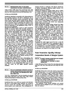
The classical model of cell cycle regulation presented in textbooks highlights growth factors as the unique stimulus to trigger proliferation. However it is now established that integrin adhesion is also an essential requirement for proliferation in metazoan cells. Normal cells either detached from ECM substrata or genetically lacking integrin subunits cannot progress into cell cycle, even in the presence of growth factors (Nikolopoulos et al., 2005; Walker and Assoian, 2005; Streuli and Akhtar, 2009; Jeanes et al., 2012). Despite the major contribution of integrins to cell cycle progression, they cannot trigger proliferation independently of GFRs. There are different mechanisms by which integrins cooperate with GFRs (Fig. 1A). One is that integrins and GFRs activate the same pathways (signal crosstalk). For proliferative responses, the major pathways regulated by GFRs and integrins are the PI3K/Akt, Mek/Erk, and small
83 84 85 86 87 88 89 90 91 92 93 94 95 96 97 98 99 100 101 102 103 104 105
111 112 113 114 115 116 117 118 119 120 121 122 123
C
81 82
E
79 80
R
77 78
R
75 76
O
73 74
C
71 72
N
69 70
U
67 68
1.1.1. Perspectives Although the basis of the synergistic relationship between integrins and GFRs in cell cycle control has been established, there are still major questions remaining. For example, what are the precise molecular connections within the adhesome that link integrin cytoplasmic tails to downstream proliferation signalling pathways? Which integrins are required for proliferation in different cell types? Are the signalling pathways downstream GFRs and integrins the same in all cell types? What adhesion complex proteins are phosphorylated or turned over by GFRs and are these GFR-specific? Are the same mechanisms activated in 2D vs 3D cultures and in vivo?
138
F
1.1. Cooperation between integrins and growth factor receptors in cell cycle regulation
66
O
108 109
64 65
R O
106 107
Integrins are widely expressed glycoprotein receptors that mediate cell adhesion and communication with the extracellular matrix (ECM). These receptors are composed of α and β subunits forming 24 heterodimers from a combination of 18 α and 8 β subunits. Integrins sense the composition and mechanical properties within the pericellular environment, and transmit this information intracellularly to both the cytoskeleton and to signals that modify cell behaviour. To achieve this, integrins recruit mediators to their cytoplasmic domains. More than 150 proteins have been identified to interact at integrin adhesion sites, forming an adhesion complex also known as the “adhesome” (Zaidel-Bar et al., 2007; Legate et al., 2009; Streuli, 2009). Adhesion complex proteins include cytoskeletal components, enzymes and adaptor proteins that collectively modulate cell shape, and control cell fate decisions including proliferation, differentiation and migration. The cell cycle is tightly regulated by a series of cyclins and cyclindependent kinases (cdks) that coordinate checkpoints for progression through each stage of the cycle. The first checkpoint to commit to cell cycle takes place during G1 phase. Both mitogenic signals and ECM adhesion activate transcription factors such as AP-1, which control the expression of cyclin D1. Cyclin D1 binds cdk4 to phosphorylate the Retinoblastoma protein (Rb), which disassociates from E2F. Relief of E2F suppression permits cyclin E transcription, formation of a cdk2/cyclin E complex and further Rb phosphorylation, thereby amplifying the production of the complex. Once the levels of this complex are sufficient, the cell passes the restriction point and enters S phase. Mitogens simultaneously suppress negative regulators such as the cyclin-dependent kinase inhibitors (CKIs) p27 and p21, which inhibit the cdk4/cyclin D complex. Thus synchronised formation of cdk/cyclin complexes together with downregulated CKIs eventually allows assembly of a replication complex and subsequent proliferation. Cell adhesion via integrins is crucial for progression through the G1/S checkpoint. Integrins indirectly recruit adaptor proteins such as talin and paxillin, and enzymes such as focal adhesion kinase (Fak) and small GTPases, which control downstream effectors in signalling cascades. These effectors regulate the levels of cyclins, CKIs, and the transcription of genes required for proliferation such as c-Jun and E2F (Walker and Assoian, 2005). Integrins also participate in mitotic spindle alignment (LaFlamme et al., 2008; Streuli, 2009). The role that integrins play in proliferation is so important that in the absence of integrinmediated adhesion, metazoan cells do not commit to enter the cell cycle and thus do not proliferate. Here, new studies on molecular mechanisms linking integrins to cell cycle entry will be discussed. We focus first on the general mechanisms of how integrins cooperate with growth factor receptors (GFRs) to control proliferation, and second on the mechanisms associated with specific integrin β subunits.
124 125
P
62 63
GTPase (Rho and Rac) pathways. Integrins also control the expression of GFRs at the transcriptional level. For example, Epidermal Growth Factor Receptor (EGFR) protein levels are decreased in human epithelial cells cultured in suspension, and the downregulation of EGFR reduces β1-integrin levels (Grassian et al., 2011). Integrins also cooperate with GFRs through physical association. Mechanisms include: activation of integrins by GFRs to enhance the mitogenic signals; activation of GFRs by integrins in the absence of GFR ligand; increased internalisation and endocytic trafficking of GFRs by integrins. However, GFR phosphorylation by integrins is only transient and less effective than that mediated by growth factors. Thus, a sustained activation of Erk mediated by GFRs and integrins together is required for the synthesis of cyclin D1. This has been discussed more in detail previously (Walker and Assoian, 2005; Streuli and Akhtar, 2009).
D
1. Introduction
1.2. Integrins control Akt and Erk signalling Akt signalling is adhesion-dependent in many cell types. This pathway can be activated by integrins via Fak, which binds to the p85 subunit of PI3K, or through a Src–vinculin complex. The Akt pathway is essential for proliferation because a dominant negative mutant of PI3K prevents cyclin D1 expression. However proliferation also requires Erk signalling. An inducible activated Mek transfected into suspended fibroblasts rescues the proliferation defect arising from ECM detachment, but when treated with a specific PI3K inhibitor the transfected cells fail to enter S phase (Walker and Assoian, 2005). Both Akt and Erk phosphorylation are required to induce cell growth when stimulated with mitogens (Dellinger and Brekken, 2011; Fournier et al., 2012; Zhang et al., 2013). Moreover, some studies suggest that Akt and Erk both regulate Elk1 activity (Mut et al., 2012). Thus, adhesion-activated Akt and Erk pathways contribute to cyclin D1 induction via different mechanisms, which complement each other to allow transition through G1/S (Fig. 1B). Besides regulating Elk1, Akt controls Foxo3a transcriptional activity by phosphorylating three of its Ser/Thr residues (Zhang et al., 2013). FoxO3a normally induces transcription of the CKIs p27 and p21. When phosphorylated by Akt, FoxO3a becomes inactive and is translocated to the cytosol. Akt also activates the mammalian target of rapamycin complex 2 (mTorc2), a kinase which stimulates the expression of Skp2, forming part the ubiquitin ligase complex (Shanmugasundaram et al., 2013). Together, these Akt-regulated events lead to p27 and FoxO3a degradation, thereby permitting cell cycle progression. Integrins also cooperate with EGFR to suppress FoxO1 via Akt signalling, thereby promoting transcription of the early response gene, Egr-1 (Cabodi et al., 2009). The CKIs are regulated by other mechanisms that permit integration of cell cycle control by GF and adhesion signals. On the GF arm, Erk can also phosphorylate Foxo3a at S294, S344 and S425, causing Foxo3a cytoplasmic localization and degradation via the E3 ubiquitin ligase MDM2 (Yang et al., 2008). In the absence of degradation, nuclear Foxo represses cyclin D1 expression in the absence of p27. Additionally, β1 integrins are required to maintain low levels of p27 or p21 in neurons and epithelial cells, respectively (Blaess et al., 2004; Li et al., 2005).
T
61
E
2
Please cite this article as: Moreno-Layseca, P., Streuli, C.H., Signalling pathways linking integrins with cell cycle progression, Matrix Biol. (2013), http://dx.doi.org/10.1016/j.matbio.2013.10.011
126 127 128 129 130 131 132 133 134 135 136 137
139 140 141 142 143 144 145 146 147 148 149 150 151 152 153 154 155 156 157 158 159 160 161 162 163 164 165 166 167 168 169 170 171 172 173 174 175 176 177 178 179 180 181 182 183 184 185
P. Moreno-Layseca, C.H. Streuli / Matrix Biology xxx (2013) xxx–xxx
197
In most cell types, the Erk pathway is one of the key axes required to trigger proliferation. Erk is transiently induced by GFs, but integrinmediated adhesion sustains Erk activation for long enough to induce cyclin D expression. To exert its mitogenic effects, Erk needs to translocate
B
S phase
P
Cdk4
Cdk2
cyclinD1
Elk1
cyclinE
Akt p21,p27
Ekr
Erk
E3 ligase
P Foxo3a
skp2
C
T
Akt
p27
E
Foxo3a
PI3K
Pak1
E
Paxillin
Src Fak
Mek Raf Grb2 Akt Ras
Torc2
Integrin
PIP3 Pdk1
GFR
ECM
ECM
A
C
cytoskeleton
Catenin
Proliferation
Physical association
Akt
Src Fak PI3K
R
200
R
198 199
N C O
193 194
U
192
F
1.3. Erk activation versus nuclear translocation by integrin signals
190 191
O
196
188 189
into the nucleus. Some studies have shown that integrin adhesion to the ECM mediates Erk nuclear translocation and the phosphorylation of the transcription factor Elk1 (Nikolopoulos et al., 2005; Streuli and Akhtar, 2009). Overexpression of activated Erk with a mutation that prevents its trafficking to the cytosol partially rescues proliferation in suspended fibroblasts. Erks do not possess a conventional nuclear localisation signal, and require anchoring proteins to regulate its subcellular distribution. A novel nuclear translocation signal has been identified, comprised by a Ser-Pro-Ser sequence, which, when phosphorylated, allows Erk nuclear shuttling through Importin7 binding. It is not known yet whether integrin-mediated adhesion could regulate phosphorylation of this sequence to promote Erk translocation. Erk anchoring proteins such as tubulin and Mek1 are also possible integrin targets (Zehorai et al., 2010). Rac activity is also required for Erk nuclear translocation in fibroblasts and epithelial cells. The mechanisms are poorly understood, but may involve Pak1 activation and crosstalk with the MAPK pathway
R O
195
1.2.1. Perspectives Integrins cooperate with Akt and Erk by jointly suppressing p21/p27 and shifting the balance of Foxo3a from the nucleus to cytosol. However, although adhesome proteins such as Fak are involved in linking integrins with Akt and Erk, the detailed mechanisms are not fully resolved. Interestingly, gene deletion studies show that Fak is necessary for proliferation of mammary epithelial cells in 2D culture, but not in mammary tissue in vivo. Thus, other proteins mediate the proximal integrin downstream signals in 3D cultures and in vivo, though these are poorly characterised at present.
D
186 187
3
Erk LIMK
Jun
Arp2/3 ROS
Jnk p27
Signal crosstalk
POSH
Pak1
Wave
p67phox adhesion complex
GFR
Integrin
Skp2 adhesion complex proteins
Rac
Integrin
ECM
ECM
Fig. 1. Integrin links to serine/threonine kinase and Rac pathways. (A) Integrins and GFRs cooperate to regulate cell cycle, either through physical association or via indirect signalling crosstalk. (B) Some of the signalling pathways linking the integrin spatial checkpoint with the temporal checkpoint for cell cycle provided by GFs are shown. Their combined activity via both Akt and Erk signalling results in both promotion of the cyclin/Cdk complexes necessary for S phase, together with the inhibition and degradation of CKIs. (C) Integrins control proliferation via Rac. Rac is a key player in cell cycle regulation, and acts through several different effectors to influence downstream signals, transcription factors and CKIs.
Please cite this article as: Moreno-Layseca, P., Streuli, C.H., Signalling pathways linking integrins with cell cycle progression, Matrix Biol. (2013), http://dx.doi.org/10.1016/j.matbio.2013.10.011
201 202 203 204 205 206 207 208 209 210 211 212 213 214 215 216 217
229
1.4. Integrin activation of Rac in cell cycle control
230 231
All 3 principal members of the GTPase family, Rac1, RhoA and Cdc42, are associated with adhesion-dependent cell cycle regulation (Welsh, 2004; Mack et al., 2011). Rac and Rho are activated by integrinmediated adhesion and they control cell cycle through different downstream mechanisms. Rac1 has a central role in linking cell adhesion to G1/S transition by activating mitogenic pathways via a range of effectors (Fig. 1C). In contrast, RhoA controls cell cycle in part via the organisation of actin stress fibres and cell shape. A key function of integrin adhesion is to recruit signalling effectors to the cell cortex, where they are activated to trigger downstream signals. One way that integrins recruit Rac1 to the membrane is via the Fak/Src/ p130Cas complex. In this complex, Src phosphorylates p130Cas, which recruits Rac1 to focal adhesions thereby activating downstream signals. Rac can be controlled by the GEF, Dock180 (Smith et al., 2008). Although Dock180 is involved in Rac-mediated motility and migration, it is not known which GEFs mediate Rac-driven cell cycle. After activation, small GTPases bind to a variety of effectors. One Rac1 effector is Pak1, which can activate the Erk pathway by phosphorylating Raf and Mek1. In turn this pathway controls nuclear translocation of Erks (Jeanes et al., 2012). Pak1 also regulates the cytoskeleton via Lim kinase, which contributes to cell cycle progression by controlling cell shape. Interestingly the Pak1-null mouse is viable and fertile while Pak2-knockout mice show early embryonic lethality (Kelly and Chernoff, 2012). This suggests that the Pak isoform involved in cell cycle regulation is Pak2. Pak may be recruited to adhesions as part of a complex that also includes Rac1, a current model including paxillin, GIT1, β-Pix, Pak and Rac1 (Zhao and Manser, 2012). A separate Rac1 effector is Jnk, which is recruited and activated via the scaffold protein, Posh, and promotes cell cycle via c-Jun. Mammary epithelial cells proliferate in response to collagen attachment and oestrogen stimulation via Rac1/Jnk/c-Jun pathway (Xie and Haslam, 2008). Rac1 also modulates the Wnt pathway via Jnk2, which phosphorylates β-catenin on S191 and S605 leading to its nuclear localization. Moreover, Rac can induce proliferation through a Pak1 interaction with β-catenin in gastric cells under gastrin stimulation (He et al., 2008). The crosstalk between Rac1 and the Wnt pathway may occur through several Rac effectors, including Jnk2 and Pak1. Further studies are needed to determine which Rac effectors enhance β-catenin nuclear effects, and the involvement of this crosstalk in cell cycle entry. The production of Reactive Oxygen Species (ROS) is required for proliferation. Rac modulates ROS production by binding to the NADPH complex. Dominant negative Rac1 decreases cell growth and superoxide production in epithelial cells that express oncogenic K-Ras (Du et al., 2011). For ROS formation, Rac recruits p67phox, a component of the NADPH complex, to the plasma membrane. This interaction is dependent on four residues present in two Rac isoforms, Rac1 and Rac2. Rac also controls proliferation by suppressing cell cycle inhibitory pathways. For example, Rac upregulates Skp2, leading to p27 degradation (Bond et al., 2008). This Skp2-p27 pathway requires Rac-mediated actin polymerisation. Integrin-mediated Rac activation therefore has a
248 249 250 251 252 253 254 255 256 257 258 259 260 261 262 263 264 265 266 267 268 269 270 271 272 273 274 275 276 277 278 279
1.5. Integrins as mechanosensors for the control of cell cycle
296
In addition to chemical signals provided by the cellular microenvironment, the physical nature of the cell niche influences proliferation. Integrins are mechanosensors, which sense changes in mechanical tension within the cellular microenvironment. Integrins convert physical signals outside the cell to chemical ones inside. Forces profoundly affect adhesion complex assembly, actin polymerisation, nuclear architecture and overall cell shape, all of which influence cell cycle progression (Walker and Assoian, 2005; DuFort et al., 2011; Eyckmans et al., 2011). In a range for cell types including mammary epithelial cells and smooth muscle cells, the elastic compliance of ECM controls Rac activation and downstream signals to produce cyclins A and D1 (Klein et al., 2009). A stiffer ECM increases adhesion complex assembly, Fak activation and Erk signalling (Paszek et al., 2005). This lowers the threshold for EGFR stimulation and Erk (but not Akt) activation, and increases the expression of numerous genes involved in the Erk signalling pathway (Kim and Asthagiri, 2011) (Yan et al., 2012). The alignment and orientation of ECM molecules also contributes to mechanosensing. Neural progenitor cells show enhanced cell cycle when cultured on aligned collagen I nanofibers compared to randomly aligned nanofibers or collagen I-coated dishes (Wang et al., 2011a,b). This effect is mediated by β1 integrin and Erk activation. The extent of cell spreading area is another factor that affects proliferation. In fully spread fibroblasts, Jnk phosphorylation mediates Elk4 activation and early gene transcription. However, when fibroblasts are plated on a restricted area that limits adhesion and spreading, p38 is activated instead, leading to Elk3 phosphorylation and the repression of early gene expression (Wozniak et al., 2012). Thus, subtle changes in the extent of adhesion can determine cell fate. One adhesion complex protein that may mediate tension-dependent proliferation signals is the scaffold protein, talin, which connects integrins to the cytoskeleton. In 2D cultures of mammary epithelial cells and breast cancer cells, the C-terminus of talin's rod domain, corresponding to its tail, is necessary for DNA synthesis, Fak phosphorylation and downregulation of p21 (Wang et al., 2011a,b). Talin-deficient CD4+ T cells are not able to proliferate normally, due to the lack of stable contacts with antigen-presenting cells resulting in defective F-actin polymerisation (Wernimont et al., 2011). In this latter example, phosphorylation of Akt, Erk and p38 are not affected by talin ablation. Downstream of adhesion complexes, the cytoskeleton mediates proliferation signals. For example, synemin can localise to focal adhesions, mediate actin polymerisation and sequester the phosphatase PP2A to the nucleus. In this way synemin prevents Akt dephosphorylation and allows G1/S transition (Pitre et al., 2012). The small GTPase, Rho, has a key role sensing tension changes and transmitting them to the cytoskeleton. Rho sustains Erk activation and stimulates cyclin D1 expression (Welsh, 2004; Park et al., 2011a,b).
297 298
T
246 247
C
244 245
E
242 243
R
240 241
R
238 239
O
236 237
C
234 235
N
232 233
U
225 226
F
227 228
1.3.1. Perspectives Although the Erk's mitogenic activity is adhesion-regulated, several questions remain to be answered. For example how do integrins control Erk nuclear translocation? Is the control on Erk by integrins microenvironment and cell type specific?
282 283
O
224
1.4.1. Perspectives Although Rac is a central downstream mediator of integrin-dependent proliferation, the detailed mechanisms of how it is recruited to adhesion complexes and which GEFs trigger its proliferative activity are not well understood. It is not known if these mechanisms are similar or not in different cell types and 3D and in vivo contexts. Also not addressed yet is whether Rac is activated in a different spatial location to exert its proliferative stimulus versus its effects on other phenotypes such as migration, differentiation, and phagocytosis. The role of Rac isoforms, Rac2, Rac3 and RhoG, in adhesion-mediated cell cycle entry is not fully understood and the isoforms may have differential effects in different cell types. For example, Rac1/2 are required for bone marrow erythropoiesis, but are dispensable for megakaryocyte and erythrocyte proliferation in the spleen (Kalfa et al., 2010).
R O
222 223
nodal role in cell cycle control by influencing both mitogenic signals 280 and suppressing CKIs. 281
P
220 221
(Jeanes et al., 2012). Paxillin and actin interact with Erk, supporting the idea that cytoskeleton rearrangements upon adhesion allow Erk nuclear shuttling. Paxillin can transit between adhesion complexes and the nucleus (Dong et al., 2009). Since Erk phosphorylates paxillin at serine residues, this might allow paxillin to act as a chaperone for Erk nuclear translocation (Sen et al., 2012).
D
218 219
P. Moreno-Layseca, C.H. Streuli / Matrix Biology xxx (2013) xxx–xxx
E
4
Please cite this article as: Moreno-Layseca, P., Streuli, C.H., Signalling pathways linking integrins with cell cycle progression, Matrix Biol. (2013), http://dx.doi.org/10.1016/j.matbio.2013.10.011
284 285 286 287 288 289 290 291 292 293 294 295
299 300 301 302 303 304 305 306 307 308 309 310 311 312 313 314 315 316 Q2 317 318 319 320 321 322 323 324 325 326 327 328 329 330 Q3 331 332 333 334 335 336 337 338 339 340 341 342 Q4
P. Moreno-Layseca, C.H. Streuli / Matrix Biology xxx (2013) xxx–xxx
360 361 362 363 364 365 366 367 368 369 370 371 372 373 374 375 376 377 378 379
t1:4 t1:5
β1 integrin β1 integrin
t1:6
β1 integrin
t1:7
β1 integrin
t1:8 t1:9 t1:10
β1 integrin β1 integrin Transgenic mice expressing a β1 integrin chimera with a mutated extracellular domain. Mice expressing a mutated β1 integrin-cytoplasmic domain. α8β1 integrin
Chondrocytes Cerebellar granule cell precursors Mammary epithelial cells
Systemic
t1:15
Mutant mice with a deletion of the cytoplasmic domain of β4 integrin β4 integrin mutant (deletion of the C-terminal portion of the integrin tail) αvβ8 integrin
t1:16
α9 integrin
t1:17 t1:18
α10 integrin α11 integrin
Skin keratinocytes and corneal epithelial cells Chondrocytes Fibroblasts from mouse embryos
t1:12 t1:13 t1:14
U
The initial studies addressing the role of integrins were mostly done by comparing suspended cells without any cell adhesion to those adhered to an ECM. However, these approaches do not distinguish between the direct role of integrin receptors in cell cycle regulation with effects that are a consequence of altered cytoskeletal and cell shape, or tension constraints. The development of transgenic mice with tissue-targeted deletion of specific integrin genes has provided definitive evidence linking integrin heterodimers to proliferation (Table 1). Genetic approaches have also been used to study integrinmediated cell cycle progression in vitro. Here we examine new studies aimed at dissecting the mechanism linking β-integrin heterodimers to cell cycle.
392
1.7. β1 integrins in cell proliferation
404
F
Integrin
N C O
t1:3
t1:11
391
O
358 Q6 359
1.6. The role of specific integrin β subunits in cell cycle progression
R O
356 Q5 357
C
354 355
E
352 353
R
350 351
R
349
382 383
P
Table 1 The effect of conditional integrin deletion on proliferation in mouse models.
347 348
1.5.1. Perspectives The development of new approaches to study how integrin adhesion controls cell cycle in 3D cultures constitutes a major challenge to the field. Emerging alternatives to 3D cultures are the polyethylene glycolhydrogel networks, in which the desired soluble factors or molecules such as the RGD motif can be incorporated (Bott et al., 2010). The advantages of this system need to be tested in cell types other than fibroblasts, but this approach is a promising way to study the effect of specific components of the ECM on proliferation.
D
t1:1 t1:2
345 346
T
380 381
Several mechanisms are involved, including the accurate timing of cyclin D1 expression during mid-G1, where Rho suppresses Rac-induced D1 expression in Go and early G1, but permits it subsequently. RhoA also suppresses the stimulatory effects of the Ras pathway on p21 expression, and it promotes p27 phosphorylation and degradation via cyclin E and cdk2. This occurs through both of Rho's effectors mDia and Rock. Interestingly, the actin cytoskeleton is not required for cell cycle progression under conditions where the levels of cyclin D are maintained, arguing that the key effect of integrin signals and cell shape in proliferation control is to regulate cyclin D (Margadant et al., 2013). Adhesion-activated Rock phosphorylates Tsc2, which increases protein translation by mTorc1 and allows production of cyclins and progression to S phase (Park et al., 2011a,b). In embryonic stem cells, Rock promotes proliferation through the PI3K/Akt and Ras/Erk pathways (Park et al., 2011a,b). Rock requires an intact actin cytoskeleton to activate Ras and induce cyclin D1 production. The involvement of Rock in tension-mediated proliferation has significant implications in cancer progression. Activated Rock in skin promotes ECM deposition, which feeds back on epidermal cells to promote β-catenin-mediated proliferation and tumour growth (Samuel et al., 2011). Apart from RhoA, its isoforms RhoE and RhoD are also associated with cell cycle. RhoD promotes G1/S transition via mDia1, and altered RhoD causes centrosome duplication (Kyrkou et al., 2013). RhoE, which lacks GTPase activity and is constitutively bound to GTP, arrests cell cycle in G1, upstream of the pRb checkpoint. This GTPase inhibits cyclin D1 translation by preventing dissociation of eIF4E from 4E-BP1, and also inhibits transcription of the eIF4E target, Myc (Villalonga et al., 2009). An overall picture emerges in which integrin-mediated adhesion controls cell cycle by acting on both signalling enzymes and the cytoskeleton. Small GTPases may provide the control nodes for coordinating adhesion-dependent proliferation. Rac is locally activated by integrins and is essential for numerous downstream enzymes controlling cell cycle, for example Pak1, Jnk, β-catenin and Erk nuclear translocation. Rho, activated by forces and stiffness within the ECM, also has a central cell cycle role, controlling both signalling enzymes such as Rock and PTEN, as well as the actin cytoskeleton. This is an essential intracellular scaffold, which works together with molecules such as talin.
β1 integrins are expressed on most cell types and have key roles in metazoan development and physiology. They are well known to be required for proliferation, and it is now apparent that there are numerous mechanisms involved. An in situ knockout model has been used to study the effect of β1 integrin gene ablation after the cells form focal adhesion complexes, without disturbing the actin cytoskeleton or modifying cell shape. Mammary epithelial cells show reduced proliferation after deleting the β1 integrin gene using CreERTam-mediated recombination (Jeanes et al., 2012). The mechanism is via integrin activation of Rac1 leading to Erk2 nuclear translocation, which occurs in parallel with GF receptor signalling. Similarly, cell cycle defects in mouse embryonic fibroblasts expressing a β1 integrin with a mutated cytoplasmic
E
343 344
5
Cell type
Effect on proliferation
Reference
Schwann cells Keratinocytes
None Decreased
Pancreatic β-cells
Decreased proliferation, decreased p-FAK, p-Erk and cyclin D1, and increased p21 Decreased proliferation and decreased phosphorylation of FAK; increased p21cip1 levels Decreased Decreased in cerebellum, high levels of p27-kip1 Reduced proliferation in pregnancy and lactation. Decreased phosphorylation of Erk, Shc and JNK Reduced proliferation in homozygote embryos
Feltri et al. (2002) Brakebusch et al. (2000), Raghavan et al. (2000), Grose et al. (2002), López-Rovira et al. (2005) Riopel et al. (2011), Diaferia et al. (2013) Li et al. (2005)
Mammary epithelial cells
Mouse embryonic fibroblasts Kidney cells
Keratinocytes Human bronchial cells
Defective growth of the ureteric bud and the ureteric epithelium Reduced proliferation in skin and intestinal epithelium Decreased Increased proliferation, low levels of secreted TGF-β None; but decreased proliferation was observed after injury Decreased Decreased
Aszodi et al. (2003) Blaess et al. (2004) Faraldo et al. (2001) Hirsch et al. (2002) Müller et al. (1997) Murgia et al. (1998) Nikolopoulos et al. (2005) Fjellbirkeland et al. (2003) Singh et al. (2009) Bengtsson et al. (2005) Popova et al. (2007)
Please cite this article as: Moreno-Layseca, P., Streuli, C.H., Signalling pathways linking integrins with cell cycle progression, Matrix Biol. (2013), http://dx.doi.org/10.1016/j.matbio.2013.10.011
384 385 386 387 388 389 390
393 394 395 396 397 398 399 400 401 402 403
405 406 407 408 409 410 411 412 413 414 415 416
440 441 442 443 444 445 446 447 448 449 450 451 452 453
F
O
R O
438 439
P
436 437
D
434 435
T
432 433
C
430 431
E
428 429
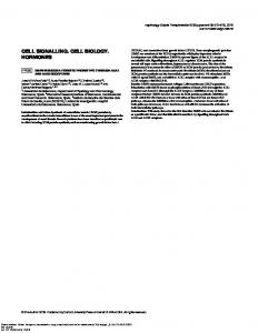
2013). β integrins also control S phase progression in the epithelia of other organisms, for example the intestinal stem cells of developing Drosophila (Lin et al., 2013). The mechanism is not known yet, but may be in parallel with Jak/Stat and EGFR signalling pathways (Beebe et al., 2010; Biteau and Jasper, 2011). In some cases, β1 integrins are low affinity receptors for mitogens themselves. For example, β1 integrin induces DNA synthesis during organogenesis by cooperating with Sonic Hedgehog in a laminindependent manner, while nerve growth factor can bind to α9β1 integrin to promote proliferation (Blaess et al., 2004). GF-integrin interactions are specific and another ligand, VEGF-A, directly binds α9β1 integrin via its EYP motif, located on the opposite side of an exposed loop on the surface of VEGF-A to the VEGFR interaction site of the ligand (Oommen et al., 2011). Although β1 integrin is generally known to promote proliferation, it can also be inhibitory, constituting a dominant negative signal over the stimulatory effects (Fig. 2A). For example, integrin α1β1 inhibits EGFR signalling in kidney cells by activating the protein tyrosine phosphatase TCPTP, while α2β1 activates PP2A, leading to Akt dephosphorylation. TCPTP dephosphorylates caveolin-1, a scaffolding protein for integrins and GF receptors, leading to reduced EGFR activation (Borza et al., 2010; Chen et al., 2010). This might provide a mechanism to protect damaged tissues following oxidative stress. In a separate example, emilin-1 is an elastic microfibril-binding ECM protein that binds to α4β1 and α9β1 integrins. It antagonises proliferation in dermal and epidermal cells to maintain homeostasis within the skin, and deletion of emilin results in hyperproliferation and accelerated closure of wounds. The inhibitory effects of emilin may occur through α9β1mediated activation of PTEN, which prevents Akt signalling (Danussi et al., 2011). The presence of different splicing variants also affects the proliferative outcome of integrin signals (Fig. 2A). Different isoforms for β1 integrin can either block or allow cell cycle entry. For example, the β1A subunit promotes proliferation, while β1C and β1D block the transition between G1 and S phase. Differential splicing of α subunits can also exert opposite proliferative effects. For example, the α6A integrin subunit maintains proliferation in colonic epithelium, while the α6B
R
426 427
A
ECM, mitogens
Ligand interaction
R
424 425
O
423
6A, 1A Proliferation
Splicing variants
Emilin-1, phosphatases
1C, 1D, 6B Heterodimer specificity
C
421 422
N
419 420
domain have been traced to defective Rac1 activity and Erk nuclear translocation (but not its activation) (Hirsch et al., 2002). In both these cases, the adhesion complex proteins linking integrin tails with Rac have not been identified. The work in mammary epithelia also reveals the specificity of function exerted by different integrins: β3 integrins remain following β1 integrin gene deletion and, although they still mediate collective cell migration, β3 integrins are unable to drive proliferation. It is not known why β3 integrins cannot compensate for the proliferation defect but possibilities include sequestration of Erk1 at adhesion sites by αvβ3 integrin, or alternatively the composition of αvβ3-containing adhesomes is different to those assembled by β1-integrins and lacks key proliferation-specific components (Schiller et al., 2013). Pancreatic epithelial β-cells also require β1-integrins for cell cycle in vivo, most likely through a control on p21 — again, β3 integrins are expressed in β-cells but are unable to compensate for the loss of β1 integrins (Diaferia et al., 2013). β1-Integrin cooperates with IL-3Rβc in endothelial cells to control proliferation, which is important in a tumour microenvironment where IL-3 is secreted and may contribute to tumour angiogenesis (Uberti et al., 2010). In fibroblasts cultured on collagen, β1 integrin signals lead to PP2A inhibition and PTEN degradation, which inactivates the transcription factor FoxO3a via the Akt pathway and provides a permissive environment for cell cycle (Nho and Kahm, 2010). In some cell types such as osteoblasts, the adhesion complex protein, kindlin-2 signals via β1 integrin to Rac1, which initiates AP-1 dependent transcription via Akt signalling, and thereby promotes proliferation (Jung et al., 2011). However other cells e.g. gastric carcinoma, are more dependent on kindlin-2 for migration than proliferation (Shen et al., 2013). One mechanism by which β1 integrins cooperate with GF receptors in cell cycle control is via other transmembrane proteins such as tetraspannins. In Her2 positive breast cancer, CD151 forms a complex with α3β1 integrin, which regulates the ErbB2 signalling axis (Novitskaya et al., 2013). Integrins can also cooperate with other types of cell surface receptors. In colorectal cancer cells, the nonclassical cadherin-17 binds α2β1 integrin and regulates its signalling to control proliferation for metastasis to the liver (Bartolomé et al.,
U
417 418
P. Moreno-Layseca, C.H. Streuli / Matrix Biology xxx (2013) xxx–xxx
E
6
B
Proliferation
Transcriptional regulation of adhesion or mitogen-related elements
Signals ECM ligand synthesis
Integrin
ECM
Fig. 2. Integrins control proliferation via different modes. (A) Different ECM or non-ECM proteins can antagonise each other to influence proliferative outcome. Similarly, proliferation can be controlled oppositely by different splicing variants of the same integrin subunits, or by different heterodimers. (B) Integrins can establish feedback loops for cell cycle, by regulating expression of mitogens themselves or ECM proteins.
Please cite this article as: Moreno-Layseca, P., Streuli, C.H., Signalling pathways linking integrins with cell cycle progression, Matrix Biol. (2013), http://dx.doi.org/10.1016/j.matbio.2013.10.011
454 455 456 457 458 459 460 461 462 463 464 465 466 467 468 469 470 471 472 473 474 475 476 477 478 479 480 481 482 483 484 485 486 487 488 489 490
P. Moreno-Layseca, C.H. Streuli / Matrix Biology xxx (2013) xxx–xxx
502 503 504 505 506 507 508 509 510 511 512 513
525
1.8. β3 integrins in cell cycle regulation
526
αvβ3 integrin binds to RGD motifs present in the ECM proteins fibronectin, vitronectin, thrombospondin, osteopontin and α-thrombin. This integrin has well-established roles in cancer cell invasion and angiogenesis (Robinson and Hodivala-Dilke, 2011). αvβ3 is also involved with proliferation in some cell types. Currently, few studies have confirmed this genetically and its role is cell type-restricted because in mammary epithelial cells, αvβ3 integrin is not able to compensate for proliferation following genetic deletion of the β1 integrin gene (Jeanes et al., 2012). An established mechanism for αvβ3 control of cell cycle in fibroblasts, endothelial and smooth muscle cells is by direct association or signalling crosstalk with GFRs such as VEGFR, PDGFRβ, EGFR, and IGFR1. αvβ3 integrin contributes to several diseases through its effects on proliferation. In hepatic stellate cells, blocking αvβ3 integrin induces the expression of the metalloproteinase MMP9 and decreases the expression of the metalloproteinase inhibitor TIMP-1. Reduced proliferation following such ECM remodelling might help to repair liver fibrosis. Knockout studies have shown that αvβ3 integrin contributes to vascular smooth muscle cell proliferation in diabetic mice, via elevated production of its ligand thrombospondin-1, though whether or not targeting this integrin to prevent restenosis is not yet clear (Panchatcharam et al., 2010). αvβ3 integrin levels are elevated in several cancers, which in some cases directly contributes to tumorigenesis (Schittenhelm et al., 2013). The mechanisms linking αvβ3 integrin to tumour cell proliferation involve the Erk pathway. Sustained Raf-Erk signalling also increases cell-surface levels of αvβ3 integrin, thereby creating a positive mitogenic feedback loop (Fig. 2B). αvβ3's contribution to proliferation may
527 528 529 530 531 532 533 534 535 536 537 538 539 540 541 542 543 544 545 546 547 548 549 550 551 552 553
C
E
R
522 523
R
520 521
N C O
518 519
U
516 517
T
524
1.7.1. Perspectives β1 integrins have a central role in the proliferation of many metazoan cell types. However, the proliferative response to integrin signalling is both heterodimer- and cell type-specific. To add more complexity, β1 integrin ligands not only include ECM components, but also soluble factors. Important challenges are to understand how integrin-signalling specificity is achieved, and how integrin signals are transmitted across the adhesome to link with S/T kinases and RhoGTPases, both of which are required to coordinate progress through the cell cycle. The reward will be clearer insights in both how different tissue types coordinate their growth and function, and how defects in cell-matrix interactions contribute to disease processes such as fibrosis and cancer.
514 515
1.8.1. Perspectives Since αvβ3 integrin expression and signalling are upregulated in cancer, this adhesion receptor is a potential therapeutic target to lower proliferation (Goodman and Picard, 2012). Some resistance mechanisms induced by MAPK and PI3K inhibitors are characterised by upregulation and redistribution of αvβ3 integrin. Thus, cyclic peptides containing the RGD motif are being tested in clinical trials, potentially reducing both proliferation and invasion. Although αvβ3 collaborates with GFRs in the proliferation of some cell types, genetic analyses to confirm the links in 3D cultures and in vivo, and dissecting the molecular basis for the integrin-GFR crosstalk, remain future challenges.
587 588
1.9. β4 integrins in cell cycle regulation
598
Integrin α6β4 is a laminin receptor that is mainly expressed in the basal layer of epithelial tissues. The β4-integrin is unique because of its long cytoplasmic tail, which interacts with the intermediate filament network rather than actin. This interaction contributes to the ability of α6β4 to maintain the integrity of tissues normally exposed to shear stress. α6β4 integrin assembles its own signalling platform. In the skin, keratinocytes within the basal epidermal layer constantly proliferate to provide the daughter cells that make up the upper squamous layers. Integrin α6β4 is required for this function, though it is not necessary for proliferation in every type of epithelial cell (Nikolopoulos et al., 2005). For example β4 integrin knockout in mammary epithelia does not prevent ductal development. We have previously discussed how α6β4 integrin controls cell cycle in normal epithelia and in cancer (Streuli and Akhtar, 2009). More recent studies continue to support this role for β4 integrin. For example in retinal pigment epithelia, overexpression of α6 or β4 integrin increases proliferation, which can help to regenerate damaged tissues (Fang et al., 2009). Transcriptional regulators of β4 integrin such as
599
F
500 501
O
498 499
R O
497
554 555
P
495 496
alternatively occur via crosstalk with other pathways. Studies using conditional knockout have revealed that in the developing tooth, β3 integrin controls the proliferation of transit-amplifying epithelial cells called pre-ameloblasts, via the stem cell marker, Lgr5 (Yoshida et al., 2013). Accumulation of extracellular ligands provides a further mechanism of elevating αvβ3 integrin levels and proliferation. In breast cancer cells, the EGF family member heregulin induces synthesis of the ECM protein, cysteine-rich protein 61 (CYR61), which leads to elevated αvβ3 integrin, Erk activation and enhanced proliferation. CYR61 lacks an RGD motif, but contains a 20 amino-acid sequence called V2 through which binds to αvβ3 integrin, and it has been linked to proliferation control in both normal and cancer cells (Franzen et al., 2009; Su et al., 2010). It is possible that upregulation of ligands in the ECM could also elevate the expression of αvβ3 integrin in nearby endothelial cells and thereby promote angiogenesis. In addition to RGD-containing ECM ligands, αvβ3 integrin can be bound by the thyroid hormones L-thyroxine (T4) and L-thyronine (T3), which stimulate proliferation independently of thyroid hormone receptor. The T3 and T4 binding sites in αvβ3 integrin are at the RGD recognition site (Freindorf et al., 2012). Competition for RGD binding stimulates Erk phosphorylation and translocation, thereby promoting proliferation in fibroblasts and osteoblast-like cells. The same effect with T3 and T4 has been observed in glioma and myeloma (Cohen et al., 2011). In some cases there is the involvement of additional receptors, for example T4 drives proliferation of lung carcinoma cells through crosstalk between αvβ3 integrin and oestrogen receptor-α (Meng et al., 2011). It is not yet known whether thyroid hormones also control proliferation in normal, non-transformed cells, or if this is restricted to cancer. Interestingly some other αv-integrin heterodimers might work in a different way to αvβ3 integrin, and have opposite effects on proliferation. For example, αvβ6 integrin binds and activates TGFβ, which serves as a negative regulator of proliferation in keratinocytes (Xie et al., 2012).
D
493 494
subunit inhibits cell cycle (Dydensborg et al., 2009). α6B can be downregulated in colon cancer, correlating with a hyper-proliferative phenotype. The generality of this regulatory mechanism has not been studied, and the mechanism for opposite fates arising from different integrin splicing is not known. Presumably it involves recruitment of different adhesion complex proteins. Integrins can exert an inhibitory effect on cell cycle entry by regulating other integrins. In endothelial cells, the α5β1 fibronectin receptor is the main integrin responsible for the proliferative response, which is triggered by Rac1 activation. However, in cells plated on laminin, α1β1 promotes quiescence instead of cell cycle progression (Cailleteau et al., 2010). If the ECM contains ligands for both integrins, α1β1 has a dominant inhibitory effect over α5β1 integrin. The mechanism may be via an interaction between the α1 subunit and the tetraspanin CD9, which prevents the assembly of focal adhesions by α5β1 integrin. As a consequence, this inhibits signalling downstream of the fibronectin receptor. This mechanism is physiologically important, because endothelial cells normally reside on a laminin-rich basement membrane, where proliferation is not required. However, during angiogenesis or vessel repair, the cells come into contact with fibronectin-containing stromal ECM, thereby providing a suitable proliferation-promoting microenvironment.
E
491 492
7
Please cite this article as: Moreno-Layseca, P., Streuli, C.H., Signalling pathways linking integrins with cell cycle progression, Matrix Biol. (2013), http://dx.doi.org/10.1016/j.matbio.2013.10.011
556 557 558 559 560 561 562 563 564 565 566 567 568 569 570 571 572 573 574 575 576 577 578 579 580 581 582 583 584 585 586
589 590 591 592 593 594 595 596 597
600 601 602 603 604 605 606 607 608 609 610 611 612 613 614 615 616
626 627 628 629 630 631 632 633 634 635 636 637 638 639 640 641 642 643 644 645 646 647
656
1.10. Final perspectives
657 658
Adhesion of cells to their ECM microenvironment via integrin receptors constitutes an important checkpoint for cell cycle entry. The diversity of mechanisms employed by β integrins to control proliferation underscores the importance of the cellular microenvironment for tissue homeostasis. The mechanistic differences most likely reflect the pathways for evolution of the various tissue types that have so far been studied in this context. Perhaps they constitute a part of the zip code that specifies the positional identity of cells within the body. Only the correct microenvironmental signals coupled with the right adhesion receptor allow the engine of cell cycle to be activated in response to soluble, temporal growth signals. Integrin adhesion complexes directly activate several different signalling cascades that allow the cells to progress to the S phase, including the Mek/Erk, PI3K/Akt, and the small GTPase Rac pathways. They also provide sustained activation to signals from growth factors. Notably however, while many downstream pathways linking integrins to proliferation have been identified, the precise components transmitting proximal integrin signals across adhesion complexes have yet to be worked out. In some cases, scaffold proteins such as talin participate in proliferation, providing the link between mechanical signals and nuclear responses (Wang et al., 2011a,b). However the mechanisms linking actin polymerisation and cytoskeleton changes in proliferation have not been fully elucidated.
661 662 663 664 665 666 667 668 669 670 671 672 673 674 675 676 677 Q7 678 679
C
E
R
R
O
C
659 660
N
652 653
U
650 651
Acknowledgements
682 683 684 685 686 687 688 689 690 691 692 693
We thank Prof Andy Sharrocks for reading the manuscript. The Wellcome Trust Centre for Cell-Matrix Research is supported by core funding from the Wellcome Trust (Grant 088785/Z/09/Z). PML was supported by the Mexican National Council for Science and Technology (CONACyT) and the General Direction of International Relations of the Mexican Ministry of Public Education (SEP).
695
References
701
Aszodi, A., Hunziker, E.B., Brakebusch, C., Fässler, R., 2003. Beta1 integrins regulate chondrocyte rotation, G1 progression, and cytokinesis. Genes Dev. 17, 2465–2479. Bartolomé, R.A., Barderas, R., Torres, S., Fernandez-Aceñero, M.J., Mendes, M., GarcíaFoncillas, J., Lopez-Lucendo, M., Casal, J.I., 2013. Cadherin-17 interacts with α2β1 integrin to regulate cell proliferation and adhesion in colorectal cancer cells causing liver metastasis. Oncogene. Beebe, K., Lee, W.-C., Micchelli, C.A., 2010. JAK/STAT signaling coordinates stem cell proliferation and multilineage differentiation in the Drosophila intestinal stem cell lineage. Dev. Biol. 338, 28–37. Bengtsson, T., Aszodi, A., Nicolae, C., Hunziker, E.B., Lundgren-Akerlund, E., Fässler, R., 2005. Loss of α10β1 integrin expression leads to moderate dysfunction of growth plate chondrocytes. J. Cell Sci. 118, 929–936. Biteau, B., Jasper, H., 2011. EGF signaling regulates the proliferation of intestinal stem cells in Drosophila. Development 138, 1045–1055. Blaess, S., Graus-Porta, D., Belvindrah, R., Radakovits, R., Pons, S., Littlewood-Evans, A., Senften, M., Guo, H., Li, Y., Miner, J.H., Reichardt, L.F., Müller, U., 2004. Beta1integrins are critical for cerebellar granule cell precursor proliferation. J. Neurosci. 24, 3402–3412. Bon, G., Di Carlo, S.E., Folgiero, V., Avetrani, P., Lazzari, C., D'Orazi, G., Brizzi, M.F., Sacchi, A., Soddu, S., Blandino, G., Mottolese, M., Falcioni, R., 2009. Negative regulation of beta4 integrin transcription by homeodomain-interacting protein kinase 2 and p53 impairs tumor progression. Cancer Res. 69, 5978–5986. Bond, M., Wu, Y.-J., Sala-Newby, G.B., Newby, A.C., 2008. Rho GTPase, Rac1, regulates Skp2 levels, vascular smooth muscle cell proliferation, and intima formation in vitro and in vivo. Cardiovasc. Res. 80, 290–298. Borza, C.M., Chen, X., Mathew, S., Mont, S., Sanders, C.R., Zent, R., Pozzi, A., 2010. Integrin α1β1 promotes caveolin-1 dephosphorylation by activating T cell protein–tyrosine phosphatase. J. Biol. Chem. 285, 40114–40124. Bott, K., Upton, Z., Schrobback, K., Ehrbar, M., Hubbell, J.A., Lutolf, M.P., Rizzi, S.C., 2010. The effect of matrix characteristics on fibroblast proliferation in 3D gels. Biomaterials 31, 8454–8464. Brakebusch, C., Grose, R., Quondamatteo, F., Ramirez, A., Jorcano, J.L., Pirro, A., Svensson, M., Herken, R., Sasaki, T., Timpl, R., Werner, S., Fässler, R., 2000. Skin and hair follicle integrity is crucially dependent on beta 1 integrin expression on keratinocytes. EMBO J. 19, 3990–4003. Cabodi, S., Morello, V., Masi, A., Cicchi, R., Broggio, C., Distefano, P., Brunelli, E., Silengo, L., Pavone, F., Arcangeli, A., Turco, E., Tarone, G., Moro, L., Defilippi, P., 2009. Convergence of integrins and EGF receptor signaling via PI3K/Akt/FoxO pathway in early gene Egr1 expression. J. Cell. Physiol. 218, 294–303. Cailleteau, L., Estrach, S., Thyss, R., Boyer, L., Doye, A., Domange, B., Johnsson, N., Rubinstein, E., Boucheix, C., Ebrahimian, T., Silvestre, J.-S., Lemichez, E., Meneguzzi, G., Mettouchi, A., 2010. α2β1 integrin controls association of Rac with the membrane and triggers quiescence of endothelial cells. J. Cell Sci. 123, 2491–2501. Chen, X., Whiting, C., Borza, C., Hu, W., Mont, S., Bulus, N., Zhang, M.-Z., Harris, R.C., Zent, R., Pozzi, A., 2010. Integrin α1β1 regulates epidermal growth factor receptor activation by controlling peroxisome proliferator-activated receptor γ-dependent caveolin-1 expression. Mol. Cell. Biol. 30, 3048–3058. Cohen, K., Ellis, M., Khoury, S., Davis, P.J., Hercbergs, A., Ashur-Fabian, O., 2011. Thyroid hormone is a MAPK-dependent growth factor for human myeloma cells acting via αvβ3 integrin. Mol. Cancer Res. 9, 1385–1394.
T
654 655
1.9.1. Perspectives The β4 subunit has a unique position in integrin biology because of its restricted cellular distribution, and because it contains a complex cytoplasmic tail. While this integrin controls both tissue integrity and proliferation, the extent of its involvement in diseases and in stem cell biology has not been fully explored. Moreover there are possibilities for uniquely targeting the interactions between the β4 cytoplasmic tail with interacting GFRs and signalling kinases, in order to treat hyperproliferative diseases.
648 649
694
F
624 625
O
623
680 681
R O
621 622
The field has progressed from studying integrin signalling in nonadherent (which is not physiological) vs substrate attached cells, to the use of genetic approaches. However to truly understand the mechanisms linking integrins to cell cycle, more sophisticated culture models need to be used. It is well established that the proliferative responses to activated GFRs are completely different in 2D vs 3D culture (Muthuswamy et al., 2001). Taking this cue of 3D culture models for proliferation studies will provide a better understanding of how integrin-mediated adhesion integrates the signals from the microenvironment, matrix stiffness and growth factors in order to control cell cycle in vivo. The emergence of new insights into how integrins coordinate protein interactions to transduce proliferative signals offers valuable opportunities for discovering novel targets to regulate cell cycle in disease processes.
P
619 620
p63 and HIPK2 also have a role, for example inactivation of HIP2K increases β4 expression and promotes cancer progression (Bon et al., 2009). In breast and lung adenocarcinoma cells α6β4 integrin associates with ErbB2 and ErbB3. β4 integrin knockdown in the latter cell type reduces proliferation induced by the ErbB3 ligand, heregulin (Kawano et al., 2010). A novel ligand for α6β4 integrin, netrin-4, has been identified in glioblastoma using mass spectrometry (Hu et al., 2012). Netrins are neuronal guidance molecules, and high concentrations normally inhibit cell cycle. However, in cancer patients, netrin-4 levels decrease, while β4 increases. This sensitises the cancer cells to β4 integrin-mediated Akt/Tor signalling and elevated proliferation. Human leukocyte antigens (HLAs) are transmembrane proteins involved with antigen presentation, but they can also signal to promote cell cycle in endothelial cells. HLA-1 binds to β4 integrin via its cytoplasmic domain, and the integrin is required for HLA-induced Akt, Erk and Src activation to induce proliferation (Zhang et al., 2010). This cisinteraction between HLA and integrins to transduce proliferative signals may have a role in the rejection of grafts in transplant recipients. Advanced prostate tumours have high levels of β4 integrin (Yoshioka et al., 2013). In mouse models of prostate cancer, genetic deletion of the signalling region of the β4 integrin cytoplasmic domain significantly reduces tumour growth and metastases, without altering adhesion between the integrin and laminin. The mechanism linking β4 integrin to proliferation is via the ability of α6β4 integrin to amplify GF signalling from the ErbB2 and Met receptors. In addition, β4 integrin deletion results in the reduction of cancer stem cells in vivo and transit amplifying cells in culture models. Thus, as with breast cancer, α6β4 has a central role in the progression of prostate cancer by expanding the progenitor pool (Guo et al., 2006).
D
617 618
P. Moreno-Layseca, C.H. Streuli / Matrix Biology xxx (2013) xxx–xxx
E
8
Please cite this article as: Moreno-Layseca, P., Streuli, C.H., Signalling pathways linking integrins with cell cycle progression, Matrix Biol. (2013), http://dx.doi.org/10.1016/j.matbio.2013.10.011
696 697 698 699 700
702 703 704 705 706 707 Q8 708 709 710 711 712 713 714 715 716 717 718 719 720 721 722 723 724 725 726 727 728 729 730 731 732 733 734 735 736 737 738 739 740 741 742 743 744 745 746 747 748 749 750 751
P. Moreno-Layseca, C.H. Streuli / Matrix Biology xxx (2013) xxx–xxx
N C O
R
R
E
C
D
P
R O
O
F
Kyrkou, A., Soufi, M., Bahtz, R., Ferguson, C., Bai, M., Parton, R.G., Hoffmann, I., Zerial, M., Fotsis, T., Murphy, C., 2013. RhoD participates in the regulation of cell-cycle progression and centrosome duplication. Oncogene 32, 1831–1842. LaFlamme, S.E., Nieves, B., Colello, D., Reverte, C.G., 2008. Integrins as regulators of the mitotic machinery. Curr. Opin. Cell Biol. 20, 576–582. Legate, K.R., Wickström, S.A., Fässler, R., 2009. Genetic and cell biological analysis of integrin outside-in signaling. Genes Dev. 23, 397–418. Li, N., Zhang, Y., Naylor, M.J., Schatzmann, F., Maurer, F., Wintermantel, T., Schuetz, G., Mueller, U., Streuli, C.H., Hynes, N.E., 2005. Beta1 integrins regulate mammary gland proliferation and maintain the integrity of mammary alveoli. EMBO J. 24, 1942–1953. Lin, G., Zhang, X., Ren, J., Pang, Z., Wang, C., Xu, N., Xi, R., 2013. Integrin signaling is required for maintenance and proliferation of intestinal stem cells in Drosophila. Dev. Biol. 377, 177–187. López-Rovira, T., Silva-Vargas, V., Watt, F.M., 2005. Different consequences of beta1 integrin deletion in neonatal and adult mouse epidermis reveal a contextdependent role of integrins in regulating proliferation, differentiation, and intercellular communication. J. Invest. Dermatol. 125, 1215–1227. Mack, N.A., Whalley, H.J., Castillo-Lluva, S., Malliri, A., 2011. The diverse roles of Rac signaling in tumorigenesis. Cell Cycle 10, 1571–1581. Margadant, C., Cremers, L., Sonnenberg, A., Boonstra, J., 2013. MAPK uncouples cell cycle progression from cell spreading and cytoskeletal organization in cycling cells. Cell. Mol. Life Sci. 70, 293–307. Meng, R., Tang, H.-Y., Westfall, J., London, D., Cao, J.H., Mousa, S.A., Luidens, M., Hercbergs, A., Davis, F.B., Davis, P.J., Lin, H.-Y., 2011. Crosstalk between integrin αvβ3 and estrogen receptor-α is involved in thyroid hormone-induced proliferation in human lung carcinoma cells. PLoS ONE 6, e27547. Müller, U., Wang, D., Denda, S., Meneses, J.J., Pedersen, R.A., Reichardt, L.F., 1997. Integrin α8β1 is critically important for epithelial–mesenchymal interactions during kidney morphogenesis. Cell 88, 603–613. Murgia, C., Blaikie, P., Kim, N., Dans, M., Petrie, H.T., Giancotti, F.G., 1998. Cell cycle and adhesion defects in mice carrying a targeted deletion of the integrin beta4 cytoplasmic domain. EMBO J. 17, 3940–3951. Mut, M., Lule, S., Demir, O., Kurnaz, I.A., Vural, I., 2012. Both mitogen-activated protein kinase (MAPK)/extracellular-signal-regulated kinases (ERK) 1/2 and phosphatidylinositide-3OH kinase (PI3K)/Akt pathways regulate activation of E-twenty-six (ETS)-like transcription factor 1 (Elk-1) in U138 glioblastoma cells. Int. J. Biochem. Cell Biol. 44, 302–310. Muthuswamy, S.K., Li, D., Lelievre, S., Bissell, M.J., Brugge, J.S., 2001. ErbB2, but not ErbB1, reinitiates proliferation and induces luminal repopulation in epithelial acini. Nat. Cell Biol. 3, 785–792. Nho, R.S., Kahm, J., 2010. beta1-Integrin–collagen interaction suppresses FoxO3a by the coordination of Akt and PP2A. J. Biol. Chem. 285, 14195–14209. Nikolopoulos, S.N., Blaikie, P., Yoshioka, T., Guo, W., Puri, C., Tacchetti, C., Giancotti, F.G., 2005. Targeted deletion of the integrin beta4 signaling domain suppresses laminin-5-dependent nuclear entry of mitogen-activated protein kinases and NF-kappaB, causing defects in epidermal growth and migration. Mol. Cell. Biol. 25, 6090–6102. Novitskaya, V., Romanska, H., Kordek, R., Potemski, P., Kusińska, R., Parsons, M., Odintsova, E., Berditchevski, F., 2013. Integrin α3β1-CD151 complex regulates dimerization of ErbB2 via RhoA. Oncogene. Oommen, S., Gupta, S.K., Vlahakis, N.E., 2011. Vascular endothelial growth factor A (VEGF-A) induces endothelial and cancer cell migration through direct binding to integrin α9β1 identification of a specific α9β1 binding site. J. Biol. Chem. 286, 1083–1092. Panchatcharam, M., Miriyala, S., Yang, F., Leitges, M., Chrzanowska-Wodnicka, M., Quilliam, L.A., Anaya, P., Morris, A.J., Smyth, S.S., 2010. Enhanced proliferation and migration of vascular smooth muscle cells in response to vascular injury under hyperglycemic conditions is controlled by beta3 integrin signaling. Int. J. Biochem. Cell Biol. 42, 965–974. Park, J., Arakawa-Takeuchi, S., Jinno, S., Okayama, H., 2011a. Rho-associated kinase connects a cell cycle-controlling anchorage signal to the mammalian target of rapamycin pathway. J. Biol. Chem. 286, 23132–23141. Park, J.H., Ryu, J.M., Han, H.J., 2011b. Involvement of caveolin-1 in fibronectin-induced mouse embryonic stem cell proliferation: role of FAK, RhoA, PI3K/Akt, and ERK 1/2 pathways. J. Cell. Physiol. 226, 267–275. Paszek, M.J., Zahir, N., Johnson, K.R., Lakins, J.N., Rozenberg, G.I., Gefen, A., Reinhart-King, C.A., Margulies, S.S., Dembo, M., Boettiger, D., Hammer, D.A., Weaver, V.M., 2005. Tensional homeostasis and the malignant phenotype. Cancer Cell 8, 241–254. Pitre, A., Davis, N., Paul, M., Orr, A.W., Skalli, O., 2012. Synemin promotes AKT-dependent glioblastoma cell proliferation by antagonizing PP2A. Mol. Biol. Cell 23, 1243–1253. Popova, S.N., Barczyk, M., Tiger, C.-F., Beertsen, W., Zigrino, P., Aszodi, A., Miosge, N., Forsberg, E., Gullberg, D., 2007. α11β1 integrin-dependent regulation of periodontal ligament function in the erupting mouse incisor. Mol. Cell. Biol. 27, 4306–4316. Raghavan, S., Bauer, C., Mundschau, G., Li, Q., Fuchs, E., 2000. Conditional ablation of beta1 integrin in skin. Severe defects in epidermal proliferation, basement membrane formation, and hair follicle invagination. J. Cell Biol. 150, 1149–1160. Riopel, M., Krishnamurthy, M., Li, J., Liu, S., Leask, A., Wang, R., 2011. Conditional β1integrin-deficient mice display impaired pancreatic β cell function. J. Pathol. 224, 45–55. Robinson, S.D., Hodivala-Dilke, K.M., 2011. The role of β3-integrins in tumor angiogenesis: context is everything. Curr. Opin. Cell Biol. 23, 630–637. Samuel, M.S., Lopez, J.I., McGhee, E.J., Croft, D.R., Strachan, D., Timpson, P., Munro, J., Schröder, E., Zhou, J., Brunton, V.G., Barker, N., Clevers, H., Sansom, O.J., Anderson, K.I., Weaver, V.M., Olson, M.F., 2011. Actomyosin-mediated cellular tension drives increased tissue stiffness and β-catenin activation to induce epidermal hyperplasia and tumor growth. Cancer Cell 19, 776–791.
E
T
Danussi, C., Petrucco, A., Wassermann, B., Pivetta, E., Modica, T.M.E., Del Bel Belluz, L., Colombatti, A., Spessotto, P., 2011. EMILIN1-α4/α9 integrin interaction inhibits dermal fibroblast and keratinocyte proliferation. J. Cell Biol. 195, 131–145. Dellinger, M.T., Brekken, R.A., 2011. Phosphorylation of Akt and ERK1/2 is required for VEGF-A/VEGFR2-induced proliferation and migration of lymphatic endothelium. PLoS ONE 6, e28947. Diaferia, G.R., Jimenez-Caliani, A.J., Ranjitkar, P., Yang, W., Hardiman, G., Rhodes, C.J., Crisa, L., Cirulli, V., 2013. β1 integrin is a crucial regulator of pancreatic β-cell expansion. Development. Dong, J.-M., Lau, L.-S., Ng, Y.-W., Lim, L., Manser, E., 2009. Paxillin nuclear-cytoplasmic localization is regulated by phosphorylation of the LD4 motif: evidence that nuclear paxillin promotes cell proliferation. Biochem. J. 418, 173–184. Du, J., Liu, J., Smith, B.J., Tsao, M.S., Cullen, J.J., 2011. Role of Rac1-dependent NADPH oxidase in the growth of pancreatic cancer. Cancer Gene Ther. 18, 135–143. DuFort, C.C., Paszek, M.J., Weaver, V.M., 2011. Balancing forces: architectural control of mechanotransduction. Nat. Rev. Mol. Cell Biol. 12, 308–319. Dydensborg, A.B., Teller, I.C., Groulx, J.-F., Basora, N., Paré, F., Herring, E., Gauthier, R., Jean, D., Beaulieu, J.-F., 2009. Integrin α6Bβ4 inhibits colon cancer cell proliferation and c-Myc activity. BMC Cancer 9, 223. Eyckmans, J., Boudou, T., Yu, X., Chen, C.S., 2011. A hitchhiker's guide to mechanobiology. Dev. Cell 21, 35–47. Fang, I.-M., Yang, C.-H., Yang, C.-M., Chen, M.-S., 2009. Overexpression of integrin α6 and β4 enhances adhesion and proliferation of human retinal pigment epithelial cells on layers of porcine Bruch's membrane. Exp. Eye Res. 88, 12–21. Faraldo, M.M., Deugnier, M.A., Thiery, J.P., Glukhova, M.A., 2001. Growth defects induced by perturbation of beta1-integrin function in the mammary gland epithelium result from a lack of MAPK activation via the Shc and Akt pathways. EMBO Rep. 2, 431–437. Feltri, M.L., Graus Porta, D., Previtali, S.C., Nodari, A., Migliavacca, B., Cassetti, A., Littlewood-Evans, A., Reichardt, L.F., Messing, A., Quattrini, A., Mueller, U., Wrabetz, L., 2002. Conditional disruption of beta 1 integrin in Schwann cells impedes interactions with axons. J. Cell Biol. 156, 199–209. Fjellbirkeland, L., Cambier, S., Broaddus, V.C., Hill, A., Brunetta, P., Dolganov, G., Jablons, D., Nishimura, S.L., 2003. Integrin αvβ8-mediated activation of transforming growth factor-beta inhibits human airway epithelial proliferation in intact bronchial tissue. Am. J. Pathol. 163, 533–542. Fournier, N.M., Lee, B., Banasr, M., Elsayed, M., Duman, R.S., 2012. Vascular endothelial growth factor regulates adult hippocampal cell proliferation through MEK/ERK- and PI3K/Akt-dependent signaling. Neuropharmacology 63, 642–652. Franzen, C.A., Chen, C.-C., Todorović, V., Juric, V., Monzon, R.I., Lau, L.F., 2009. Matrix protein CCN1 is critical for prostate carcinoma cell proliferation and TRAIL-induced apoptosis. Mol. Cancer Res. 7, 1045–1055. Freindorf, M., Furlani, T.R., Kong, J., Cody, V., Davis, F.B., Davis, P.J., 2012. Combined QM/ MM study of thyroid and steroid hormone analogue interactions with αvβ3 integrin. J. Biomed. Biotechnol. 2012, 959057. Goodman, S.L., Picard, M., 2012. Integrins as therapeutic targets. Trends Pharmacol. Sci. 33, 405–412. Grassian, A.R., Schafer, Z.T., Brugge, J.S., 2011. ErbB2 stabilizes epidermal growth factor receptor (EGFR) expression via Erk and Sprouty2 in extracellular matrix-detached cells. J. Biol. Chem. 286, 79–90. Grose, R., Hutter, C., Bloch, W., Thorey, I., Watt, F.M., Fässler, R., Brakebusch, C., Werner, S., 2002. A crucial role of beta 1 integrins for keratinocyte migration in vitro and during cutaneous wound repair. Development 129, 2303–2315. Guo, W., Pylayeva, Y., Pepe, A., Yoshioka, T., Muller, W.J., Inghirami, G., Giancotti, F.G., 2006. Beta 4 integrin amplifies ErbB2 signaling to promote mammary tumorigenesis. Cell 126, 489–502. He, H., Shulkes, A., Baldwin, G.S., 2008. PAK1 interacts with beta-catenin and is required for the regulation of the beta-catenin signalling pathway by gastrins. Biochim. Biophys. Acta 1783, 1943–1954. Hirsch, E., Barberis, L., Brancaccio, M., Azzolino, O., Xu, D., Kyriakis, J.M., Silengo, L., Giancotti, F.G., Tarone, G., Fässler, R., Altruda, F., 2002. Defective Rac-mediated proliferation and survival after targeted mutation of the beta1 integrin cytodomain. J. Cell Biol. 157, 481–492. Hu, Y., Ylivinkka, I., Chen, P., Li, L., Hautaniemi, S., Nyman, T.A., Keski-Oja, J., Hyytiäinen, M., 2012. Netrin-4 promotes glioblastoma cell proliferation through integrin β4 signaling. Neoplasia 14, 219–227. Jeanes, A.I., Wang, P., Moreno-Layseca, P., Paul, N., Cheung, J., Tsang, R., Akhtar, N., Foster, F.M., Brennan, K., Streuli, C.H., 2012. Specific β-containing integrins exert differential control on proliferation and two-dimensional collective cell migration in mammary epithelial cells. J. Biol. Chem. 287, 24103–24112. Jung, G.-Y., Park, Y.-J., Han, J.-S., 2011. Mediation of Rac1 activation by kindlin-2: an essential function in osteoblast adhesion, spreading, and proliferation. J. Cell. Biochem. 112, 2541–2548. Kalfa, T.A., Pushkaran, S., Zhang, X., Johnson, J.F., Pan, D., Daria, D., Geiger, H., Cancelas, J.A., Williams, D.A., Zheng, Y., 2010. Rac1 and Rac2 GTPases are necessary for early erythropoietic expansion in the bone marrow but not in the spleen. Haematologica 95, 27–35. Kawano, S., Mizutani, K., Miyata, M., Ikeda, W., Takai, Y., 2010. Interaction of integrin α6β4 with ErbB3 and implication in heregulin-induced ErbB3/ErbB2-mediated DNA synthesis. Genes Cells 15, 995–1001. Kelly, M.L., Chernoff, J., 2012. Mouse models of PAK function. Cell Logist. 2, 84–88. Kim, J.-H., Asthagiri, A.R., 2011. Matrix stiffening sensitizes epithelial cells to EGF and enables the loss of contact inhibition of proliferation. J. Cell Sci. 124, 1280–1287. Klein, E.A., Yin, L., Kothapalli, D., Castagnino, P., Byfield, F.J., Xu, T., Levental, I., Hawthorne, E., Janmey, P.A., Assoian, R.K., 2009. Cell-cycle control by physiological matrix elasticity and in vivo tissue stiffening. Curr. Biol. 19, 1511–1518.
U
752 753 754 755 756 757 758 759 760 Q9 761 762 763 764 765 766 767 768 769 770 771 772 773 774 775 776 777 778 779 780 781 782 783 784 785 786 787 788 789 790 791 792 793 794 795 796 797 798 799 800 801 802 803 804 805 806 807 808 809 810 811 812 813 814 815 816 817 818 819 820 821 822 823 824 825 826 827 828 829 830 831 832 833 834 835 836 837
9
Please cite this article as: Moreno-Layseca, P., Streuli, C.H., Signalling pathways linking integrins with cell cycle progression, Matrix Biol. (2013), http://dx.doi.org/10.1016/j.matbio.2013.10.011
838 839 840 841 842 843 844 845 846 847 848 849 850 851 852 853 854 855 856 857 858 859 860 861 862 863 864 865 866 867 868 869 870 871 872 873 874 875 876 877 878 879 880 881 882 883 884 885 886 887 888 Q10 889 890 891 892 893 894 895 896 897 898 899 900 901 902 903 904 905 906 907 908 909 910 911 912 913 914 915 916 917 918 919 920 921 922 923
D
P
R O
O
F
randomly oriented collagen nanofibers through β1 integrin/MAPK signaling pathway. Biomaterials 32, 6737–6744. Welsh, C.F., 2004. Rho GTPases as key transducers of proliferative signals in g1 cell cycle regulation. Breast Cancer Res. Treat. 84, 33–42. Wernimont, S.A., Wiemer, A.J., Bennin, D.A., Monkley, S.J., Ludwig, T., Critchley, D.R., Huttenlocher, A., 2011. Contact-dependent T cell activation and T cell stopping require talin1. J. Immunol. 187, 6256–6267. Wozniak, M.A., Cheng, C.Q., Shen, C.J., Gao, L., Olarerin-George, A.O., Won, K.-J., Hogenesch, J.B., Chen, C.S., 2012. Adhesion regulates MAP kinase/ternary complex factor exchange to control a proliferative transcriptional switch. Curr. Biol. 22, 2017–2026. Xie, J.-W., Haslam, S.Z., 2008. Extracellular matrix, Rac1 signaling, and estrogen-induced proliferation in MCF-7 breast cancer cells. Breast Cancer Res. Treat. 110, 257–268. Xie, Y., McElwee, K.J., Owen, G.R., Häkkinen, L., Larjava, H.S., 2012. Integrin β6-deficient mice show enhanced keratinocyte proliferation and retarded hair follicle regression after depilation. J. Invest. Dermatol. 132, 547–555. Yan, Y., Gong, Y., Guo, Y., Lv, Q., Guo, C., Zhuang, Y., Zhang, Y., Li, R., Zhang, X., 2012. Mechanical strain regulates osteoblast proliferation through integrin-mediated ERK activation. PLoS ONE 7, e35709. Yang, J.-Y., Zong, C.S., Xia, W., Yamaguchi, H., Ding, Q., Xie, X., Lang, J.-Y., Lai, C.-C., Chang, C.-J., Huang, W.-C., Huang, H., Kuo, H.-P., Lee, D.-F., Li, L.-Y., Lien, H.-C., Cheng, X., Chang, K.-J., Hsiao, C.-D., Tsai, F.-J., Tsai, C.-H., Sahin, A.A., Muller, W.J., Mills, G.B., Yu, D., Hortobagyi, G.N., Hung, M.-C., 2008. ERK promotes tumorigenesis by inhibiting FOXO3a via MDM2-mediated degradation. Nat. Cell Biol. 10, 138–148. Yoshida, T., Iwata, T., Umemoto, T., Shiratsuchi, Y., Kawashima, N., Sugiyama, T., Yamato, M., Okano, T., 2013. Promotion of mouse ameloblast proliferation by Lgr5 mediated integrin signaling. J. Cell. Biochem. 114, 2138–2147. Yoshioka, T., Otero, J., Chen, Y., Kim, Y.-M., Koutcher, J.A., Satagopan, J., Reuter, V., Carver, B., de Stanchina, E., Enomoto, K., Greenberg, N.M., Scardino, P.T., Scher, H.I., Sawyers, C.L., Giancotti, F.G., 2013. β4 Integrin signaling induces expansion of prostate tumor progenitors. J. Clin. Invest. 123, 682–699. Zaidel-Bar, R., Itzkovitz, S., Ma'ayan, A., Iyengar, R., Geiger, B., 2007. Functional atlas of the integrin adhesome. Nat. Cell Biol. 9, 858–867. Zehorai, E., Yao, Z., Plotnikov, A., Seger, R., 2010. The subcellular localization of MEK and ERK—a novel nuclear translocation signal (NTS) paves a way to the nucleus. Mol. Cell. Endocrinol. 314, 213–220. Zhang, X., Rozengurt, E., Reed, E.F., 2010. HLA class I molecules partner with integrin β4 to stimulate endothelial cell proliferation and migration. Sci. Signal. 3, ra85. Zhang, S., Huan, W., Wei, H., Shi, J., Fan, J., Zhao, J., Shen, A., Teng, H., 2013. FOXO3a/ p27kip1 expression and essential role after acute spinal cord injury in adult rat. J. Cell. Biochem. 114, 354–365. Zhao, Z.-S., Manser, E., 2012. PAK family kinases: physiological roles and regulation. Cell Logist. 2, 59–68.
T
Schiller, H.B., Hermann, M.-R., Polleux, J., Vignaud, T., Zanivan, S., Friedel, C.C., Sun, Z., Raducanu, A., Gottschalk, K.-E., Théry, M., Mann, M., Fässler, R., 2013. β1- and αvclass integrins cooperate to regulate myosin II during rigidity sensing of fibronectinbased microenvironments. Nat. Cell Biol. 15, 625–636. Schittenhelm, J., Schwab, E.I., Sperveslage, J., Tatagiba, M., Meyermann, R., Fend, F., Goodman, S.L., Sipos, B., 2013. Longitudinal expression analysis of αv integrins in human gliomas reveals upregulation of integrin αvβ3 as a negative prognostic factor. J. Neuropathol. Exp. Neurol. 72, 194–210. Sen, A., De Castro, I., Defranco, D.B., Deng, F.-M., Melamed, J., Kapur, P., Raj, G.V., Rossi, R., Hammes, S.R., 2012. Paxillin mediates extranuclear and intranuclear signaling in prostate cancer proliferation. J. Clin. Invest. 122, 2469–2481. Shanmugasundaram, K., Block, K., Nayak, B.K., Livi, C.B., Venkatachalam, M.A., Sudarshan, S., 2013. PI3K regulation of the SKP-2/p27 axis through mTORC2. Oncogene 32, 2027–2036. Shen, Z., Ye, Y., Kauttu, T., Seppänen, H., Vainionpää, S., Wang, S., Mustonen, H., Puolakkainen, P., 2013. Novel focal adhesion protein kindlin-2 promotes the invasion of gastric cancer cells through phosphorylation of integrin β1 and β3. J. Surg. Oncol. 108, 106–112. Singh, P., Chen, C., Pal-Ghosh, S., Stepp, M.A., Sheppard, D., Van De Water, L., 2009. Loss of integrin α9β1 results in defects in proliferation, causing poor re-epithelialization during cutaneous wound healing. J. Invest. Dermatol. 129, 217–228. Smith, H.W., Marra, P., Marshall, C.J., 2008. uPAR promotes formation of the p130Cas-Crk complex to activate Rac through DOCK180. J. Cell Biol. 182, 777–790. Streuli, C.H., 2009. Integrins and cell-fate determination. J. Cell Sci. 122, 171–177. Streuli, C.H., Akhtar, N., 2009. Signal co-operation between integrins and other receptor systems. Biochem. J. 418, 491–506. Su, J.-L., Chiou, J., Tang, C.-H., Zhao, M., Tsai, C.-H., Chen, P.-S., Chang, Y.-W., Chien, M.-H., Peng, C.-Y., Hsiao, M., Kuo, M.-L., Yen, M.-L., 2010. CYR61 regulates BMP-2-dependent osteoblast differentiation through the αvβ3 integrin/integrin-linked kinase/ERK pathway. J. Biol. Chem. 285, 31325–31336. Uberti, B., Dentelli, P., Rosso, A., Defilippi, P., Brizzi, M.F., 2010. Inhibition of β1 integrin and IL-3Rβ common subunit interaction hinders tumour angiogenesis. Oncogene 29, 6581–6590. Villalonga, P., Fernández de Mattos, S., Ridley, A.J., 2009. RhoE inhibits 4E-BP1 phosphorylation and eIF4E function impairing cap-dependent translation. J. Biol. Chem. 284, 35287–35296. Walker, J.L., Assoian, R.K., 2005. Integrin-dependent signal transduction regulating cyclin D1 expression and G1 phase cell cycle progression. Cancer Metastasis Rev. 24, 383–393. Wang, P., Ballestrem, C., Streuli, C.H., 2011a. The C terminus of talin links integrins to cell cycle progression. J. Cell Biol. 195, 499–513. Wang, Y., Yao, M., Zhou, J., Zheng, W., Zhou, C., Dong, D., Liu, Y., Teng, Z., Jiang, Y., Wei, G., Cui, X., 2011b. The promotion of neural progenitor cells proliferation by aligned and
C
924 925 926 927 928 929 930 931 932 933 934 935 936 937 938 939 940 941 942 943 944 945 946 947 948 949 950 951 952 953 954 955 956 957 958 959 960 961 962 963 964 965 966
P. Moreno-Layseca, C.H. Streuli / Matrix Biology xxx (2013) xxx–xxx
E
10
U
N
C
O
R
R
E
1011
Please cite this article as: Moreno-Layseca, P., Streuli, C.H., Signalling pathways linking integrins with cell cycle progression, Matrix Biol. (2013), http://dx.doi.org/10.1016/j.matbio.2013.10.011
967 968 969 970 971 972 973 974 975 976 977 978 979 980 981 982 983 984 985 986 987 988 989 990 991 992 993 994 995 996 997 998 999 1000 1001 1002 1003 1004 1005 1006 1007 1008 1009 1010