APPLIED AND ENVIRONMENTAL MICROBIOLOGY, Oct. 1999, p. 4475–4483 0099-2240/99/$04.00⫹0 Copyright © 1999, American Society for Microbiology. All Rights Reserved.
Vol. 65, No. 10
Significance of Size and Nucleic Acid Content Heterogeneity as Measured by Flow Cytometry in Natural Planktonic Bacteria JOSEP M. GASOL,1* ULLA LI ZWEIFEL,2 FRANCESC PETERS,1 JED A. FUHRMAN,3 ¨ M2 AND ÅKE HAGSTRO Departament de Biologia Marina i Oceanografia, Institut de Cie`ncies del Mar, CSIC, E-08039 Barcelona, Catalunya, Spain1; Department of Marine Sciences, Kalmar University, Kalmar, Sweden2; and Department of Biological Sciences, University of Southern California, Los Angeles, California 90089-03713 Received 6 April 1999/Accepted 27 July 1999
Total bacterial abundances estimated with different epifluorescence microscopy methods (4ⴕ,6-diamidino2-phenylindole [DAPI], SYBR Green, and Live/Dead) and with flow cytometry (Syto13) showed good correspondence throughout two microcosm experiments with coastal Mediterranean water. In the Syto13-stained samples we could differentiate bacteria with apparent high DNA (HDNA) content and bacteria with apparent low DNA (LDNA) content. HDNA bacteria, “live” bacteria (determined as such with the Molecular Probes Live/Dead BacLight bacterial viability kit), and nucleoid-containing bacteria (NuCC) comprised similar fractions of the total bacterial community. Similarly, LDNA bacteria and “dead” bacteria (determined with the kit) comprised a similar fraction of the total bacterial community in one of the experiments. The rates of change of each type of bacteria during the microcosm experiments were also positively correlated between methods. In various experiments where predator pressure on bacteria had been reduced, we detected growth of the HDNA bacteria without concomitant growth of the LDNA bacteria, such that the percentage contribution of HDNA bacteria to total bacterial numbers (%HDNA) increased. This indicates that the HDNA bacteria are the dynamic members of the bacterial assemblage. Given how quickly and easily the numbers of HDNA and LDNA bacteria can be obtained, and given the similarity to the numbers of “live” cells and NuCC, the %HDNA is suggested as a reference value for the percentage of actively growing bacteria in marine planktonic environments.
radiography (33), formazan salts reduction (46, 49), rRNA probes (22, 53), or other fluorescent dyes (e.g., see reference 46). But consensus on what each of the above-listed methods measures has not yet been reached. Part of the problem in understanding the meaning of the concepts “active” and “inactive” when applied to bacteria arises from the fact that each method partitions bacteria in a different and often not comparable way: “active” and “live” are often used as analagous terms, and “inactive” and “dead” are also often confounded. For example, it is not known to what extent the inactive bacteria detected by some of these methods include only bacterial ghosts or cell fragments, as implicitly stated by some authors (17, 52, 55), or also include cells in a state of very low or null respiration, as suggested by Choi et al. (4). Similarly, active bacteria can be those with intact membranes (17), those with detectable respiratory activity (46, 49), those with compacted DNA (55) resistant to enzymatic attack (52), or those with enough rRNA to bind significant amounts of rRNA probes (22). Intercalibration between the methods is needed. In the past few years, flow cytometry has become a viable technique for counting natural planktonic bacteria. Earlier, researchers used large, expensive, and complicated flow cytometers with UV lasers in conjunction with the DNA stains Hoechst 33342 and DAPI (34, 45). However, the availability of relatively simple and portable flow cytometers with lasers emitting in the blue zone of the spectrum, and of DNA stains that could be excited by these lasers (5, 26, 28, 30, 31), simplified the protocols and allowed an avalanche of papers from many researchers using flow cytometry to count bacteria (e.g., see reference 13). One of the observations of the first researchers using this combination of bench-top flow cytometers and blueexcitable DNA stains was that at least two groups of bacteria
Bacteria have a very important role in planktonic marine microbial food webs (e.g., see reference 1). They comprise an important share of plankton biomass (9, 11), and their activity has a large impact on ecosystem metabolism and function (e.g., see reference 7). Thus, the accurate determination of bacterial abundance, biomass, and activity is essential for our understanding of pelagic oceanography. Epifluorescence microscopy of acridine orange- or 4⬘,6-diamidino-2-phenylindole (DAPI)-stained bacteria has been the standard method of determining bacterial abundance in plankton samples for several decades (23, 43, 54). These methods are among the few in aquatic microbial ecology where agreement had been reached in the interpretation of data obtained. However, Zweifel and Hagstro ¨m (55) presented evidence that a large fraction of what were then being counted as bacteria in DAPI-stained samples were in fact particles without a genome: dead cells, or ghosts. At about the same time, Heissenberger et al. (17) showed electron microscopic evidence that a large percentage of bacteria in marine samples had damaged intracellular integrity. These two studies gave renewed importance to a question that had been put forward before: are bacterial abundances overestimated due to the inclusion in the counts of nonbacterial particles and dead cells? It is known that a fraction of the bacteria in the ocean are not actively growing. Different methods have been used to try to estimate the relative contribution of actively growing and nongrowing bacteria to the global bacterial pools: microauto* Corresponding author. Mailing address: Departament de Biologia Marina i Oceanografia, Institut de Cie`ncies del Mar, CSIC, Passeig Joan de Borbo ´ s/n, E-08039 Barcelona, Catalunya, Spain. Phone: (3493) 2216416. Fax: (3493) 2217340. E-mail:
[email protected]. 4475
4476
GASOL ET AL.
could be differentiated based on cell-specific DNA staining (28, 31). Li et al. (28) called the high-DNA-content group the type II bacteria and the low-DNA-content group the type I bacteria. Jellett et al. (19) suggested the usage of an active cell index (ACI) to refer to the percent contribution of type II bacteria to the bacterial community. In spite of the field evidence presented by these authors, whether the ACI numbers were comparable to other estimates of active bacterial numbers was left unexplored. In this paper we compare several different methods of counting planktonic bacteria, both by epifluorescence microscopy and by flow cytometry. We further describe how the two bacterial groups based on DNA staining are related to the numbers of “live” (determined as such with the Live/Dead BacLight bacterial viability kit from Molecular Probes) and nucleoid-containing NuCC cells on the one side and to the number of “dead” cells on the other side. We conclude that the type II bacteria, renamed high-DNA (HDNA) bacteria, are the dynamic members of the bacterial community and that the ACI (renamed %HDNA) is a valid estimate of the proportion of actively metabolizing bacteria in natural plankton communities, as good as, if not better than, other methods currently in use. MATERIALS AND METHODS Experiments. Data from experiments 1 and 2 (Exp#1 and Exp#2, respectively) were used to compare the different methods of defining bacterial subgroups. Exp#3 and Exp#4 were designed to test for the effect of predators on the %HDNA bacteria. Exp#5 was used to check the growth of low-DNA (LDNA) and HDNA bacteria in the absence of predators. Comparison between methods to define bacterial subgroups was done in microcosm experiments (15-liter containers) with northwestern Mediterranean coastal water obtained between 1.5 and 2 miles offshore from the Masnou harbor in September 1997. After initial treatment (see below), the microcosms were incubated in an environmental chamber at 20°C and under a 12 h-12 h light-dark cycle (225 microeinsteins m⫺2 s⫺1). In Exp#1, water was separated into different size fractions and this treatment was combined with a turbulence treatment. In Exp#2, a combination of nutrients was added to the treatments (see reference 41 for details). The experiments were run for 5 days, and samples were taken every morning and every afternoon. While all the samples were counted by flow cytometry, half of the samples were counted with SYBR Green epifluorescence, and only at 0, 36, and 84 h were samples counted with the other protocols. Exp#3 and Exp#4 were also done with northwestern Mediterranean coastal water in microcosms and with samples from January 1997 and March 1997, respectively. The containers were 1-liter borosilicate glass beakers and were incubated at 14°C. In Exp#3, water was filtered through a 150-m Nitex net or, additionally, through a 0.8-m-pore-diameter MF-Millipore cellulose ester filter. Details are reported in the paper by Peters et al. (40). In Exp#4 the water was filtered through either a 1-m- or a 20-m-pore-diameter polycarbonate filter in order to vary the relative amount of flagellates in the samples. Exp#5 was performed in the Rı´a de Vigo estuary in the Atlantic coast of Spain during the cruise Incoce´ano-1 on board R/V Cornide de Saavedra in May 1997. Water from a midestuary station (3B4, at 42°9.05⬘N, 8°55.11⬘W) was filtered through polycarbonate 0.8-m-pore-diameter filters, placed in 2-liter Nalgene bottles, and left to grow in the dark and at the in situ temperature in a so-called dilution-growth experiment. We estimated bacterial activity as the rate of radioactive leucine incorporation by bacteria, using the method described in Kirchman (25) but in Eppendorf vials as suggested by Smith and Azam (48). We added 40 nM leucine to quadruplicate vials plus two trichloroacetic acid-killed controls. Further details can be obtained from the study by Gasol and Mora´n (12). Flow cytometry. Samples (1.2 ml) were immediately fixed with freshly prepared 1% paraformaldehyde plus 0.05% glutaraldehyde (final concentrations), incubated for 10 min at room temperature, and then stored frozen in liquid nitrogen. The samples were later thawed, stained with a dilution of dimethyl sulfoxideSyto13 (Molecular Probes) (10:1) at 2.5 M, left for ⬃10 min in the dark to complete the staining, and run through a flow cytometer. We used a Becton Dickinson FACScalibur bench cytometer with a laser emitting at 488 nm. Samples were run at low speed (approximately 18 l min⫺1), and data were acquired in log mode until around 10,000 events had been acquired. We were careful to maintain the rate of particle passage below 300 events per second by diluting the sample, if necessary, with sheath fluid (MilliQ water in this case). We added (10 l per sample) a solution of yellow-green 0.92-m Polysciences latex beads (106 beads ml⫺1) as an internal standard. The bead solution was counted daily, and the counts were cross-calibrated with those concentrations obtained from the rate of particle passage and the time of sample passage. This method is based on
APPL. ENVIRON. MICROBIOL. that published by del Giorgio et al. (5). Bacteria were detected by their signatures in a plot of 90° side light scatter (SSC) versus green fluorescence (FL1). In a plot of FL1 versus red fluorescence (FL3) we could differentiate photosynthetic prokaryotes from nonphotosynthetic prokaryotes. We separated HDNA bacteria from LDNA bacteria in the SSC versus FL1 plot. Cytometric noise (that is, particles which cannot be assigned to any population but that appear close to the position of the populations) at times interferes with the determination of LDNA bacteria. This noise can be either electronic or due to the fixative not being fresh. After delimiting the bacterial populations in the SSC versus FL1 plot, we separated cells from noise in the FL1 versus FL3 plot. In such a plot, the bacterial cells, both HDNA and LDNA, remain in a diagonal line while the beads are placed in a parallel diagonal and noise particles are placed in yet another one (see reference 13 for more details). Epifluorescence microscopy. Samples for determination of bacterial abundance with DAPI (10 to 15 ml) were stained with the dye (final concentration, 5 g ml⫺1) for 5 min and then were filtered through 0.2-m-pore-diameter black polycarbonate filters (43). Filters were then mounted on microscope slides with nonfluorescent oil (R. P. Cargille Lab., Inc.) and stored frozen until counted. Filters were counted by epifluorescence microscopy with a Nikon Labophot microscope. About 200 to 400 bacteria per sample were counted. For heterotrophic nanoflagellates (HNF), slide preparation was identical except for filtering larger volumes (up to 50 ml) and using 0.8-m-pore-diameter polycarbonate filters. Between 100 and 200 cells per filter were counted. SYBR Green I (Molecular Probes) as supplied by the manufacturer was diluted with filtered (pore diameter, 0.02 m) deionized water (1:10), and 2.5 l was added to a drop of filtered (pore diameter, 0.02 m), sterile deionized water on the bottom of a petri dish. The sample (1 to 5 ml) was filtered onto a 0.02-m-pore-diameter aluminum oxide filter, and the filter was placed side up on the stain drop left in the petri dish. After 15 min of staining in the dark, the filter was removed, dried, and mounted with 50% glycerol and 50% phosphatebuffered saline with 0.1% phenylene-diamine. Details are provided by Noble and Fuhrman (39). Live/Dead. We used the Molecular Probes Live/Dead BacLight viability kit (16), which is formed by Syto9 as a viability marker and propidium iodine (PI) as a membrane-compromised cell marker. Both stains are added simultaneously to the sample and left 10 to 15 min for staining. Thereafter, we filtered the sample onto a 0.2-m-pore-diameter polycarbonate filter and rinsed the filter with 2 ml of isopropanol. The isopropanol rinse, in our experience, does not reduce the total number of cells but does distinguish the cells as clearly red or green fluorescent cells, as opposed to leaving a large number of cells that exhibit a continuum of in-between colors. Cells with damaged membranes will be penetrated by both the live stain (Syto9) and the dead stain (PI), though the dead stain supposedly binds more strongly to DNA. The isopropanol wash possibly removes the less tightly bound stain in situations of competition for binding sites. The number of nucleoid-containing bacteria (NuCC) was quantified according to the method of Zweifel and Hagstro ¨m (55). Samples were killed with sodium azide (final concentration, 0.5 M), diluted with filtered (pore diameter, 0.2 m) MilliQ water, and then incubated for 2 h with DAPI (2 g per ml of sample) and Triton X-100 (0.1 [vol/vol]). They were then collected onto the polycarbonate filters. Ten milliliters of 2-propanol for destaining of the DAPI was then filtered, and the filter was air dried and mounted. These samples were viewed with a Zeiss Axioplan epifluorescence microscope at a magnification of ⫻1,250 with filter set 450-490 FT 510 LP 520. The number of fields (typically more than 20) was adjusted to maintain a standard error of the enumeration of ⬍5%. Data comparisons. Simultaneous determinations of cell abundance were scaled to the Syto13 total count (TC) as percentages, because the Syto13 numbers were always available. The rates of change in cell abundance were computed as follows: ⫽ ln
冉
1 ⫹
共Ct ⫺ C0兲/t C0
冊
where C0 is the cell concentration at the beginning of the time interval considered, Ct is the concentration at the end of that time interval, and t is the length of time (in days) of the interval considered. The rates of cell change are dimensionally equivalent to specific growth rates.
RESULTS Total counts. Throughout Exp#1 and Exp#2, the bacterial TC varied one order of magnitude, from between 3.5 ⫻ 105 and 4.3 ⫻ 105 ml⫺1 to between 3.7 ⫻ 106 and 4.4 ⫻ 106 ml⫺1. The detailed analyses of these variations is not the subject of this paper. Because not all samples had been counted by all the methods, the comparisons between the TC obtained with each method are uneven. On average, the different methods of estimating the TC produced very similar results, with all the log-log slopes between methods being not significantly different from 1 (Table 1). Most noticeably, the slopes of the rela-
VOL. 65, 1999
ANALYSIS OF PLANKTONIC BACTERIA BY FLOW CYTOMETRY
4477
TABLE 1. Relationships between the TCs obtained with the different counting methodsa Method for determining TC used for variable: x
y
Syto13 F Syto13 F Syto13 F DAPI E DAPI E SYBRG E
SYBRG E DAPI E Live/Dead E SYBRG E Live/Dead E Live/Dead E
n
r2
Slope (mean ⫾ SE)
F ratio
41 22 57 17 23 41
0.76 0.64 0.44 0.89 0.70 0.48
1.09 ⫾ 0.10 0.94 ⫾ 0.16 0.95 ⫾ 0.15 1.10 ⫾ 0.10 1.26 ⫾ 0.18 0.78 ⫾ 0.13
126.4 36.5 42.7 120.8 48.8 35.4
a All data from Exp#1 and Exp#2 have been pooled. All data were logarithmically transformed. All the slopes are significantly different from 0 (P ⬍ 0.0005) and not significantly different from 1. F, flow cytometry; E, epifluorescence; SYBRG, SYBR Green.
tionship between the Syto13 TC and the DAPI TC and between the SYBR Green TC and the Live/Dead TC were between 0.94 and 1.09. Some individual values were sometimes different, a fact that is reflected in relatively low correlation coefficients (Table 1). The results achieved by the Live/Dead TC differed most from those of the other methods. These relationships had lower correlation coefficients and slopes further away from 1. Flow cytometric heterogeneity. When stained samples are run through a flow cytometer, it is usually possible to separate bacteria from cytometric noise both in a plot of 90° SSC versus
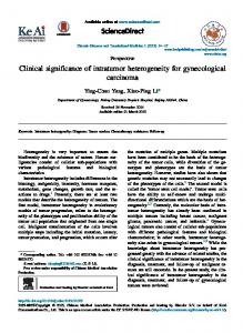
green fluorescence (normally FL1) and in a plot of FL3 versus FL1 (Fig. 1). In this last plot and with the exception of extremely stratified oceanic waters, prochlorophytes and cyanobacteria can be separated from chemotrophic bacteria (13). As shown by Fig. 1, on the basis of their SSC, FL1, and FL3 signals, it is also possible to differentiate two groups of bacteria: we have called HDNA bacteria those that have high FL1 and FL3 values and LDNA bacteria those with slightly lower SSC values and much lower fluorescence values. HDNA bacteria. In Exp#1 and Exp#2, the numbers of HDNA bacteria varied by 14-fold and 21-fold, respectively,
FIG. 1. Different ways of presenting the heterogeneity in the bacterial population as it can be detected in a flow cytometric run of a Syto13-stained sample. The dot plots correspond to the 10,000 events acquired. The lower plots present the same data in a density plot where the gray shading is proportional to the number of points in each area. The HDNA bacteria and the LDNA bacteria form discrete clusters, as do the reference 0.92-m yellow-green beads. A second peak, corresponding to the doublets of the beads, is also visible. In the upper part of the graph we included three-dimensional plots of the same data, where the two groups of bacteria and the beads can also be differentiated. Note that, in the case of the SSC versus FL1 plot, we have changed the angle of view.
4478
GASOL ET AL.
APPL. ENVIRON. MICROBIOL. TABLE 2. Bacterial counts from Exp#1 and Exp#2 No. of bacteria Exp#1
Type of bacteria
All LDNA HDNA a
Exp#2
Minimum
Maximum
Relative changea
5.3 ⫻ 105 2.5 ⫻ 105 1.7 ⫻ 105
4.4 ⫻ 106 8.4 ⫻ 105 3.6 ⫻ 106
8 3 21
Minimum
Maximum
Relative change
3.5 ⫻ 105 1.5 ⫻ 105 2.0 ⫻ 105
3.6 ⫻ 106 9.0 ⫻ 105 2.8 ⫻ 106
10 6 14
The relative change is defined as the maximum number of bacteria/the minimum number of bacteria.
while the TCs varied by 8-fold and 10-fold, respectively (Table 2). The numbers of HDNA bacteria were well correlated with those of “live” bacteria (Pearson’s correlation coefficient R ⫽ 0.52, n ⫽ 57, P ⬍ 0.0005) but not with the numbers of NuCC. However, the percentage of the bacterial population classified as HDNA, NuCC, or live was very similar in both experiments (Fig. 2), averaging 60%. HDNA bacteria and “live” bacteria probably include the same cells; this is evident from a plot in which we compared the rates of variation of HDNA bacteria throughout the different experiments with those of the “live” cells and those of NuCC (Fig. 3). Because samples for “live” and NuCC had only been collected at 0, 36, and 84 h, the plot presents the rates of variation of these types of cells between 0 and 36 h and between 36 and 84 h. The rates of change of NuCC, HDNA cells, and “live” cells were highly correlated (P was ⬍0.0005 for all) (Table 3). LDNA bacteria. In Exp#1 and Exp#2, the numbers of LDNA bacteria varied by 3-fold and 6-fold, respectively, while the TCs varied by 8-fold and 10-fold, respectively (Table 2). The numbers of LDNA bacteria were not significantly correlated with those of “dead” bacteria, but the rates of change of these two groups of bacteria were slightly correlated (P ⫽ 0.06) (Table 3). In Exp#1 the average percentage of the TC classified as LDNA was very close to the average percentage of the count classified as “dead” cells (Fig. 2), with values around 37%. However, in Exp#2, while the percentage of the count
classified as “dead” cells averaged 18%, the average percentage of the TC classified as LDNA bacteria was 41% (Fig. 2). Predators and bacterial community composition. The absence of predators, or the uncoupling of bacterial predators and larger predators in bottle experiments or in microcosms, immediately resulted in the growth of HDNA bacteria without a concomitant growth of LDNA bacteria (Fig. 4 to 6) to the extent that the %HDNA bacteria approached ⬎95% in 1 or 2 days. In Exp#3, and after a few days of incubation, HNF developed in the treatment that had not been filtered through 0.8-m-diameter pores and produced a decline in the total number of bacteria (not shown), with a dramatic decline in the %HDNA bacteria (Fig. 4). We designed Exp#4 to discern whether HNF development was associated with a decrease in HDNA bacteria, with an increase in LDNA bacteria, or with both. The different treatments (filtration through 1-m pores and filtration through 20-m pores) left many HNF in the water, but these organisms grew only in the ⬍20-m-pore-diameter filter treatment (Fig. 5). HNF growth was associated with a slight decrease in the numbers of LDNA bacteria and with no net growth of HDNA bacteria. However, in the treatment where HNF did not grow (pore diameter, ⬍1 m), a 2.5-fold increase in HDNA bacteria occurred parallel to the stability in the numbers of LDNA bacteria (Fig. 5). HNF affected HDNA bacteria in the treatment where they developed (pore diameter, ⬍20 m).
FIG. 2. Relationships between the different counts and types of bacteria. Each individual value was scaled to the TC obtained with Syto13 and flow cytometry (as those were the values that existed for all sampling times), and all values for each experiment were averaged. Error bars represent standard errors of the means. (A) Exp#1; (B) Exp#2. FC, flow cytometry.
VOL. 65, 1999
ANALYSIS OF PLANKTONIC BACTERIA BY FLOW CYTOMETRY
FIG. 3. Relationships between the rates of change (day⫺1) of HDNA bacteria, NuCC, and “live” bacteria, calculated between those sampling times for which there was data for the three types. Data of Exp#1 and Exp#2 were placed together. Statistical data for these graphs are presented in Table 3.
Bacterial growth. The HDNA bacteria were the dynamic components of the bacterial community. In dilution-growth experiments like the one whose results are presented in Fig. 6, where predators are absent or unable to compensate for bacterial growth, a slight increase in both HDNA and LDNA bacteria in the first 10 h was followed by a decrease and stabilization of the numbers of LDNA bacteria and a severalfold net increase of HDNA bacteria (Fig. 6). A large increase in the rate of leucine uptake after 10 h was followed by HDNA bacteria growth and an increase of the %HDNA to up to 90% of the community (Fig. 6).
TABLE 3. Pearson pairwise correlation coefficients between rates of variation (day⫺1)a Comparison
R
n
P
Syto13 TC, SYBR Green TC Syto13 TC, Live/Dead TC SYBR Green TC, Live/Dead TC LDNA, “dead” HDNA, “live” HDNA, NuCC Live, NuCC
0.93 0.74 0.28 0.26 0.74 0.62 0.57
31 46 25 51 47 48 48
⬍0.0005 ⬍0.0005 0.176 0.060 ⬍0.0005 ⬍0.0005 ⬍0.0005
a Rates of variation of the different numbers of bacteria throughout Exp#1 and Exp#2. Results from both experiments and both rates estimated (rate of change from 0 to 36 h and rate of change from 36 to 84 h) are included together. Most of the values obtained with DAPI were not contemporaneous to these estimates and are thus not included in the comparison.
4479
FIG. 4. Change in the %HDNA (F) in microcosm Exp#3, where water had been either filtered through 0.8-m-diameter pores (upper panel) or through 150-m-diameter pores (lower panel). Results are averages and standard errors of two replicates. After three days, we counted the number of HNF (E) in one of the replicates. Flagellates did not grow in the ⬍0.8-m-pore-diameter filtrates.
DISCUSSION Total bacterial counts. The different methods used to count total bacteria provided reasonably close counts (Table 1). This is especially true given that the different methods were analyzed by different operators and that between-operator variability is recognized as one of the main sources of error in counting planktonic bacteria (24, 36). Interuser variability in counting DAPI-stained samples usually ranges between 5 and 20% depending on the level of complexity of the sample (e.g., see reference 23). While the experiments presented here were not designed to test for good correlation of results between methods, the data nevertheless clustered around the 1:1 line and log-log slopes were very close to 1 (Table 1). Our correlations are similar to those presented by other researchers that have compared flow cytometry and epifluorescence TCs (5, 26, 28, 31). The relationship between the Syto13 TCs and SYBR Green TCs was the closest of all studied. While the DAPI counts could slightly underestimate true concentration (⬃2 to 6% of the bacteria can cross the polycarbonate 0.2-m-pore-diameter filter in these waters [12]), the SYBR Green-stained samples were counted on 0.02-m-pore-diameter filters, and it could be assumed that no bacteria were left out of the counts. Flow cytometry would also include all bacteria, because the method is so sensitive that even viruses can be seen and counted (32). The Live/Dead method provided systematically lower counts and worse correlations than all other methods considered. The protocol for the double staining, which includes displacement of one stain by the other, could affect reliability (16). Even with that in mind, however, the correlations between the Live/Dead TCs and those of the other methods were significant. In a comparison of different nucleic acid dyes, Lebaron et al. (26) recommended Syto9 (the live component of the Live/
4480
GASOL ET AL.
APPL. ENVIRON. MICROBIOL.
FIG. 6. The upper panel shows the change in the numbers of HDNA (E), LDNA (ƒ), and total bacteria (F) in a dilution-growth experiment performed in the Rı´a de Vigo estuary (Exp#5). The lower panel shows the change in the %HDNA (E) and the rate of radioactive leucine uptake by the whole community (F) in the same experiment. inc., incorporation.
FIG. 5. The upper panel shows the change in the number of HDNA bacteria and LDNA bacteria in samples of microcosm Exp#4, where water had been filtered either through 1-m pores (closed symbols) or through 20-m pores (open symbols). Results are averages and standard errors of two replicated microcosms. The lower panel shows the change in the number of HNF in the same experiment.
Dead kit) or SYBR Green II as the best stain for counting bacteria. Syto13 generated slightly lower fluorescence yields than these two stains, especially in seawater, but is unlikely that this could affect TC determinations in routine work. The fact that our Syto13 TCs correlate well with those of other methods validates the use of this stain for normal work, without dismissing the potential of other stains. Given the recognized interuser variability in counting total bacteria, we consider our Syto13 flow cytometric method a viable alternative for obtaining TCs of marine planktonic bacteria. Relationship between bacterial size and fluorescence intensity. Syto13 is assumed to stain both DNA and RNA with a similar quantum yield (16). However, Guindulain et al. (14) found it to stain mainly DNA in natural marine samples, a
result that had also been observed for other stains, such as TO-PRO and TO-TO (28). This is probably due to differences in the ability of the stains to bind to the two nucleic acids in salt water conditions, or to the relatively more protected structure of rRNA with respect to DNA, rather than being due to the cell-specific amounts of RNA in natural marine bacteria (which tend to have similar amounts of RNA and DNA [27]). Evidence has accumulated to permit the association of high bacterial fluorescence with high DNA content and large bacterial size and of low fluorescence with low DNA content and small bacterial size. (i) Filtration through filters of varying nominal pore sizes alters the composition of the bacterial community in terms of %HDNA, strongly suggesting that there is a correspondence between bacterial average size and fluorescence intensity (12). (ii) This correspondence has also been established for bacteria stained with other DNA stains, suggesting a direct relationship between fluorescence and bacterial size through the relationship between fluorescence and DNA content and that between DNA content and size (50). (iii) The average fluorescence of the bacterial population, as normalized to that of the beads, is well correlated (r2 ⫽ 0.66)
TABLE 4. Concentrations of total, HDNA, and LDNA bacteria in, and %HDNA values for, the supernatant of a plankton marine samplea Treatment
None Centrifugation Resuspension a b
No. of bacteriab LDNA
HDNA
Total
9.36 ⫻ 105 ⫾ 0.05 ⫻ 105 (100) 6.44 ⫻ 105 ⫾ 0.72 ⫻ 105 (69) 8.96 ⫻ 105 ⫾ 0.37 ⫻ 105 (96)
1.73 ⫻ 106 ⫾ 0.15 ⫻ 106 (100) 1.00 ⫻ 106 ⫾ 0.06 ⫻ 106 (57) 1.89 ⫻ 106 ⫾ 0.09 ⫻ 106 (109)
2.67 ⫻ 106 ⫾ 0.15 ⫻ 106 (100) 1.64 ⫻ 106 ⫾ 0.13 ⫻ 106 (61) 2.79 ⫻ 106 ⫾ 0.13 ⫻ 106 (105)
%HDNAb
64.8 ⫾ 1.8 (100) 60.8 ⫾ 1.3 (94) 67.9 ⫾ 0.2 (105)
Sample was centrifuged for 5 min in Eppendorf tubes and afterwards resuspended. Values shown are averages ⫾ standard errors of two replicates. Numbers in parentheses are percentages of values determined for the untreated sample.
VOL. 65, 1999
ANALYSIS OF PLANKTONIC BACTERIA BY FLOW CYTOMETRY
with bacterial size (range analyzed, 0.028 to 0.072 m3) determined empirically in a range of planktonic environments by image analysis of DAPI-stained samples (44). To further explore the relationship between size and relative fluorescence, we performed a small experiment in which we centrifuged a bacterial community for 5 min and measured the relative concentration of each group of bacteria (Table 4). HDNA bacteria sedimented faster than LDNA bacteria. When the sample was resuspended, concentrations of LDNA and HDNA cells similar to the original ones were recovered, providing an indication of the different relative densities of the two types of cells, which, even if not definitive by itself, provides evidence that supports the results cited above. In a similar experiment NuCC were found to sediment faster than the ghost cells (55). Thus, the HDNA bacteria are larger and denser cells and the LDNA bacteria are smaller and less dense cells. Two groups of marine planktonic bacteria. Sieracki and Viles (47) detected the presence of a very abundant type of small and dim particles stained both with DAPI and with acridine orange in a detailed image analysis study of bacteria in the North Atlantic. Those small and dim particles that were stained with DAPI were 45 to 85% of the DAPI TC, and in dot plots of fluorescence versus size, they were clustered separately from the high fluorescence, large size cells. These authors speculated that the low fluorescence group of bacteria could be either large viruses, microbacteria, or even DAPI-stained free DNA. A similar low DAPI fluorescence group of cells appeared in the flow cytometry analysis of Kaneohe Bay bacteria presented by Monger and Landry (34). Since the publication of that paper, the two groups of bacteria have been observed in TO-TO- and TO-PRO-stained marine bacteria (28), in DAPIstained freshwater bacteria (3), and in SYBR Green-stained marine bacteria (31). Large (0.4 m) viruses exist in the ocean (2) and could form a portion of the low fluorescence particles, but it is certainly unlikely that they would be such a large percentage of the total bacterial count. Bacteria with sizes around 0.2 m (ultramicrobacteria [29]) but completely active could also make up part of the low fluorescence particles. However, if the LDNA particles include active bacteria, why should there be a clear separation between both (high and low fluorescence, HDNA and LDNA) bacterial groups? And why did such LDNA bacteria not show any growth in the dilution-growth experiments (Fig. 6)? While bacterial subpopulations other than our LDNA and HDNA groups have been described in some studies (13, 18, 28, 31), only these two groups are consistently found in all samples. Even though the bacterial subgroups have been observed by many authors, few studies have been conducted to characterize the significance of each of the groups. Li et al. (28) found that their group II bacteria (HDNA) counts were better correlated with chlorophyll a than their group I (LDNA) bacteria and that the fluorescence difference between the two groups was positively related to chlorophyll. Also, in a dilution-growth experiment, they showed that HDNA bacteria grew three times as fast as LDNA bacteria. In a follow-up paper, Jellett et al. (19) compared the %HDNA (their ACI) to tritiated substrate uptake rates and found patterns of %HDNA and substrate uptake that were similar. They also determined that the HDNA cells had on average five times more DNA per cell than the LDNA cells. The work of Li and colleagues, which were the only studies that tried to unveil the meaning of the different subpopulations, suggested that the %HDNA values had potential for being a useful index of bacterial growth. Interestingly, Jellett et al. (19) considered the LDNA bac-
4481
teria to be inactive cells rather than bacterial ghosts on the basis of a slow growth pattern in the experiments reported and on the basis of varying concentrations in different sites. Button et al. (3) considered the dim particles (LDNA) that appeared in Lake Zu ¨rich not to be ghosts because they had normal light scatter signals. That LDNA bacteria have 90° scatter signals which are similar to those of the HDNA bacteria is also apparent from Fig. 1. However, the flow cytometric measurement of light scatter for particles below 0.5 m is problematic, cannot be considered precise, and by no means can be related to bacterial size in natural planktonic bacteria (51). We directly compared the values of HDNA bacteria with those of “live” bacteria and with those of NuCC and obtained a striking correspondence between average values (Fig. 2) and between directly estimated rates of change through time (Fig. 3). That the relationships are relatively strong is important because the range of the data was not large and, as commented above, each method bears a certain degree of inexactitude in the determinations that adds up when two methods are compared. It is now recognized that a certain percentage of all bacteria in planktonic environments are not actively growing (35). To have better estimates of the rates of activity and growth of individual bacteria in the sea, bulk parameters should be scaled to the number of bacteria that are alive or maintain cellular activity (8, 46). It is equally important that the absolute number of bacteria that rRNA or DNA probes are expected to recover be known to determine whether there are bacteria that are not recognized by the probes (42). A wide range of methods are available for the determination of this number of active (or actively growing) bacteria: microautoradiography (33), the direct viable count (20), formazan salts (46, 49), universal rRNA probes (22, 53), etc. However, the methods differ in what they are actually measuring and no consensus has been reached yet on whether the results are at all comparable. In that sense, our results include the determination of the %HDNA as a valid method for the fast determination of the number of active, actively growing, and/or live bacteria. HDNA bacteria are live bacteria, but our data do not allow us to conclude whether they are live and active or live and inactive. We also compared the abundance of “dead” (PI-stained) and LDNA bacteria and found some evidence of correspondence between those numbers (Fig. 2 and Table 3). At least three types of particles could be included in the LDNA bacteria pool: inactive cells, recently dead bacteria, and fragments of cells that still have pieces of DNA able to bind the stains. The last two of these three groups of bacteria could properly be called bacterial ghosts. Direct evidence for the presence of dead cells in seawater has been provided by Heissenberger et al. (17), who observed free-living cells by transmission electron microscopy and found that only 34% of the cells had wellpreserved internal structures while 42% of the cells exhibited cellular damage and 24% lacked any internal structure. Inactive cells can be assumed to have lower DNA content than growing cells due to their nonreplicative state. Jellett et al. (19) calculated that HDNA bacteria had on average five times more DNA per cell than LDNA bacteria, and this would seem to indicate that dead cells must be a significant component of the LDNA fraction. A fast growing cell can have two to three times more DNA than an inactive cell due to multiple replication forks. Additionally, inactive cells may lose extra DNA, such as plasmids or copies of the same gene (e.g., see reference 38). LDNA cells did not present any growth when predators had been removed in Exp#4 and Exp#5 (Fig. 5 and 6). Thus, it seems likely that dead cells (i.e., recently dead cells or cell fragments with DNA remains) are a large part of the LDNA
4482
GASOL ET AL.
fraction. Because there should be a continuum between cell fragments with no DNA and recently dead cells with still large amounts of DNA, the separation of LDNA bacteria from noise in a SSC versus FL1 plot could at times be problematic (Fig. 1). HDNA and LDNA bacteria in nature. In the experiments reported, the ratio of HDNA bacteria to total bacterial abundance (HDNA plus LDNA) ranged from ⬍40 to ⬎80%, with an average of ⬃55%. We have further unpublished data on %HDNA in field samples that range from ⬍15% (in deep central Atlantic samples) to 95% (in a eutrophic reservoir). Li et al. (28) in the Mediterranean and in the North Atlantic (10 to 90%) and Jellett et al. (19) in Bedford Basin (19 to 80%) found similar ranges. These %HDNA values compare well to those of active bacteria obtained by different methods, values that tend to cluster at around ⬃50% when autoradiography and rRNA probes are used (e.g., see reference 22) or at ⬃25% when formazan salts are used (46). The values are relatively larger than the NuCC values published to date, which tend to be below 30 to 40% (4, 15, 55), with a few exceptions approaching (10, 22, 42) or surpassing (52) 50%. What drives the variability in the relative amount of active, live, and/or HDNA bacteria and inactive, dead, and/or LDNA bacteria? Our results provide evidence that when predators are absent the %HDNA tends to approach a value close to the maximum (Fig. 4 to 6). The relaxation in predator control allows the growth of HDNA bacteria to the point that they dominate the community. The development of predators reverses the situation (Fig. 4), probably due to their preferential predation of HDNA bacteria. It has been shown that predators prefer larger-sized (21) and active (6) bacteria, consistent with our interpretation that HDNA bacteria are large and active cells. Grazing on bacteria by flagellates (37) or viral activity could be responsible for the production of LDNA bacteria if they are composed mainly of dead and damaged cells. However, if LDNA bacteria include both of these types of cells and inactive or starved cells, then nutrient availability could be the ultimate factor responsible for the variability of LDNA bacterial numbers. Conclusion. We have presented evidence of a good relationship between the numbers of “live” bacteria as measured with the Molecular Probes Live/Dead kit, of NuCC, and of HDNA cells as determined by flow cytometry of Syto13-stained plankton samples. We also show that HDNA bacteria are the dynamic members of the bacterial community, those that respond immediately to changes in predation pressure and nutrient availability. We endorse the use of the %HDNA as an index of the amount of active or live bacterial cells in plankton that can be obtained in less than a minute if a flow cytometer is available. We envision a representation of bacteria in plankton in which the total DAPI count, which has been and still is for most researchers the true number of bacteria, is composed of different particles. Ordered from less active to more active, (i) some are not even bacteria (large viruses and cell fragments, or ghosts); (ii) some are dead cells, with no potential for growth but intact with regard to shape; (iii) some are inactive because the proper conditions for their development are not present; (iv) some are growing at a very low rate; and (v) some are large, growing at a fast pace. To study the composition of the bacterial community we have many tools that will correctly identify the fifth group as highly active and live and the first and second groups as inactive and dead. The problem arises with the third and fourth groups, which probably are the most abundant ones, for which each method indicates different quantities and whose cells are assigned to an active or an inactive pool depending on the method, the protocol, and even
APPL. ENVIRON. MICROBIOL.
the researcher involved, thus generating the lack of consensus for a universal method to determine these numbers. In that framework, we suggest that the determinations of HDNA and LDNA bacteria and of the %HDNA values are a fast and simple alternative, as good as, or even better than, most of the other currently used techniques. ACKNOWLEDGMENTS We thank Ce`lia Marrase´, who brought us all together for the TUR MED experiments, and we thank all the other researchers that participated in the workshop. Data of the Incoce´ano cruise was collected with the help of Carlos Pedro ´s-Alio ´, who also supported this work in many other ways. R. Massana and M. Schauer contributed to the estimation of the DAPI count variability in our lab. We also appreciate the usual help of S. Canut and the comments and encouragement of P. A. del Giorgio, C. Marrasse´, and R. Massana. This work was supported by two EU grants, MAS3-CT95-0016 (MEDEA) and MAR95-1901-C03-03 (MIDAS). REFERENCES 1. Azam, F., T. Fenchel, J. G. Field, J. S. Gray, L.-A. Meyer-Reil, and F. Thingstad. 1983. The ecological role of water-column microbes in the sea. Mar. Ecol. Prog. Ser. 10:257–263. 2. Bratbak, G., O. H. Haslund, M. Heldal, A. Næs, and T. Røeggen. 1992. Giant marine viruses? Mar. Ecol. Prog. Ser. 85:201–202. 3. Button, D. K., B. R. Robertson, and F. Ju ¨ttner. 1996. Microflora of a subalpine lake: bacterial populations, size and DNA distributions, and their dependence on phosphate. FEMS Microbiol. Ecol. 21:87–101. 4. Choi, J. W., E. B. Sherr, and B. F. Sherr. 1996. Relation between presenceabsence of a visible nucleoid and metabolic activity in bacterioplankton cells. Limnol. Oceanogr. 41:1161–1168. 5. del Giorgio, P. A., D. F. Bird, Y. T. Prairie, and D. Planas. 1996. Flow cytometric determination of bacterial abundance in lake plankton with the green nucleic acid stain SYTO 13. Limnol. Oceanogr. 41:783–789. 6. del Giorgio, P. A., J. M. Gasol, D. Vaque´, P. Mura, S. Agustı´, and C. M. Duarte. 1996. Protistan control of the proportion of metabolically active cells in coastal marine bacterioplankton. Limnol. Oceanogr. 41:1169–1179. 7. del Giorgio, P. A., J. J. Cole, and A. Cimbleris. 1997. Respiration rates in bacteria exceed phytoplankton production in unproductive aquatic systems. Nature 385:148–151. 8. del Giorgio, P. A., Y. T. Prairie, and D. F. Bird. 1997. Coupling between rates of bacterial production and the abundance of metabolically active bacteria in lakes, enumerated using CTC reduction and flow cytometry. Microb. Ecol. 34:144–154. 9. Fuhrman, J. A., T. D. Sleeter, C. A. Carlson, and L. M. Proctor. 1989. Dominance of bacterial biomass in the Sargasso Sea and its ecological implications. Mar. Ecol. Prog. Ser. 57:207–217. 10. Gasol, J. M., M. D. Doval, J. Pinhassi, J. I. Caldero´n-Paz, N. Guixa-Boixereu, D. Vaque´, and C. Pedro ´s-Alio ´. 1998. Diel variations in bacterial heterotrophic production in the northwestern Mediterranean Sea. Mar. Ecol. Prog. Ser. 164:125–133. 11. Gasol, J. M., P. A. del Giorgio, and C. M. Duarte. 1997. Biomass distribution of marine planktonic communities. Limnol. Oceanogr. 42:1353–1363. 12. Gasol, J. M., and X. A. G. Mora ´n. 1999. Effects of filtration on bacterial activity and picoplankton community structure as assessed by flow cytometry. Aquat. Microb. Ecol. 16:251–264. 13. Gasol, J. M., and P. A. del Giorgio. Using flow cytometry for counting natural planktonic bacteria and understanding the structure of planktonic bacterial communities. Sci. Mar, in press. 14. Guindulain, T., J. Comas, and J. Vives-Rego. 1997. Use of nucleic acid dyes SYTO-13, TOTO-1, and YOYO-1 in the study of Escherichia coli and marine prokaryotic populations by flow cytometry. Appl. Environ. Microbiol. 63:4608–4611. 15. Hagstro ¨m, Å., J. Pinhassi, and U. L. Zweifel. Unpublished results. 16. Haugland, R. P. 1996. Handbook of fluorescent probes and research chemicals. Molecular Probes Inc., Eugene, Oreg. 17. Heissenberger, A., G. G. Leppard, and G. J. Herndl. 1996. Relationship between the intracellular integrity and the morphology of the capsular envelope in attached and free-living marine bacteria. Appl. Environ. Microbiol. 62:4521–4528. 18. Jacquet, S., J.-F. Lennon, D. Marie, and D. Vaulot. 1998. Picoplankton population dynamics in coastal waters of the northwestern Mediterranean Sea. Limnol. Oceanogr. 43:1916–1931. 19. Jellett, J. F., W. K. W. Li, P. M. Dickie, A. Boraie, and P. E. Kepkay. 1996. Metabolic activity of bacterioplankton communities assessed by flow cytometry and single carbon substrate utilization. Mar. Ecol. Prog. Ser. 136:213– 225. 20. Joux, F., and P. Lebaron. 1997. Ecological implications of an improved direct
VOL. 65, 1999
21. 22. 23. 24.
25.
26. 27. 28. 29. 30. 31.
32. 33. 34. 35. 36.
37. 38.
ANALYSIS OF PLANKTONIC BACTERIA BY FLOW CYTOMETRY
viable count method for aquatic bacteria. Appl. Environ. Microbiol. 63: 3643–3647. Ju ¨rgens, K., and H. Gu ¨de. 1994. The potential importance of grazing-resistant bacteria in planktonic systems. Mar. Ecol. Prog. Ser. 112:169–188. Karner, M., and J. A. Fuhrman. 1997. Determination of active marine bacterioplankton: a comparison of universal 16S rRNA probes, autoradiography, and nucleoid staining. Appl. Environ. Microbiol. 63:1208–1213. Kepner, R. L., Jr., and J. R. Pratt. 1994. Use of fluorochromes for direct enumeration of total bacteria in environmental samples: past and present. Microbiol. Rev. 58:603–615. Kirchman, D. L. 1993. Statistical analysis of direct counts of microbial abundance, p. 117–119. In P. F. Kemp, B. F. Sherr, E. B. Sherr, and J. J. Cole (ed.), Handbook of methods in aquatic microbial ecology. Lewis Publishers, Boca Raton, Fla. Kirchman, D. L. 1993. Leucine incorporation as a measure of biomass production by heterotrophic bacteria, p. 509–512. In P. F. Kemp, B. F. Sherr, E. B. Sherr, and J. J. Cole (ed.), Handbook of methods in aquatic microbial ecology. Lewis Publishers, Boca Raton, Fla. Lebaron, P., N. Parthuisot, and P. Catala. 1998. Comparison of blue nucleic acid dyes for flow cytometric enumeration of bacteria in aquatic systems. Appl. Environ. Microbiol. 64:1725–1730. Lee, S.-H., and P. F. Kemp. 1994. Single-cell RNA content of natural marine planktonic bacteria measured by hybridization with multiple 16S rRNAtargeted fluorescent probes. Limnol. Oceanogr. 39:869–879. Li, W. K. W., J. F. Jellett, and P. M. Dickie. 1995. DNA distributions in planktonic bacteria stained with TOTO or TO-PRO. Limnol. Oceanogr. 40:1485–1495. MacDonell, M. T., and M. A. Hood. 1982. Isolation and characterization of ultramicrobacteria from a Gulf Coast estuary. Appl. Environ. Microbiol. 43:566–571. Marie, D., D. Vaulot, and F. Partensky. 1996. Application of the novel nucleic acid dyes YOYO-1, YO-PRO-1, and PicoGreen for flow cytometric analysis of marine prokaryotes. Appl. Environ. Microbiol. 62:1649–1655. Marie, D., F. Partensky, S. Jacquet, and D. Vaulot. 1997. Enumeration and cell cycle analysis of natural populations of marine picoplankton by flow cytometry using the nucleic acid stain SYBR Green I. Appl. Environ. Microbiol. 63:186–193. Marie, D., C. P. D. Brussard, R. Thyrhaug, G. Bratbak, and D. Vaulot. 1999. Enumeration of marine viruses in culture and natural samples by flow cytometry. Appl. Environ. Microbiol. 65:45–52. Meyer-Reil, L.-A. 1978. Autoradiography and epifluorescence microscopy combined for the determination of number and spectrum of actively metabolizing bacteria in natural waters. Appl. Environ. Microbiol. 36:506–512. Monger, B. C., and M. R. Landry. 1993. Flow cytometric analysis of marine bacteria with Hoechst 33342. Mar. Ecol. Prog. Ser. 59:905–911. Morita, R. Y. 1997. Bacteria in oligotrophic environments. Starvation-survival lifestyle. Chapman & Hall, New York, N.Y. Nagata, T., T. Someya, T. Konda, M. Yamamoto, K. Morikawa, M. Fukui, N. Kuroda, T. Takahashi, S. Oh, M. Mori, S. Araki, and K. Kato. 1989. Intercalibration of the acridine orange direct count method of aquatic bacteria. Bull. Jpn. Soc. Microb. Ecol. 4:89. Nagata, T., and D. L. Kirchman. 1992. Release of macromolecular organic complexes by heterotrophic marine flagellates. Mar. Ecol. Prog. Ser. 83:233– 240. Neidhardt, F. C., J. L. Ingraham, and M. Schaechter. 1990. Physiology of the
39. 40. 41. 42. 43. 44. 45. 46. 47. 48. 49. 50. 51. 52.
53.
54. 55.
4483
bacterial cell. A molecular approach. Sinauer Associates, Inc., Sunderland, Mass. Noble, R. T., and J. A. Fuhrman. 1998. Use of SYBR Green I for rapid epifluorescence counts of marine viruses and bacteria. Aquat. Microb. Ecol. 14:113–118. Peters, F., C. Marrase´, J. M. Gasol, M. M. Sala, and L. Arin. 1998. Food-web mediated effects of turbulence on bacterial production and growth. Mar. Ecol. Prog. Ser. 172:293–303. Peters, F., C. Marrase´, H. Havskum, F. Rassoulzadegan, J. Dolan, M. Alcaraz, and J. M. Gasol. Unpublished results. Pinhassi, J., U. L. Zweifel, and Å. Hagstro ¨m. 1997. Dominant marine bacterioplankton species found among colony-forming bacteria. Appl. Environ. Microbiol. 63:3359–3366. Porter, K. G., and Y. S. Feig. 1980. The use of DAPI for identification and enumeration of bacteria and blue-green algae. Limnol. Oceanogr. 25:943– 948. Prairie, Y. T., P. A. del Giorgio, D. F. Bird, and J. M. Gasol. Unpublished results. Robertson, B. R., and D. K. Button. 1989. Characterizing aquatic bacteria according to population, cell size, and apparent DNA content by flow cytometry. Cytometry 10:70–76. Sherr, B. F., P. A. del Giorgio, and E. B. Sherr. 1999. Estimating abundance and single-cell characteristics of actively respiring bacteria via the redox dye CTC. Aquat. Microb. Ecol. 18:117–131. Sieracki, M. E., and C. L. Viles. 1992. Distributions and fluorochromestaining properties of submicrometer particles and bacteria in the North Atlantic. Deep-Sea Res. 39:1919–1929. Smith, D. C., and F. Azam. 1992. A simple, economical method for measuring bacterial protein synthesis rates in seawater using 3H-leucine. Mar. Microb. Food Webs 6:107–114. Tabor, P. S., and R. A. Neihof. 1982. Improved method for determination of respiring individual microorganisms in natural waters. Appl. Environ. Microbiol. 43:1249–1255. Veldhuis, M. J. W., T. L. Cucci, and M. E. Sieracki. 1997. Cellular DNA content of marine phytoplankton using two new fluorochromes: taxonomic and ecological implications. J. Phycol. 33:527–541. Vives-Rego, J., R. Lo ´pez-Amoro´s, and J. Comas. 1994. Flow cytometric narrow-angle light scatter and cell size during starvation of Escherichia coli in artificial sea water. Lett. Appl. Microbiol. 19:374–376. Vosjan, J. H., and G. J. van Noort. 1998. Enumerating nucleoid-visible marine bacterioplankton: bacterial abundance determined after storage of formalin fixed samples agree with isopropanol rinsing method. Aquat. Microb. Ecol. 14:149–154. Williams, S. C., Y. Hong, D. C. A. Danavall, M. H. Howard-Jones, D. Gibson, M. E. Frischer, and P. G. Verity. 1998. Distinguishing between living and nonliving bacteria: evaluation of the vital stain propidium iodine and its combined use with molecular probes in aquatic samples. J. Microbiol. Methods 32:225–236. Zimmermann, R., and L.-A. Meyer-Reil. 1974. A new method for fluorescence staining of bacterial populations on membrane filters. Kieler Meeresforsch. 30:24–27. Zweifel, U. L., and Å. Hagstro ¨m. 1995. Total counts of marine bacteria include a large fraction of non-nucleoid-containing bacteria (ghosts). Appl. Environ. Microbiol. 61:2180–2185.