RESEARCH ARTICLE
Simple Detection of the IS6110 Sequence of Mycobacterium tuberculosis Complex in Sputum, Based on PCR with Graphene Oxide Sang-Hyun Hwang1,2*, Dong-Eun Kim3, Heungsup Sung4, Byeong-Min Park1, MiJeong Cho1, Ok-Jin Yoon1, Do-Hoon Lee1 1 Department of Laboratory Medicine, Center for Diagnostic Oncology, Research Institute and Hospital, National Cancer Center, Goyang-si, Gyeinggi-do, 410–769, Republic of Korea, 2 Hematologic Malignancy Branch, Research Institute and Hospital, National Cancer Center, Goyang-si, Gyeinggi-do, 410–769, Republic of Korea, 3 Department of Bioscience and Biotechnology, Konkuk University, Seoul, 143–701, Republic of Korea, 4 Department of Laboratory Medicine, University of Ulsan College of Medicine and Asan Medical Center, 388–1 Pungnap-dong, Songpa-gu, Seoul, 138–736, Republic of Korea *
[email protected]
Abstract OPEN ACCESS Citation: Hwang S-H, Kim D-E, Sung H, Park B-M, Cho M-J, Yoon O-J, et al. (2015) Simple Detection of the IS6110 Sequence of Mycobacterium tuberculosis Complex in Sputum, Based on PCR with Graphene Oxide. PLoS ONE 10(8): e0136954. doi:10.1371/ journal.pone.0136954 Editor: Olivier Neyrolles, Institut de Pharmacologie et de Biologie Structurale, FRANCE Received: April 5, 2015 Accepted: August 10, 2015 Published: August 31, 2015 Copyright: © 2015 Hwang et al. This is an open access article distributed under the terms of the Creative Commons Attribution License, which permits unrestricted use, distribution, and reproduction in any medium, provided the original author and source are credited.
Graphene oxide (GO) has proven to be a satisfactory DNA-sensor platform for applications in enzyme-free signal amplification, fluorescence-based amplification, and nanoparticle-based platforms because of its excellent electrical, thermal, and optical properties. In this study, we designed a novel platform for the fluorescence detection of biomolecules, using a fluorescent dye-labeled primer and GO. We applied this system for the detection of the IS6110 insertion sequence of the Mycobacterium tuberculosis complex (MTB) and evaluated its feasibility for use in molecular diagnostics. Fifty-four sputum specimens were collected at our institution from October 2010 to March 2012. To detect MTB in the samples, we performed PCR amplification of the IS6110 DNA sequence using FAM-labeled primers, after which the PCR amplicon was incubated with GO and the fluorescence was measured. The results were compared with those obtained by conventional real-time quantitative PCR (RQ-PCR). The fluorescence intensity observed increased in a concentration-dependent manner with the FAM-labeled IS6110 amplicon. The results of the PCR-GO system for detecting IS6110 DNA were in good agreement with those obtained with conventional RQ-PCR (kappa statistic = 0.925). The PCR-GO system detected MTB DNA in 23 of 25 RQ-PCR-positive sputum samples (92.0%; 95% CI, 75.0–98.0%), but not in 29 of 29 RQ-PCR-negative sputum samples (100%; 95% CI, 88.1– 100.0%). These results indicate the utility of the PCR-GO system in molecular diagnostics.
Data Availability Statement: All relevant data are within the paper and its Supporting Information files. Funding: This research was supported by the National Cancer Center, Grant NCC-1510100. The funders had no role in study design, data collection and analysis, decision to publish, or preparation of the manuscript. Competing Interests: The authors have declared that no competing interests exist.
Introduction Graphene is a one-atom-thick planar sheet of sp2-bonded carbon atoms that forms a honeycomb crystal lattice [1]. Graphene is an attractive nanomaterial because of its unique optical, physical, and electrochemical properties, which make it promising for biosensing applications [2,3] and nucleic acid detection [4,5]. Optically, graphene shows light absorption from the
PLOS ONE | DOI:10.1371/journal.pone.0136954 August 31, 2015
1/8
Graphene-Based PCR for Detecting the IS6110 Sequence
ultraviolet to the near-infrared regions. In addition, graphene is an efficient fluorescence quencher that is useful in the optical-based detection of biomolecules [6–9]. Graphene oxide (GO) is a water-soluble derivative of graphene that preferentially associates with singlestranded (ss) nucleic acids by π-π stacking interactions, with a lower affinity for doublestranded (ds) nucleic acids [3,10]. Therefore, GO could potentially be used as a fluorescence quencher for the detection of DNA sequences instead of conventional fluorescent probes, such as TaqMan or Molecular Beacon probes. Using GO as a quencher may be advantageous in that it more efficiently quenches fluorescence than other commonly used quenchers. In addition, GO can serve as a universal quencher, in contrast to conventional fluorescent probes such as TaqMan and Molecular Beacon probes, which are FRET-based donor-acceptor (quencher) dye pairs that must be prepared for each target sequence being detected [6]. Tuberculosis (TB) is caused by gram-positive bacteria that are members of the Mycobacterium tuberculosis (MTB) complex, including such human pathogens as M. tuberculosis and M. africanum. TB poses a serious global health threat, with 9 million new cases and 1.5 million deaths reported worldwide in 2013 [11]. Multidrug-resistant (MDR) TB strains, with which patients fail to respond to the first-line drugs isoniazid and rifampin, are spreading rapidly and threaten to undermine the control of TB [12]. Reliable, early, and sensitive diagnosis of the MTB complex is required for the effective treatment of patients and disease control [12]. The sensitivity of sputumsmear microscopy is low, and the culture method, which is generally used to confirm diagnoses made by microscopy, typically takes several weeks due to the slow growth of MTB, although the use of liquid culture media has reduced the time required for MTB detection [13]. Nucleic-acid amplification tests based on methods such as real-time quantitative PCR (RQ-PCR) or isothermal amplification are currently the most promising methods for clinical diagnostics [14–17]. In this study, we developed a simple, separation-free method for MTB DNA detection based on the quenching of GO-bound fluorophores. During the amplification step, a 6-carboxyfluorescein (FAM)-labeled primer is incorporated into the amplicon. In the absence of the target, it remains free in solution. Once GO is added, the single-stranded primer adsorbs to GO, resulting in quenching of the FAM fluorescence. Adsorption of the double-stranded amplicon to GO is less favorable; thus, fluorescence is detected in the presence of the target amplicon. A low-cost fluorometer was used for detection purposes. In recent studies, non-amplified single-stranded RNA sequences such as microRNAs were captured and detected using GO [18]. However, we first amplified the target DNA by PCR and then measured the fluorescence from dye-labeled primers that were incorporated into the target amplicons, resulting in desorption from GO and fluorescence signaling. In addition, we evaluated the feasibility of our PCR-GO method for detecting MTB and compared its performance with that of a commercial RQ-PCR test.
Materials and Methods Sample collection and DNA extraction We used 54 sputum specimens that had been stored after completing a previous study for MTB detection [19]. Out of the 54 specimens tested, 46.3% (25/54) were MTB-positive. The sputum samples (1 mL) were inactivated by heating at 80°C for 60 min, and 200 μL of each sample was used for DNA extraction with the QIAamp DNA Stool Mini Kit (QIAGEN, Valencia, CA, USA), according to the manufacturer’s instructions.
IS6110 PCR Gene-specific primers targeting the IS6110 sequence (GenBank Accession No. X52471) were designed with the Primer3 program [20]. The nucleotide sequences of the forward and reverse primers were 50 -FAM-GCCTACGTGGCCTTTGTCAC-30 and 50 -C3-GTCCAGATGGCTTG
PLOS ONE | DOI:10.1371/journal.pone.0136954 August 31, 2015
2/8
Graphene-Based PCR for Detecting the IS6110 Sequence
CTCGAT-30 , respectively, where C3 is a 3-carbon spacer. The amplicon size was 113 bp. Primers were synthesized by Integrated DNA Technologies (Coralville, Iowa, USA). Amplification was performed using the HotStarTaq Plus Master Mix Kit (QIAGEN), as described previously [19]. Reaction mixtures contained 0.25 μM of each dNTP, 0.4 μM forward primer, 0.3 μM reverse primer, and 5 μL of sample DNA. A GeneAmp PCR System 9700 thermal cycler (Applied Biosystems, Foster City, USA) was used for DNA amplification. The thermal cycling conditions used were as follows: 5 min at 95°C; 40 cycles at 94°C for 30 s, 65°C for 30 s, and 72°C for 30 s; and 72°C for 10 min. All experiments were performed in duplicate. For comparison purposes, conventional RQ-PCR was performed using the AdvanSure TB/NTM Real Time PCR Kit (LG Lifesciences, Seoul, Korea) on the ABI Prism 7000 Sequence Detection System (Applied Biosystems), as described previously [19]. For serial dilution sensitivity testing, we generated plasmids containing the IS6110 DNA sequence (p-IS6110) as described previously [19], and a 10-fold dilution series (101–106 copies/ μL) was prepared.
Detection of amplification using GO as a fluorescence quencher The principle of the GO-based amplification detection system is illustrated in Fig 1. Briefly, after PCR amplification, 20 μL of FAM-labeled PCR amplicon was mixed with 20 μL of 1 mg/mL
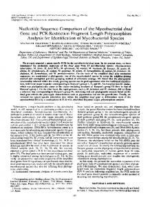
Fig 1. Illustration describing the GO-based sensing system for homogenous detection of the IS6110 DNA amplicon. In the presence of the IS6110 DNA amplicon, the fluorescent dye-labeled primers are incorporated into the amplicon causing the primers to separate from GO, leading to increased fluorescence (upper part of the figure). In the absence of the target DNA, the fluorescent dye-labeled primers remain adsorbed to the GO layer, resulting in fluorescence quenching (lower part of the figure). doi:10.1371/journal.pone.0136954.g001 PLOS ONE | DOI:10.1371/journal.pone.0136954 August 31, 2015
3/8
Graphene-Based PCR for Detecting the IS6110 Sequence
Fig 2. Fluorescence signaling after PCR-GO is proportional to the amplicon concentration. Fluorescence in tubes containing 0 (NTC) to 105 copies/ reaction of a plasmid encoding the IS6110 sequence is shown. Twenty microliters of a 1 mg/mL stock of GO was added at a final concentration 0.2 mg/mL. The fluorescence images were obtained using the IVIS Lumina Series Ⅲ imaging system. NTC, no-template control. doi:10.1371/journal.pone.0136954.g002
stock of GO (Graphene Square, Seoul, South Korea) and 60 uL of distilled water for a final GO concentration of 0.2 mg/mL, and the mixture was incubated for 15 min at room temperature. The fluorescence intensities of the samples were measured with the Qubit 2.0 Fluorometer (Invitrogen, Eugene, OR, USA), using the blue LED (max ~470 nm) excitation light source. The fluorescent signals were recorded as relative fluorescent units (RFUs) using a green fluorescence emission filter (510–580 nm), and the RFU (sample)/RFU (control) ratios were calculated.
Results The fluorescent signal detected using the PCR-GO system is proportional to the amplicon concentration As shown in Fig 2, the fluorescence intensity displayed gradual increases with increasing concentrations of fluorescent dye-labeled amplicons.
PLOS ONE | DOI:10.1371/journal.pone.0136954 August 31, 2015
4/8
Graphene-Based PCR for Detecting the IS6110 Sequence
Sensitivity assay of PCR-GO detection To determine the detection sensitivity of the PCR-GO method, the p-IS6110 plasmid was serially diluted and amplified using a primer pair, one of which was FAM-labeled. Subsequently, the PCR amplicons were incubated with GO, after which the fluorescence intensity was measured. The fluorescence intensity at 543 nm and the RFU ratios increased proportionally to the increase in p-IS6110 amplicon concentration (Fig 3).
Performance of the PCR-GO system with clinical samples Finally, we evaluated the performance of the PCR-GO system for detecting IS6110 DNA in clinical samples. The results were in good agreement with those obtained using a conventional RQ-PCR kit (kappa statistic = 0.925). The PCR-GO system detected MTB DNA in 23 out of 25 RQ-PCR-positive samples (92.0%; 95% CI: 75.0–98.0%) but not in 29 out of 29 RQ-PCR-negative samples (100%; 95% CI: 88.1–100.0%).
Discussion In this study, we explored the feasibility of using GO as a DNA biosensor for detecting the IS6110 sequence of MTB and prototyped a PCR-GO detection system for potential applications in molecular diagnostics. We evaluated the performance of the PCR-GO system in comparison to the commercial AdvanSure RQ-PCR kit that is approved for in vitro diagnostics by the Korean Food and Drug Administration. In a previous study, we used this kit to detect TB in 129 culture–positive TB specimens. We found that 82 out of 82 AFB-stain-positive specimens (100%) and 35 of 47 (74.5%) AFB-stain-negative specimens were detected using this kit, indicating a sensitivity of 90.7% and a specificity of 100% [21]. To the best of our knowledge, no previous studies have been performed to compare the PCR-GO method with current clinical molecular-detection techniques. The PCR-GO results showed strong agreement with the results obtained using conventional RQ-PCR (96.3% agreement; kappa = 0.925). The PCR-GO-based fluorometric assay is cost-effective and does not need spectrally matched reporter and quencher dyes, as used in TaqMan assays.
Fig 3. Detection sensitivity of the PCR-GO system. Left, The RFU ratios (sample/control) increased in proportion to the concentration of p-IS6110 used for PCR amplification (101–106 copies/reaction). Negative group (no template control) was significantly different from positive groups (101, 102, 103, 104, 105, 106 copies/reaction) (Tukey-Kramer multiple-comparison test, p < 0.05). 10 replicates were performed at each concentration. Right, Gel electrophoresis of the 113-bp amplicons after PCR amplification using 101–106 copies of the p-IS6110 plasmid per reaction, as indicated. Right, C, blank; NC, no-template control. doi:10.1371/journal.pone.0136954.g003
PLOS ONE | DOI:10.1371/journal.pone.0136954 August 31, 2015
5/8
Graphene-Based PCR for Detecting the IS6110 Sequence
Some MTB strains contain do not encode the target DNA IS6110 sequence in their genomes, and this assay would not be suitable in such cases [22]. One of advantages of GO is that it more efficiently quenches the fluorescence of common fluors. GO can also serve as an effective universal quencher, without the need for designing new dual-labeled probes for each target, as is done with conventional fluorescent probes such as TaqMan and Molecular Beacon probes [6–9]. Moreover, primers labeled with different fluors in combination with GO as quencher can be used for multiplexing, in contrast to detection via intercalating dyes such as SYBR green that do not enable multiplexing. However, GO could be used for multiplexing if the primers were labeled with different fluors. In addition, the cost per sample of PCR-GO is less expensive. The cost per sample of a FAM-labeled primer was ~ $0.2 and that of GO was ~$0.02. In contrast, the cost per sample of dual-labeled probes including a quencher dye was ~$0.7 in this study. The appropriate design of DNA probes and optimization of GO concentrations was important for improving the specificity of detection of IS6110 DNA based on the GO platform. DNA absorption/desorption from GO is critically affected by salts, pH, temperature, and organic solvents such as ethanol [9,23]. The amplicon and primer/probe sizes have also been shown to affect DNA absorption/desorption from GO [9]. In agreement with previous findings, we observed that fluorescent signal intensities varied according to amplicon sizes (data not shown). The optimal GO concentration for quenching fluorescence emission was found to be 0.2 mg/mL (S1 Fig). Similar concentrations (0.25–0.4 mg/mL) of GO were used in other studies [24,25]. One drawback of our PCR-GO system is that primer-dimers can produce false positive signals. Another disadvantage is that the end point-detection format requires opening of the tube containing amplicon to add the GO. This introduces significant risk of amplicon carryover, and subsequent false positives. Real-time detection formats provide a key advantage in that tubes containing amplicon do not have to be opened up again. However, lab-on-a-chip technologies including microfluidics could add amplicons to the GO in closed system in the future. However, unresolved issues and technical problems remain, such as optimizing the probe size, as well as the surface modification and structure of GO, which could enhance the performance of the GO-based DNA detection platform. Therefore, further studies should be performed to improve the sensitivity of the PCR-GO system. The PCR-GO assay was developed using the Qubit 2.0 fluorometer, which is a small, lowcost, easy-to-use analytical instrument for DNA, RNA, and protein quantitation. This fluorometer has 2 light sources (blue and red LEDs), 2 excitation filters (blue: 430–495 nm and red: 600–645 nm), and 2 emission filters (green: 510–580 nm and red: 665–720 nm). FAM has an excitation/emission peak at 495/520 nm. Therefore, we could detect the fluorescence signal from the FAM-labeled primers with the Qubit 2.0 fluorometer. In conclusion, we developed a novel and simple PCR-GO-based system and demonstrated its application in detecting MTB. In this study, we showed that the performance of our detection system using a simple fluorometer was comparable to that of conventional RQ-PCR. Improvements and optimization of the PCR-GO system are challenges that merit further attention. In the future, biosensors, lab-on-chip technologies, and microfluidics systems are just some of the technologies in which GO-based analysis can be integrated to generate highly sensitive in vitro diagnostic devices.
Supporting Information S1 Fig. Optimization of the GO concentration for FAM-fluorescence quenching. (DOCX)
PLOS ONE | DOI:10.1371/journal.pone.0136954 August 31, 2015
6/8
Graphene-Based PCR for Detecting the IS6110 Sequence
Author Contributions Conceived and designed the experiments: SHH DEK HS DHL. Performed the experiments: BMP MJC OJY. Analyzed the data: DHL. Contributed reagents/materials/analysis tools: SHH. Wrote the paper: SHH.
References 1.
Artiles MS, Rout CS, Fisher TS (2011) Graphene-based hybrid materials and devices for biosensing. Adv Drug Deliv Rev 63: 1352–1360. doi: 10.1016/j.addr.2011.07.005 PMID: 21867736
2.
Zhang M, Le HN, Ye BC (2013) Graphene oxide-based fluorescent "on/off" switch for visual bioassay using "molecular beacon"-hosted Hoechst dyes. ACS Appl Mater Interfaces 5: 8278–8282. doi: 10. 1021/am402429n PMID: 23968374
3.
Chang H, Tang L, Wang Y, Jiang J, Li J (2010) Graphene fluorescence resonance energy transfer aptasensor for the thrombin detection. Anal Chem 82: 2341–2346. doi: 10.1021/ac9025384 PMID: 20180560
4.
Lee J, Park IS, Jung E, Lee Y, Min DH (2014) Direct, sequence-specific detection of dsDNA based on peptide nucleic acid and graphene oxide without requiring denaturation. Biosens Bioelectron 62: 140– 144. doi: 10.1016/j.bios.2014.06.028 PMID: 24997367
5.
Kang T, Choi H, Joo SW, Lee SY, Yoon KA, Lee K. (2014) Peptide nucleic acid-mediated aggregation of reduced graphene oxides and label-free detection of DNA mutation. J Phys Chem B 118: 6297– 6301. doi: 10.1021/jp501820j PMID: 24821658
6.
Dong H, Gao W, Yan F, Ji H, Ju H (2010) Fluorescence resonance energy transfer between quantum dots and graphene oxide for sensing biomolecules. Anal Chem 82: 5511–5517. doi: 10.1021/ ac100852z PMID: 20524633
7.
Liu F, Choi JY, Seo TS (2010) Graphene oxide arrays for detecting specific DNA hybridization by fluorescence resonance energy transfer. Biosens Bioelectron 25: 2361–2365. doi: 10.1016/j.bios.2010. 02.022 PMID: 20299201
8.
Loh KP, Bao Q, Eda G, Chhowalla M (2010) Graphene oxide as a chemically tunable platform for optical applications. Nat Chem 2: 1015–1024. doi: 10.1038/nchem.907 PMID: 21107364
9.
Wu M, Kempaiah R, Huang PJ, Maheshwari V, Liu J (2011) Adsorption and desorption of DNA on graphene oxide studied by fluorescently labeled oligonucleotides. Langmuir 27: 2731–2738. doi: 10.1021/ la1037926 PMID: 21302946
10.
Park JS, Goo NI, Kim DE (2014) Mechanism of DNA adsorption and desorption on graphene oxide. Langmuir 30: 12587–12595. doi: 10.1021/la503401d PMID: 25283243
11.
Organization WH (2014) Global Tuberculosis Report 2014. Geneva: WHO.
12.
Zignol M, van Gemert W, Falzon D, Sismanidis C, Glaziou P, Floyd K, et al. (2012) Surveillance of antituberculosis drug resistance in the world: an updated analysis, 2007–2010. Bull World Health Organ 90: 111–119D. doi: 10.2471/BLT.11.092585 PMID: 22423162
13.
Wallis RS, Kim P, Cole S, Hanna D, Andrade BB, Maeurer M, et al. (2013) Tuberculosis biomarkers discovery: developments, needs, and challenges. Lancet Infect Dis 13: 362–372. doi: 10.1016/S14733099(13)70034-3 PMID: 23531389
14.
Antonenka U, Hofmann-Thiel S, Turaev L, Esenalieva A, Abdulloeva M, Sahalchyk E, et al. (2013) Comparison of Xpert MTB/RIF with ProbeTec ET DTB and COBAS TaqMan MTB for direct detection of M. tuberculosis complex in respiratory specimens. BMC Infect Dis 13: 280. doi: 10.1186/1471-233413-280 PMID: 23786563
15.
Lim J, Kim J, Kim JW, Ihm C, Sohn YH, Cho HJ, et al. (2014) Multicenter evaluation of Seegene Anyplex TB PCR for the detection of Mycobacterium tuberculosis in respiratory specimens. J Microbiol Biotechnol 24: 1004–1007. PMID: 24786527
16.
Liu Y, Guo YL, Jiang GL, Zhou SJ, Sun Q, Chen X, et al. (2013) Application of hyperbranched rolling circle amplification for direct detection of mycobacterium tuberculosis in clinical sputum specimens. PLoS One 8: e64583. doi: 10.1371/journal.pone.0064583 PMID: 23750210
17.
Veigas B, Pedrosa P, Couto I, Viveiros M, Baptista PV (2013) Isothermal DNA amplification coupled to Au-nanoprobes for detection of mutations associated to Rifampicin resistance in Mycobacterium tuberculosis. J Nanobiotechnology 11: 38. doi: 10.1186/1477-3155-11-38 PMID: 24274610
18.
Zhu D, Zhang L, Ma W, Lu S, Xing X (2014) Detection of microRNA in clinical tumor samples by isothermal enzyme-free amplification and label-free graphene oxide-based SYBR Green I fluorescence platform. Biosens Bioelectron 65C: 152–158. doi: 10.1016/j.bios.2014.10.019 PMID: 25461151
PLOS ONE | DOI:10.1371/journal.pone.0136954 August 31, 2015
7/8
Graphene-Based PCR for Detecting the IS6110 Sequence
19.
Hwang SH, Im SG, Sung H, Hah SS, Cong VT, Lee DH, et al. (2014) Upconversion nanoparticle-based Forster resonance energy transfer for detecting the IS6110 sequence of Mycobacterium tuberculosis complex in sputum. Biosens Bioelectron 53: 112–116. doi: 10.1016/j.bios.2013.09.011 PMID: 24135541
20.
Rozen S, Skaletsky H (2000) Primer3 on the WWW for general users and for biologist programmers. Methods Mol Biol 132: 365–386. PMID: 10547847
21.
Kim YJ, Park MY, Kim SY, Cho SA, Hwang SH, Kim HH, et al. (2008) [Evaluation of the performances of AdvanSure TB/NTM real time PCR kit for detection of mycobacteria in respiratory specimens]. Korean J Lab Med 28: 34–38. doi: 10.3343/kjlm.2008.28.1.34 PMID: 18309253
22.
Lok KH, Benjamin WH Jr., Kimerling ME, Pruitt V, Lathan M, Razeq J, et al. (2002) Molecular differentiation of Mycobacterium tuberculosis strains without IS6110 insertions. Emerg Infect Dis 8: 1310–1313. PMID: 12453362
23.
Huang PJ, Liu J (2012) Molecular beacon lighting up on graphene oxide. Anal Chem 84: 4192–4198. doi: 10.1021/ac300778s PMID: 22489847
24.
Zhang C, Yuan Y, Zhang S, Wang Y, Liu Z (2011) Biosensing platform based on fluorescence resonance energy transfer from upconverting nanocrystals to graphene oxide. Angew Chem Int Ed Engl 50: 6851–6854. doi: 10.1002/anie.201100769 PMID: 21656878
25.
Alonso-Cristobal P, Vilela P, El-Sagheer A, Lopez-Cabarcos E, Brown T, Muskens OL, et al. (2015) Highly Sensitive DNA Sensor Based on Upconversion Nanoparticles and Graphene Oxide. ACS Appl Mater Interfaces.
PLOS ONE | DOI:10.1371/journal.pone.0136954 August 31, 2015
8/8