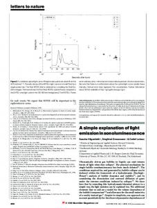
Feb 14, 2017 - lipidomics studies by Ellen et al. (2009 Extremophiles 13:67-79) showed that exosomes from S. acidocaldarius were made of > 29 proteins and ...
Tuesday, February 14, 2017 provide a more detailed mechanism of how a cell controls its raft -nonraft compartmentalization. 1859-Pos Board B179 Oxidation of Cholesterol Changes the Permeability of Lipid Membranes Waldemar T. Kulig1,2, Heikki Mikkolainen2, Agnieszka Olzynska3, Piotr Jurkiewicz3, Lukasz Cwiklik3,4, Tomasz Rog1,2, Martin Hof3, Ilpo Vattulainen1,2, Pavel Jungwirth2,4. 1 Department of Physics, University of Helsinki, Helsinki, Finland, 2 Department of Physics, Tampere University of Technology, Tampere, Finland, 3J. Heyrovsky Institute of Physical Chemistry, Academy of Sciences of the Czech Republic, v. v. i., Prague, Czech Republic, 4Institute of Organic Chemistry and Biochemistry, Academy of Sciences of the Czech Republic, Prague, Czech Republic. The function of living systems is inherently based on cells, which are the key building blocks of living organisms. Each cell in a human body is surrounded by a cell membrane, which essentially is a functional interface separating the cell from its surroundings. Biological membranes consist of lipid bilayers, in which a plethora of different proteins is embedded. Cholesterol molecules commonly present in lipid bilayers are largely susceptible to oxidation. This oxidation leads to oxysterols which play a crucial role in many regulatory processes in a human body. They are produced naturally from cholesterol during either enzymatic side chain hydroxylation (catalysed by cytochrome P450) or non-enzymatic oxidation (due to direct exposure of cholesterol to reactive oxygen species). Oxysterols differ from cholesterol by the presence of additional polar groups that are typically hydroxyl, keto, hydroperoxy, epoxy, or carboxyl moieties. Like cholesterol, many oxysterols are hydrophobic and hence confined to cell membranes. However, small chemical differences between the sterols can significantly affect how they interact with other membrane components, and this in turn can have a substantial effect on membrane properties. Using several experimental techniques, including dynamic light scattering and time-resolved fluorescence spectroscopy, together with atomistic molecular dynamics simulations, we characterized the behaviour of oxysterols in phospholipid membranes and compared the resulting data to that of cholesterol. We found that the permeability of lipid bilayers changes drastically (as compared to cholesterol) when tail-oxidized sterols are present, meanwhile this effect was not observed in systems containing ring-oxidized sterols. Here, we rationalize the different behaviour of various oxysterol classes based on both experimental data and molecular dynamics simulations. 1860-Pos Board B180 Pre-Transition Effects Mediate Forces of Assembly between Transmembrane Proteins: Recent Results on the Orderphobic Effect Shachi Katira, Kranthi K. Mandadapu, David Chandler. Chemistry, University of California, Berkeley, Berkeley, CA, USA. We have recently shown a mechanism for a generic and powerful force of assembly and mobility for transmembrane proteins in model lipid bilayers, referred to as the orderphobic effect [1]. The force is a pre-transition effect related to the first-order order–disorder transition in lipid bilayers and is modulated by proximity to the phase transition and structural features of transmembrane proteins such as hydrophobic mismatch. We present recent developments related to the orderphobic effect in the liquid-disordered phase. We also discuss the implications of this effect on the physical mechanism of formation of lipid rafts. [1] Katira, S., Mandadapu, K. K., Vaikuntanathan, S., Smit, B., & Chandler, D. (2016). Pre-transition effects mediate forces of assembly between transmembrane proteins. Elife, 5, e13150. 1861-Pos Board B181 Molecular Dynamics Simulations Reveal the Impact of Compositional Asymmetry in Lipid Membranes on Phase Behavior and Leaflet Interactions Michael D. Weiner1, Gerald W. Feigenson2. 1 Field of Physics, Cornell University, Ithaca, NY, USA, 2Field of Biophysics, Cornell University, Ithaca, NY, USA. The plasma membrane of eukaryotic cells is compositionally asymmetric, meaning that different lipids are found in the cytosolic and exoplasmic leaflets. Since compositionally asymmetric bilayers are difficult to form in vitro, studies in silico play an especially important role. Model membranes embody the behavior of the exoplasmic leaflet with coexisting liquid-ordered and liquid-disordered phases. We use Molecular Dynamics simulations to study the effects of compositional asymmetry and to examine the interactions between the two leaflets. Coarse-grained simulations are employed to produce phase separation. In the outer leaflet, a phase-separating mixture of phosphatidylcholines and cholesterol is chosen, while the inner leaflet contains phosphatidylethanolamines, phosphatidylserines, and cholesterol. We consider the
377a
impact of asymmetry on the liquid-liquid demixing phase transition, on order, and on clustering. Partitioning and clustering across from ordered and disordered phases are studied. The effect of varying cholesterol concentration is explored. 1862-Pos Board B182 The Mechanism of Copper-Induced Peroxidation Dov A. Lichtenberg, Ilya Pinchuk. Tel Aviv University, Tel Aviv, Israel. In spite of the great interest and intensive research effort, the actual mechanism of copper-catalyzed peroxidation is debatable. It might have been initiated via consecutive reduction of Copper(II) by lipid hydroperoxide (r.1) followed by a Fenton-like reaction of Copper(I) with hydroperoxide (r.2). The sum of reactions r.1 and r.2 gives r.3 2LOOH / LO2,þ LO,þ H2O r.3 However, reaction r.1 is ‘‘thermodynamically unfavorable’’ because the reduction potential of LO2, (0.77V - 1.44V), is higher than the reduction potential of Cu2þ/Cu1þ (0.16V). The commonly accepted paradigm is therefore that the 2 steps mechanism (r.1; r.2} is inconsistent with the experimentally observed peroxidation and that some additional agent(s) are involved in reducing Copper(II) to Copper(I). Nevertheless, this argument is not valid for systems far from equilibrium state. Furthermore, the overall dismutation process (r.3) is thermodynamically favorable, so that the reaction may occur either via the two steps mechanism or through an intermediate complex that undergoes intramolecular rearrangement without formation of intermediate Copper(I). Notably, the two mechanisms of r.3 may be expected to result in different overall reaction orders. Initiation via consecutive reactions r.1 and r.2 is expected to result in a first order kinetics of initiation with respect to hydroperoxide concentration, whereas formation of copper-hydroperoxide complex(es) may result in higher order initiation reactions. Experimentally, the dependence of peroxidation on the concentration of hydroperoxides accords with second order kinetic law far better than with the first order. This is consistent with the observation that continuous peroxidation of lipids within the given lipoprotein particle requires the presence of two hydroperoxide molecules per lipoprotein. It is also consistent with the effects of deuteration of LDL on its peroxidation. 1863-Pos Board B183 F4,5GWALP Partitioning, Orientation, and Effect on Bending Moduli in Models of the Plasma Membrane Rebecca D. Usery1, Thais A. Enoki1, Vanessa P. Nguyen2, Barrera N. Francisco2, Gerald W. Feigenson1. 1 Molecular Biology and Genetics, Cornell University, Ithaca, NY, USA, 2 Biochemistry & Cellular and Molecular Biology, University of Tennessee, Knoxville, TN, USA. Ternary mixtures of high-melting lipid, low-melting lipid, and cholesterol exhibit a region of liquid-ordered (Lo) þ liquid-disordered (Ld) phase coexistence analogous to raft þ non-raft behavior in cells. Curvature in vitro can induce sorting of both protein and lipid components resulting in accommodation by the less rigid phase. However a number of highly-curved membranes in vivo have more rigid, raft-like composition. We have measured membrane rigidity by fluctuation analysis of giant unilamellar vesicles (GUVs) with phase contrast microscopy. We find that including transmembrane helical WALP peptides rigidifies the liquid-disordered phase. This rigidifying effect has been examined at various cholesterol fractions. A change in rigidity was not observed for Lo phases. Electron paramagnetic resonance (EPR) and oriented circular dichroism (OCD) studies of the location and orientation of the peptide show that it is largely excluded from the Lo phase at high concentrations. Based on the partition coefficient of the peptide determined by FRET-based methods, the effect of WALP on Lo rigidity has been examined for low but relevant fractions. Our findings indicate the disordered phase could be more rigid than the raft phase when transmembrane proteins are present. 1864-Pos Board B184 Non-Monotonic Effect of Solutes on Miscibility Transition Temperatures; Simple Explanation of Closed-Loop Phase Diagrams Michael Schick1, David W. Allender2. 1 Physics, University of Washington, Seattle, WA, USA, 2Physics, Kent State University, Kent, OH, USA. There has been renewed interest in the effect of adding a solute to a lipid mixture which can undergo a miscibility transition to coexisting liquid-ordered (lo) and liquid-disordered (ld) phases. Most solutes in sufficient quantity decrease the transition temperature, Tc, simply by diluting the components that bring about the phase separation. Differences in solutes are manifest at low and intermediate concentrations. If the solute is an amphiphile that likes
378a
Tuesday, February 14, 2017
to sit at the interface of such phases then, as is well known, the amphiphile lowers the transition temperature at all concentrations. More interesting is the solute which prefers one phase or the other. If it prefers the ld phase, which has the larger entropy per particle of the two phases, the solute initially lowers Tc. At intermediate concentrations, the solute can increase Tc. At large concentrations it must lower Tc by simple dilution of the system. On the other hand, if the solute prefers the lo phase, then its initial addition raises Tc. This continues at intermediate concentrations. At large concentrations it must, again, lower Tc. This latter case provides a very simple explanation for the occurrence of closed loop ternary phase diagrams in systems in which the solute is cholesterol. The above behavior is illustrated in a simple model calculation. 1865-Pos Board B185 Physiochemical Characterization of Archaeal Exosomes Alexander P. Bonanno, Michelle Jiang, Noah Gilly, Parkson L.-G. Chong. Temple University, Philadelphia, PA, USA. Exosomes (microvesicles) secreted from thermoacidophilic archaea are believed to play an important role in cell division and biofilm formation and hold great promise for technological applications. However, there have been very few studies characterizing the exosomes from thermoacidophiles. In the present study, we have isolated exosomes (~130-170 nm in diameter) from the thermoacidophilic archaeon Sulfolobus acidocaldarius (optimum growth: 78-80oC, pH 2-3) and used dynamic light scattering, fluorescence spectroscopy and conventional biochemical methods to characterize them. Proteomics and lipidomics studies by Ellen et al. (2009 Extremophiles 13:67-79) showed that exosomes from S. acidocaldarius were made of > 29 proteins and tetraether lipids. Using the steady-state polarization and the average lifetimes of intrinsic protein fluorescence (excited at 285 nm from an LED) obtained by phase-modulation fluorometry, we found that the rotational relaxation time of proteins in S. acidocaldarius exosomes (buffer: 50 mM Tris containing 10 mM EDTA and 0.02% NaN3 at pH 7.2) underwent a steady decrease from 0.30 to 0.06 ns when the temperature increased from 20.0 to 50.4oC. Thereafter, to our surprise, the trend was reversed and the rotational relaxation time increased with increasing temperature, to 0.14 ns at 63oC. The biphasic change in exosome protein fluorescence at 50.4oC also occurs in the cooling mode and could be attributed to temperature-induced protein conformational changes within the exosomes or a temperature-induced phase transition in exosome tetraether lipid membranes. The possible mechanism underlying the decrease in rotational rate with increasing temperature above 50.4oC will be discussed. (Support by NSF). 1866-Pos Board B186 Water Dynamics at the Bilayer Interface is Similar to that within the SecY Translocon Venkatramanan Krishnamani1, Sara Capponi2, Stephen H. White3. 1 Department of Molecular Physiology and Biophysics, University of Iowa, Iowa City, IA, USA, 2Cardiovascular Research Institute, University of California in San Francisco, San Francisco, CA, USA, 3Department of Physiology and Biophysics and Center for Biomembrane Systems, University of California, Irvine, Irvine, CA, USA. The membrane bilayer interface is a chemically complex region composed of water, lipid head groups, and acyl chain carbonyls. This interface facilitates the formation of secondary structure, which is a necessary step before in vitro insertion of transmembrane helices in lipid bilayers [1, 2]. Hence, the energetics of partitioning of peptides into the bilayer interface is an important step towards understanding the thermodynamics of protein folding. Exactly how the hydrophobic effect drives the partitioning of peptides into the membrane interface is unclear, because the atomic solvation parameter (s) for interface partitioning is one half the value observed for the bulk-phase partitioning of peptides [2, 3]. Because the hydrophobic effect is directly related to properties of water, we have examined water dynamics at the POPC bilayer interface. Our findings reveal that the water dynamics deviate significantly from bulk behavior at the bilayer interface, similar to the behavior inside the translocon [4]. This suggests that the energetics of folding of helices inside the translocon might be similar to the energetics at the membrane interface. [1] Ladokhin AS, White SH. 1999. Folding of amphipathic a-helices on membranes: energetics of helix formation by melittin. J. Mol. Biol. 285:1363-69 [2] Wimley WC, White SH. 1996. Experimentally determined hydrophobicity scale for proteins at membrane interfaces. Nat. Struct. Biol. 3:842-48 [3] Jacobs, RE. and White, SH. 1989. The nature of the hydrophobic binding of small peptides at the bilayer interface: Implications for the insertion of transbilayer helices. Biochemistry 28:3421-3437 [4] Capponi S, Heyden M, Bondar A-N, Tobias DJ, White SH. 2015. Anomalous behavior of water inside the SecY translocon. Proc. Natl. Acad. Sci. USA. 112:9016-9021
1867-Pos Board B187 Progress Towards a Membrane-Bound Structure of the ITK/NEF Complex Rebecca Eells1, Kindra Whitlatch2, Frank Heinrich1,3, Mathias Lo¨sche1,3, Thomas E. Smithgall2. 1 Department of Physics, Carnegie Mellon University, Pittsburgh, PA, USA, 2 Department of Microbiology and Molecular Genetics, University of Pittsburgh School of Medicine, Pittsburgh, PA, USA, 3Center for Neutron Research, National Institute of Standards and Technology, Gaithersburg, MD, USA. Human immunodeficiency virus-1 (HIV-1) Nef is a membrane-associated accessory protein essential for viral replication, immune evasion, and AIDs progression. Nef functions via interactions with numerous host cell proteins involved in signal transduction and trafficking. Nef interaction with Src family kinases in HIV-1 target cells leads to constitutive kinase activity that enhances infectivity and viral replication while down-regulating immune receptors. Recently, Nef was also shown to interact selectively with Tec kinases in HIV-1 target cells. In particular, interaction with Itk promotes infectivity and replication (Tarafdar et al, J. Biol. Chem. 2014). Engagement of the two proteins at the inner plasma membrane is mediated by Itk’s SH3 domain, but the detailed mechanism and contribution of other domains to Nef engagement remain unclear since most studies so far are solution-based thus do not account for the influence of the membrane on the interaction between the binding partners. In addition, detailed structures of the membrane-associated complexes have yet to be elucidated, and little is known about the structure of active Itk. Sparsely-tethered bilayer lipid membranes (stBLMs) are resilient biomimetic bilayers for the surface-sensitive characterization of protein binding to functionalized membranes and structural determination of the resulting protein-membrane complexes. We use surface plasmon resonance and neutron reflection to characterize Itk/Nef interactions at the membrane interface, both in terms of the conformation of the individual proteins on the bilayer and, eventually, as a complex. Here we will discuss the binding of Nef to anionic membranes and the specific interaction of Itk with stBLMs that contain phosphoinositolphosphates (PIPs). 1868-Pos Board B188 Viscoelastic Measurements of Hydroxycholesterol Phospholipid Monolayers Nikki Hoang1, Lyle H. Nyberg2, Benjamin L. Stottrup1. 1 Physics, Augsburg College, Minneapolis, MN, USA, 2Chemistry, Augsburg College, Minneapolis, MN, USA. The rheological characteristics of multi-component cholesterol/phospholipid monolayers have received less attention than measurements of phase behavior, domain morphology, and pressure-area isotherms. The liquid-liquid coexistence observed in these systems is a compelling model to investigate both biological membranes as well as for the physical chemistry of two dimensional phase separated systems in their own right. We use a pulsating drop module for an optical tensiometer to measure the elastic and viscous moduli for lipid monolayers exhibiting unique liquid-liquid phase behavior. Robustly, hydroxycholesterol isomers induce a kink in pressure-area isotherms for monolayers over a wide range of compositions and phospholipids. Likewise, monolayers of 27-hydroxycholesterol and 1,2-Dimysristoyl-sn-glycero-3-phosphocholine (DMPC) system exhibit the nucleation and growth of a sterol rich phase which increases in area fraction over a range of surface pressures. The role of sterol concentration, phase behavior, and droplet curvature are investigated in determining the viscoelastic properties of these Langmuir films.
Membrane Active Peptides and Toxins I 1869-Pos Board B189 Interaction of Aurein 1.2 and Charged Lipid Bilayers Shuo Qian, Durgesh Rai. Oak Ridge National Laboratory, Oak Ridge, TN, USA. Aurein 1.2 is one of more active antibacterial peptides secreted by tree frog Litoria aurea. As a short membrane-active peptide with only 13 amino acids in sequence, it has been found to be residing on the surface of lipid bilayer but it permeabilizes bacterial membranes at high concentrations. However the molecular level detail is largely unknown. In this study, we investigated the action of Aurein 1.2 in DMPC/DMPG lipid bilayers in the forms of unilamellar vesicles and multilamellar bilayers. The oriented circular dichroism (OCD) results show the peptide is on the surface of lipid bilayer regardless the charged lipid ratio. Only at very high peptide-to-lipid ratio (~1/10), the peptide is found to be inserted. To further understand how it interacts with charged lipids, we also applied Small-Angle Neutron Scattering (SANS) to study lipid vesicles bilayer structures under the influence of Aurein 1.2.