Nov 20, 1989 - JOHN C. ROYER AND J. P. NAKAS*. Department ofEnvironmental and Forest Biology, College ofEnvironmental Science and Forestry,.
APPLIED AND ENVIRONMENTAL MICROBIOLOGY, June 1990, p. 1516-1517
Vol. 56, No. 6
0099-2240/90/061516-02$02.00/0 Copyright © 1990, American Society for Microbiology
Simple, Sensitive Zymogram Technique for Detection of Xylanase Activity in Polyacrylamide Gels JOHN C. ROYER AND J. P. NAKAS* Department of Environmental and Forest Biology, College of Environmental Science and Forestry, State University of New York, Syracuse, New York 13210 Received 20 November 1989/Accepted 16 March 1990
A method capable of detecting as little as 0.11 U of xylanase activity in polyacrylamide gels was developed. The method entails incubation of protein gels in contact with substrate gels containing unmodified xylan, followed by immersion of substrate gels in 95% ethanol. Resulting zymograms contain transparent bands corresponding to enzymatic activity against an opaque background.
appeared to be pure (Royer, Ph.D. thesis) when 2.0 mg was subjected to sodium dodecyl sulfate electrophoresis (4) and silver staining (6). Cathodic discontinuous gel electrophoresis. Nondenaturing cathodic disk gels (7) were run as described by Hames and Rickwood (2). Enzyme was concentrated by acetone precipitation and redissolved in KOH (0.03 M)-acetic acid (0.03 M), pH 6.8. Prior to electrophoresis, glycerol and basic fuchsin were added to final concentrations of 10.0 and 0.002%, respectively. The resolving gel acrylamide concentration was 10.0%, and gels were run at approximately 2,000 V with cooling by circulating tap water. Zymogram analyses. Two xylanase zymogram techniques were utilized. The first used unmodified oat spelt xylan (Sigma) as the substrate, while the second was a modification of the procedure of Biely et al. (1) and used remazol brilliant blue (RBB)-dyed xylan (Sigma) as the substrate. To prepare substrate gel (11 by 12.5 cm), 150 mg of xylan was dissolved in 10 ml of deionized water (heated to 50°C for RBB-dyed xylan) and mixed with 20 ml of melted 2% agarose (Sigma Type I) in sodium citrate-HCl (0.2 M, pH 4.8). The mixture was cooled to approximately 55°C and poured on a sheet of agarose Gel Bond (FMC Corp.). After hardening, the substrate gel was carefully overlaid with a protein gel. The gels were next smoothed to remove bubbles, wrapped in plastic, and incubated at 55°C for 20 min. The gels were finally separated, and the protein gel was fixed and silver stained. Substrate gels containing oat spelt xylan were immersed in 95% ethanol for 30 to 60 min and were photographed while raised approximately 12 in. (ca. 30 cm) above a black background. Gels could be preserved for several days by soaking them in 2:1 ethanol-sodium citrate (0.05 M, pH 4.8) overnight, spreading a thin layer of glycerol over their surfaces, and storing them in plastic wrap at 4°C. Substrate gels containing RBB-dyed xylan were immersed in ethanol-sodium citrate-HCl (2:1) for 48 h to enhance zones of clearing against a blue background. RBB-dyed gels were photographed on a light table.
Zymograms, or activity stains, are useful for visualizing the activity of enzymes in nondenaturing protein gels. A method of detecting xylanase activity in polyacrylamide gels by using chemically modified xylan has been reported (1). The present paper is an extension of previous research on the extracellular enzymes of Trichoderma longibrachiatum (8-10) and reports a simple, rapid, and inexpensive method of determining xylanase activity in gels. This procedure utilizes unmodified xylan and is based on ethanol precipitation of undegraded xylan. MATERIALS AND METHODS Enzyme and assays. Xylanase was generated from T. longibrachiatum and assayed as described by Royer and Nakas (8). Protein was measured by the method of Lowry et al. (5) with bovine serum albumin (Sigma Chemical Co.) as standard. Xylanases A and B were purified from crude enzyme as described by J. C. Royer (Ph.D. thesis, State University of New York, Syracuse, 1988). Cultures were initially centrifuged (12,000 x g, 20 min, 4°C) to remove mycelia, and the resulting supernatant was subjected to ethanol fractionation at -20°C (3). Considerable contaminating protein was removed by centrifugation (6,000 x g, 20 min, 20°C) after addition of 2.5 volumes of 95% ethanol. Xylanase activity was precipitated by an additional 7.5 volumes of ethanol. After centrifugation and air drying (-20°C), the pellet was dissolved in sodium acetate (100 mM, pH 5.0) and applied to a column (150 by 1.5 cm) packed with Bio-Gel P-60 (Bio-Rad Laboratories). The column was eluted at 7 ml/min, and 2-ml fractions were collected and analyzed for xylanase activity. Two peaks of xylanase activity were observed, and fractions were combined into two pools, designated xylanase A and xylanase B. After dialysis in sodium acetate (20 mM, pH 5.0), the two enzymes were further purified by chromatography on carboxymethyl Bio-Gel (Bio-Rad). Both enzymes bound to the column under the initial conditions (sodium acetate, 20 mM, pH 5.0) and were eluted from the column by NaCl gradients (0 to 500 mM). Xylanase B was further purified by chromatography on Phenyl Sepharose CL-4B (Pharmacia). After elution with 2 volumes of start buffer (Tris, 20 mM, pH 7.5), xylanase activity was eluted from the column with a gradient of sodium thiocyanate (0 to 1.0 M). Flow rate during ionexchange and hydrophobic-interaction chromatography was 20 mlIh, and 2.0-ml fractions were collected. Both enzymes *
RESULTS AND DISCUSSION Faint bands of clearing were visible at high enzyme concentrations on oat spelt zymograms immediately after the incubation period (data not shown). Immersion of the substrate gel in ethanol resulted in precipitation of undegraded xylan, which enhanced contrast and revealed additional clear bands against an opaque background. After ethanol precipitation, the new zymogram technique dis-
Corresponding author. 1516
ZYMOGRAMS FOR DETECTING XYLANASE ACTIVITY
VOL. 56, 1990
B.
A. 1 2 3 4
5
1
2 3 4 5
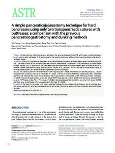
FIG. 1. Comparison of xylanase zymogram techniques. A cathodic disk gel containing two sets of a range (0.02 to 1.70 ,ug) of xylanase A from T. longibrachiatum was run as described in Materials and Methods. The gel was then sliced in half, and each half was used to generate a zymogram by using one of the techniques described in Materials and Methods, ethanol precipitation (A) or RBB-dyed xylan (B). The following amounts of enzyme were used (micrograms): lane 1, 0.02; lane 2, 0.07; lane 3, 0.20; lane 4, 0.57; lane 5, 1.70.
played a sensitivity similar to that of the method using RBB-dyed xylan and was capable of clearly detecting as little as 200 ng (0.11 U) of purified xylanase A of T. longibrachiatum (Fig. 1). In previous work, standard xylanase assays based on reducing sugar formation revealed two xylanase fractions (xylanase A, M, = 21,500; xylanase B, M, = 33,000) after
1517
gel filtration of either crude or ethanol-precipitated T. longibrachiatum culture supernatant (Royer, Ph.D. thesis). Zymogram analysis after nondenaturing polyacrylamide gel electrophoresis (Fig. 2) confirmed the presence of these two enzymes and revealed at least one additional xylanolytic enzyme of lower electrophoretic mobility. Zymograms which use ethanol precipitation of unmodified xylan are equally applicable to nondenaturing polyacrylamide gel electrophoresis and to isoelectric focusing gels (Royer, Ph.D. thesis). They develop quickly, are easily photographed, and are considerably less expensive than those which use RBB-dyed xylan. The basis of enzyme detection is hydrolysis of ethanolprecipitable xylan. Therefore, the method may prove useful in identification of enzymes not easily detected by standard assays based on reducing sugar formation. ACKNOWLEDGMENTS This study was supported in part by funds provided through the McIntire-Stennis Cooperative Forestry Research Program of the U.S. Department of Agriculture. We give special thanks to Penny Weiman for her assistance in preparing the manuscript. LITERATURE CITED 1. Biely, P., 0. Markovic, and C. Mislovicova. 1985. Sensitive detection of endo-1,4-,B-glucanases and endo-1,4-p-xylanases in gels. Anal. Biochem. 144:147-151. 2. Hames, B., and D. Rickwood. 1981. Gel electrophoresis of proteins; a practical approach. IRL Press, Ltd., Oxford. 3. Kaufmnan, S. 1971. Fractionation of protein mixtures with organic solvents. Methods Enzymol. 22:233-238. 4. Laemmli, U. K. 1970. Cleavage of structural proteins during the assembly of the head of bacteriophage T4. Nature (London)
227:680-685. 5. Lowry, 0. H., N. J. Rosebrough, A. L. Farr, and R. J. Randall. 1951. Protein measurement with the Folin phenol reagent. J. Biol. Chem. 193:265-275. 6. Nielsen, B. L., and L. R. Brown. 1984. The basis for colored silver-protein complex formation in stained polyacrylamide gels. Anal. Biochem. 141:311-315. 7. Reisfeld, R. A., U. J. Lewis, and D. E. Williams. 1962. Disk electrophoresis of basic proteins and peptides on polyacrylamide gels. Nature (London) 195:281-283. 8. Royer, J. C., and J. P. Nakas. 1989. Xylanase production by Trichoderma longibrachiatum. Enzyme Microb. Technol. 11:
405-410. FIG. 2. Ethanol precipitation
zymogram
of crude
enzyme and were
purified xylanases A and B of T. longibrachiatum. Samples
subjected to cathodic disk gel electrophoresis and ethanol precipitation zymogram techniques as described in Materials and Methods. Lane 1, Crude enzyme (150 jig); lane 2, xylanase A (0.5 ,ug); lane 3, xylanase B (2.0 ,ug).
9. Sharma, A., and J. P. Nakas. 1987. Preliminary characterization of laminarinase from Trichoderma longibrachiatum. Enzyme Microb. Technol. 9:89-93. 10. Tangarone, B., J. C. Royer, and J. P. Nakas. 1989. Purification and characterization of an endo-(1,3)-P-D-glucanase from Trichoderma longibrachiatum. Appl. Environ. Microbiol. 55:177184.