1 381
SIMULATING IN VITRO EPITHELIAL MORPHOGENESIS IN MULTIPLE ENVIRONMENTS M. R. Grant, S. H. J. Kim, C. A. Hunt* Joint UCSF/UCB Bioengineering Graduate Group and The Biosystems Group, Department of Biopharmaceutical Sciences, The University of California, San Francisco, CA 94143, USA * Email:
[email protected] •
[email protected] •
[email protected] In vitro studies of epithelial cell morphogenesis have demonstrated the influence of environment composition and orientation in the development of multicellular epithelial structures such as tubules and cysts. We have constructed a low resolution, discrete event simulation model and report on its use to explore how experimentally observed morphogenetic phenomena under four growth conditions might be generated and controlled. We identified simulation attributes that may have in vitro counterparts. We studied how changes in the logic governing simulated epithelial cell behavior might cause abnormal growth. Simulation results support the importance of a polarized response to the environment to the generation of a normal epithelial phenotype and show how disruptions of tight mechanistic control lead to aberrant growth characteristics.
1. INTRODUCTION* Epithelial cells are studied in vitro in order to better understand the mechanisms that lead to normal and disregulated epithelial cell morphogenesis1. It has been observed that epithelial cells are capable of sensing the presence of matrix in the local environment through specific cell surface receptors such as integrins, and that decisions about cell survival are made at least in part based on attachment to matrix2. Attachment to neighboring cells is important for specification of an apical surface, and to the directed transport of factors to the apical and basal surfaces3. Finally, the presence of an apical surface free of matrix contact appears to be an important requirement for the stability of an epithelial monolayer4. Epithelial development in vitro leads ultimately to structures in which cells have a basal surface associated with matrix, lateral surfaces associated with other epithelial cells, and an apical surface adjacent to an environment free of cell or matrix content. Epithelial cell behavior is thus hypothesized to be governed by processes, including environment sensing, that lead ultimately to the acquisition of a stable structure in which each cell has a three-surface environment5. We report on the behaviors of low-resolution discrete space, discrete event simulation models based on the above hypothesis. Each action taken by a simulated cell is strictly determined by environment-focused rules. Actions are mandated in response to type and location in the local environment of any combination of three components: cells, matrix, or free space. One model
*
Corresponding author.
provides strong support for the above hypothesis. It mimics key in vitro epithelial cell phenotypic attributes in four different in vitro conditions: in surface, embedded, suspension, and overlay cultures. The model stands as a testable analogue for the high-level mechanisms driving epithelial cell behavior in vitro. However, we hypothesized that it is unlikely that every cell always adheres to a set of genome-controlled mandates. To test that hypothesis we placed selected rules under stochastic control. We report results of in silico experiments conducted to determine how much a rule can be relaxed without significant erosion of targeted behaviors. We also report the initial results of altering selected rules to identify potential conditions that may lead to aberrant cell growth in vitro.
2. MODEL STRUCTURE We use the agent-based, simulation modeling package 6 MASON . It provides tools for event scheduling, controlling event execution, and visualization of simulation components. The simulations consist of 2D hexagonal grids, three simulation component specifications, and visualizations of grid states. In order to avoid confusion, simulated components, environments, and outcomes will hereafter be labeled in small caps in order to distinguish them from their real-world referents. FREE SPACE and MATRIX components do not exhibit any behaviors. All interaction with the environments occurs through the actions of individual CELLS on their immediate environment.
2 382
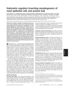
Fig. 1. Examples of each of the nine transition rules. Two rules were altered to simulate mutations resulting in altered epithelial cell behavior: M1: mutated rule 1; M8: mutated rule 8.
We identified twenty-one out of a potential sixtyfour distinct local environment configurations that could give distinctive epithelial cell behaviors. Final states of the local environment for each of these environment configurations were assigned based on observations from in vitro experiments, and the outcomes were simplified into eight rules that generate a complete mapping from any initial configuration of the local environment to a final configuration. The derived rules, examples of which are pictured in Fig. 1, are as follows. During each simulation cycle (time step), each CELL determines if it has one, two, or three types of neighbors. Only one type: follow Rule 1, 2, or 3. Two types: follow Rule 4, 5, 6, or 7. Three types: follow Rule 8 or 9. Rules 1-3: if the environment contains only: 1) CELLS : die; 2) FREE SPACE : die; 3) MATRIX : divide and replace a MATRIX with a daughter to maximize CELL neighbors. Rules 4-7: when there is 4) FREE SPACE, no MATRIX, and just one CELL : add MATRIX between self and that CELL; 5) FREE SPACE , no MATRIX , and at least two CELLS : die; 6) at least one CELL and MATRIX (no FREE SPACE): divide and replace a MATRIX with a daughter to maximize CELL neighbors; 7) M A T R I X and at least one FREE SPACE: divide and place the daughter in a FREE SPACE that has a matrix neighbor. Rule 8: when there is at least one CELL plus MATRIX neighbored by an adjacent FREE SPACE: divide and place the daughter in a FREE SPACE that neighbors MATRIX. Otherwise, 9) do nothing: mandates achieved. Four simulated environments were constructed in order to provide a range of environments to analyze simulation outcomes. Each simulated environment corresponds to a particular condition frequently used to study epithelial cells in vitro. Surface culture was simulated as two 2D square grids, the lower grid filled with MATRIX
and the upper grid with FREE SPACE components. To initiate a simulation a single CELL is placed in the center of the FREE SPACE grid. Simulated embedded culture was represented using a MATRIX-filled, 2D hexagonal grid, with the center of the grid at the start of a simulation occupied by a single CELL . Overlay culture was simulated by placing a row of CELLS on a horizontal plane through the center of a 2D grid filled with MATRIX. Finally, suspension culture was simulated by placing two CELLS adjacent to one another in a 2D hexagonal grid filled with FREE SPACE. To explore the consequences of perturbations in CELL behavior, modifications to the rules were implemented. We altered the rules for CELL survival (mutated Rule 1), and the rules for orientation of CELL division (mutated Rule 8) (Fig. 1). Also, probabilistic option was added to Rule 1 to allow for CELL survival in the presence of a CELL -only environment. When a CELL event is executed and the CELL is in a CELL-only environment, there is a probability that the CELL will die. If the probability is not met, the CELL survives. That probability is a parameter value shared by all of the CELLS in a particular simulation run. At each event, a CELL simulates sampling from a uniform distribution using an implementation of the Mersenne Twister random number generation algorithm provided with MASON. For each simulation experiment, CELL numbers were recorded at each step, for fifty steps, for at least 25 simulation runs per simulated condition.
3. RESULTS A simulation model in which epithelial cell behavior is determined by the types and locations of components in the local environment produces a range of phenotypes exhibited by epithelial cells during growth in vitro, under a range of different conditions. The 2D simulation successfully generated stable monolayers in simulated surface culture, stable structures resembling lumenfilled cysts in simulated embedded culture, stable inverted cysts in simulated suspension culture, and tubule-like structures in simulated overlay culture (not shown). An example of a growth sequence is presented in Fig. 2. Similarity to additional characteristics of in vitro cultures was observed as well, including the development of a stable CYSTS after 10 simulated days (Fig. 3A), the distribution of CYST sizes (Fig. 3B), and the continuation of CELL death and division during the growth of a CYST (Fig. 3C).
3 383
Fig. 2. Results of a typical simulation of growth in embedded culture. The numbers refer to the simulation step at which the snapshot was taken. In this example, a stable structure formed at simulation step 13. The rules are applied once to each CELL at each simulation step, with random ordering of CELL scheduling each step. As a consequence a wide variety of stable CYSTS are formed.
To insure that the collection of rules in Fig. 1 is necessary for forming the targeted attributes, simulations were conducted using a rule set in which one of the first eight rules were broken or weakened. Breaking or weakening any one of these rules caused a dramatic change in growth characteristics. Two examples are presented. The first rule change simulated the consequences of a loss in the ability of the CELL to die. The simulated CELLS having the altered rule set generated normal phenotypes in simulated surface culture and suspension culture, but abnormal attributes in simulated embedded and simulated overlay conditions. A second rule change simulated a disruption in the direction in which CELL division was implemented. Both altered rule sets caused a loss in the ability to form stable structures in simulated embedded culture (Fig. 3D). Reduction in the probability of CELL death as a consequence of following Rule 1 resulted in an increase in the number of CELLS found in the structures formed in simulated embedded culture (Fig. 4), but not in uncontrolled growth. We also observed that despite a reduction in CELL death probability of 80% and more, stable structures were still formed, although their morphology varied.
4. DISCUSSION The model described simulates outcomes under four different growth conditions where the key features are remarkably similar to observations of epithelial cell growth under corresponding in vitro conditions. Simulations also generated additional in silico system-level attributes (not shown) under other conditions. Some of these may merit in vitro exploration as a means of further model validation, possibly invalidation. This model provides a foundation for the design of increasingly representative and predictive simulation models of epithelial cell morphogenesis. This model and its descendants will have three important uses: 1) it will provide a
framework for the systematic reverse engineering of the mechanisms underlying the targeted behaviors. The process will lead to causal linkages between known molecular level events and system level events and system level phenotype. 2) It will provide means to posit causes of disregulated epithelial cell growth, and 3) it will also enable in silico experimental methods to discover and analyze potential treatments of disregulated growth.
Fig. 3. Characteristics of simulated epithelial cell morphogenesis under different conditions (n > 24). A: Comparison of simulation and in vitro epithelial cell growth rates in embedded culture; error bars: 1 SD; two simulation steps represents about 24 hours in vitro. B: The frequency distribution of CELL number per CYST is similar to in vitro referent data from embedded culture. C: Analysis of division and death events in simulated embedded culture. D: Consequences of rule changes described in the text.
Although two fundamentally different rule changes were identified that both give rise to disregulated growth in simulated embedded culture, others are possible and are being explored to identify additional changes in simulated cell behavior that could result in altered growth patterns under different conditions. We anticipate that some of these will merit in vitro follow-up. Despite the ability of the model to simulate targeted outcomes under a range of conditions, there are in vitro observations that are evident and measurable at the current level of resolution that the model currently is not capable of representing. For example, not all cysts formed in 3D embedded culture are lumen-filled, monolayer structures. In the case of MDCK cells, a kidney epithelial cell line, nearly thirty percent of cysts that develop contain either no lumenal space or have partially filled 7 lumens after day ten . Another unmatched in vitro observation is the polarity inversion that takes place in the transfer of a cyst grown in suspension culture into an
4 384 8
embedded matrix . It is straightforward to add rules representing additional mechanistic detail with the goal of extending model behavior to cover one or more of these additional attributes. It requires iterative in silico experimentation to also adjust existing mechanistic rules, as new ones are added, so that the behavior of the resulting new model validates against the behavior of the original model.
Acknowledgments This work was abstracted in part from material assembled by MRG for his Ph.D dissertation. We are grateful for the funding provided by the CDH Research Foundation, and for the support provided by our cell biology collaborators and their groups: Profs. Keith Mostov (tetrad.ucsf.edu/faculty.php?ID=45) and Thea Tlsty (tetrad.ucsf.edu/faculty.php?ID=78). Our dialogues with Glen Ropella, Hal Berman, and Nancy Dumont covering many technical, biological, and theoretical issues proved important. For their support helpful advice, we thank the members of the BioSystems Group (biosystems.ucsf.edu).
References
Fig. 4. Consequences of stochastic application of Rule 1 in simulated embedded culture (n = 50). In addition to p = 1.0, five probabilities were tested: 0.8, 0.6, 0.4, 0.2, and 0. When p = 0.4, Rule 1 is followed 40% (average) of the time when applied. The plateau level, the number of cells in the final, stable structure, was highly dependent on the early form of that structure. Consequently, variance is large.
We had such additional attributes in mind when we changed the model so that rule application would become stochastic. The results in Fig. 4 for the stochastic application of Rule 1 were unexpected: a gradual shift from formation of stable structures to unregulated growth was judged a possible outcome. Application of Rule 1 as infrequently as 1% of the time will result in formation of stable (yet large) structures. Furthermore, the morphology resulting from different rule application probabilities was different. Such results suggest that tight control and regulation by currently unknown mechanisms are both necessary and essential for proper growth and morphogenesis.
5. CONCLUSION Modeling and simulating epithelial cell morphogenesis with a focus on the types and locations of components in the extracellular environment result in a representation of epithelial cell behavior that has remarkably high fidelity at this low level of resolution. This model serves as a reference point for more complex models of epithelial cell morphogenesis that are at least as predictive as the model presented here.
1. Zegers MMP, O'Brien LE, Yu W, Datta A, Mostov KE. Epithelial polarity and tubulogenesis in vitro. Trends Cell Biol 2003; 13: 169-176. 2. Meredith JEJ, Fazeli B, Schwartz MA. The extracellular matrix as a cell survival factor. Mol Biol Cell 1993; 4: 953-961. 3. Lipschutz JH, Guo W, O'Brien LE, Nguyen YH, Novick P, et al. Exocyst is involved in cystogenesis and tubulogenesis and acts by modulating synthesis and delivery of basolateral plasma membrane and secretory proteins. Mol Biol Cell 2000; 11: 42594275. 4. Hall HG, Farson DA, Bissel MJ. Lumen formation by epithelial cell lines in response to collagen overlay: a morphogenetic model in culture. Proc Natl Acad Sci 1982; 79: 4672-4676. 5. O'brien LE, Zegers MM, Mostov KE. Opinion: Building epithelial architecture: insights from threedimensional culture models. Nat Rev Mol Cell Biol 2002; 3: 531-537. 6. Luke S, Balan GC, Panait L, Cioffi-Revilla C, Paus S. MASON: A Java Multi-Agent Simulation Library; 2003. 7. Lin H-H, Yang T-P, Jian S-T, Yang H-Y, Tang MJ. Bcl-2 overexpression prevents apoptosis-induced Madin-Darby canine kidney simple epithelial cyst formation. Kidney International 1999; 55: 168-178. 8. Wang AZ, Ojakian GK, Nelson WJ. Steps in the morphogenesis of a polarized epithelium I. Uncoupling the roles of cell-cell and cell-substratum contact in establishing plasma membrane polarity in multicellular epithelial (MDCK) cysts. J Cell Sci 1990; 95: 137-151.