Clinical Chemistry 47:3 486 – 490 (2001)
Molecular Diagnostics and Genetics
Simultaneous Absolute Quantification of Target and Control Templates by Real-Time Fluorescence Reverse Transcription-PCR Using 4-(4⬘-Dimethylaminophenylazo)benzoic Acid as a Dark Quencher Dye Karl-Anton Kreuzer,* Alexander Bohn,* Joachim Lupberger, Jerome Solassol, Philipp le Coutre, and Christian Andreas Schmidt†
Results: For M-bcr/abl duplex RT-PCR, the correlation coefficient (r) for starting template amounts and threshold cycle values was 0.99; for m-bcr/abl, r ⴝ 0.96, indicating a precise log-linear relation for 10 –105 copies/ 100 ng of cDNA. In the same PCR reactions, r ⴝ 0.99 for -actin (coamplified with M-bcr/abl or m-bcr/abl) for 103–107 copies/100 ng cDNA. The linear correlation coefficient for single and duplex measurements was 0.98 for M- and m-bcr/abl in patient samples. Conclusions: DABCYL can be used as dark quencher fluorophore in real-time fluorescence PCR. The duplex fluorescence RT-PCR assay for bcr/abl and -actin transcripts allows monitoring of bcr/ablⴙ leukemias.
Background: Despite the many advantages of real-time fluorescence reverse transcription-PCR (RT-PCR) as a quantitative analytical tool, simultaneous quantification of target and reference templates within one reaction has not been reported. We developed such an assay with an internal reference template. Methods: For quantification of target and reference sequences, we used two fluorescent probes in one reaction vessel on an ABI PRISM 7700 SDS instrument. Fluorescent probes were labeled with either 6-carboxyfluorescein or hexachloro-6-carboxy-fluorescein as reporter dye and 4-(4ⴕ-dimethylaminophenylazo)benzoic acid (DABCYL) as a dark quencher fluorophore. To test the sensitivity and specificity of this assay, serial dilutions of reference and target templates were analyzed in one PCR reaction. In the presence of 10 -actin molecules as control templates, 105 bcr/abl molecules were amplified, and 105 -actin molecules were amplified in the presence of 10 bcr/abl copies. We also performed single and duplex measurements on samples from five patients with documented Philadelphia chromosomepositive chronic myelogenous leukemia disease courses (72 samples) and three with minor bcr/ablⴙ acute myelogenous leukemias (26 samples).
© 2001 American Association for Clinical Chemistry
The recently introduced real-time fluorescent PCR allows the rapid and reliable quantification of target sequences in a closed-tube format, which has led to a breakthrough in generating quantitative PCR results for diagnostic purposes (1, 2 ). However, for real-time fluorescent PCR as for conventional PCR, a control reaction is necessary to correct quantitative results with respect to the total amount of amplifiable material in the investigated sample (3 ). This is of special importance in reverse transcription-PCR (RT-PCR)1 because RNA preparation and reverse tran-
Abteilung fu¨r Innere Medizin und Poliklinik m.S. Ha¨matologie und Onkologie, Campus Virchow-Klinikum, Medizinische Fakulta¨t Charite´ der Humboldt-Universita¨t zu Berlin, Augustenburger Platz 1, 13353 Berlin, Germany. *These authors contributed equally to the present work. †Author for correspondence. Fax 49-30-45053929; e-mail christian.
[email protected]. Received August 9, 2000; accepted December 26, 2000.
1 Nonstandard abbreviations: RT-PCR, reverse transcription-PCR; TAMRA, 6-carboxy-tetramethylrhodamine; DABCYL, 4-(4⬘-dimethylaminophenylazo)benzoic acid; M- and m-bcr/abl, major and minor bcr/abl, respectively; HEX, hexachloro-6-carboxy-fluorescein; FAM, 6-carboxy-fluorescein; CML, chronic myelogenous leukemia; and AML, acute myelogenous leukemia.
486
Clinical Chemistry 47, No. 3, 2001
scription reactions usually produce various quantities of cDNA. A control can be performed by quantification of an external standard in a separate assay. Nevertheless, because this requires a second reaction and each PCR has its own kinetics, internal standardization is thought to be more reliable (4 ). In fluorescence PCR, this can be done only if the simultaneously used reporter dye of the control reaction signal can be quantitatively distinguished from the fluorescence generated by target-specific probes. To date, quantification of an internal standard with the ABI PRISM 7700 SDS instrument has been limited to fluorescent quencher fluorophores, such as 6-carboxytetramethylrhodamine (TAMRA), that themselves produce substantial fluorescence at wavelengths close to those of the reporter dyes. Thus, depending on the reporter fluorophore used, TAMRA can produce measurable fluorescence signals within the reporter dye channels, which would again decrease the specificity and sensitivity of the assays. Moreover, if TAMRA is used as a quencher dye, it blocks a broad wavelength range that could be otherwise be used for incorporation of additional reporter dyes (5 ). This is of relevance for future development of multicolor-multiplex real-time fluorescence PCR techniques. In an attempt to facilitate development of such techniques, we investigated whether 4-(4⬘-dimethylaminophenylazo)benzoic acid (DABCYL), a quencher dye with no or minimal autofluorescence within the wavelengths measured in the ABI PRISM 7700 SDS, can be used for this purpose. To demonstrate the clinical applicability of this approach, we quantified bcr/abl fusion transcripts in two cases of Philadelphia chromosome-positive leukemias by single and duplex RT-PCR.
487
design of fluorescent probes The fluorescent probes for M-bcr/abl and m-bcr/abl were designed to hybridize within the abl terminus of the amplicons as published previously (7 ). Probes were covalently labeled with hexachloro-6-carboxy-fluorescein (HEX) at the 5⬘ end, representing the reporter dye, and DABCYL was incorporated as amidite after deprotection at the 3⬘ end of the probe sequences. To prevent probe elongation, a phosphate group was attached. Probes were purified by HPLC. -actin was used as reference gene, and fluorescent probe and primer sequences were as published elsewhere (8 ). The -actin probe was labeled at the 5⬘ end with 6-carboxy-fluorescein (FAM) phosphoramidite and at the 3⬘ terminus with DABCYL. To prevent probe extension, phosphate groups were attached to the 3⬘ ends of all probes (TIB Molbiol).
primers and pcr conditions PCR of bcr/abl was performed using a single pair of primers located in exon 1 (m-bcr/abl: 5⬘-CAgATCTggCCCAACgATg-3⬘) or exon 2 (M-bcr/abl: 5⬘-CTgACCAACTCgTgTgTgAAAC-3⬘) of bcr and in exon 3 (5⬘-
Materials and Methods preparation of templates e1a2 and -actin transcripts from K562 cells and b2a3 fusion transcripts from a patient known to carry this specific breakpoint were amplified by RT-PCR and cloned into the pCR2.1 vector (TOPO TA Cloning Kit; Invitrogen). After clones were digested with HindIII and XbaI (Boehringer Mannheim), reamplified, and purified (PCR Purification Kit; Qiagen), they were measured in a photometer, and molecule concentrations were calculated. Serial dilutions were then prepared at concentrations of 0.5 ⫻ 107 to 0.5 ⫻ 10⫺2 molecules/L of a mixture (1:1 by volume) of distilled water and Tris-EDTA buffer (pH 8.0).
blood samples The total RNA of peripheral blood samples was extracted by a guanidinium isothiocyanate-acid phenol procedure (6 ), and 2000 ng of the extracted RNA was reversetranscribed into cDNA with random hexamer primers. cDNA samples were stored at ⫺20 °C until assayed.
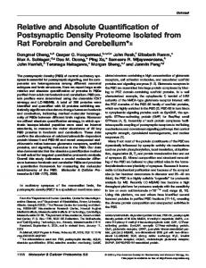
Fig. 1. Fluorescence emission spectra pre- and post-duplex PCR. (A), changes in fluorescence during cycling of 105 M-bcr/abl copies (detected by a HEX/DABCYL probe) in the presence of 10 -actin transcripts (detected by a FAM/DABCYL probe). (B), increase in the FAM fluorescence signal in a sample containing 105 -actin copies in the presence of 10 m-bcr/abl copies (HEX/ DABCYL probe).
488
Keuzer et al.: Quantitative Duplex Real-Time Fluorescence RT-PCR
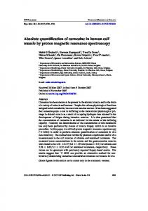
Fig. 2. TaqMan duplex RT-PCR of M-bcr/abl, m-bcr/abl, and -actin. (A and C), calibration curves. All experiments were performed in triplicate. Maximum SDs: (A), 1.14 for M-bcr/abl, 0.94 for -actin; (C), 1.75 for m-bcr/abl, 1.65 for -actin. (B and D), 3% agarose gel electrophoresis and ethidium bromide staining of the PCR products. The expected sizes were 103 bp for b2a3, 258 bp for e1a2, and 175 bp for -actin. MW, molecular weight marker (123 bp). NTC, non-template control.
CCCCATTgTgATT-ATAgCCTAAgA-3⬘) of abl. Thus, all four M-bcr/abl fusions could be amplified with a single set of oligonucleotides. An ABI PRISM 7700 Sequence Detector (PE Applied Biosystems) was used for PCR reactions and fluorescence measurements. The 50-L PCR reaction mixture for single measurements of bcr/abl or -actin contained 5 L of 10⫻ PCR buffer, 4.5 mM MgCl2, 0.8 mM dNTPs (Life Technologies), 1 M 5,6-carboxy-x-rhodamine, 0.8 L of Tris-EDTA buffer (pH 8.0), 0.5 M each primer, 1 L of probe, 1.25 U of Taq DNA polymerase (Platinum DNA polymerase; Life Technologies), and 100 ng of sample cDNA. PCR amplification began with a 5-min denaturation step at 95 °C, followed by 45 cycles of denaturation at 95 °C for 30 s and annealing/extension at 63 °C (m-bcr/abl), 65 °C (M-bcr/ abl), or 67 °C (-actin) for 1 min. Duplex PCR measurements were performed with a few modifications. The PCR reaction mixture included two primer sets, two fluorescence probes, and sample cDNA in the same concentrations used in the single assay, except for -actin primers, which were limited to 0.05 M each. Duplex real-time PCR was performed for 5 min at
95 °C and 45 cycles of 95 °C for 30 s and 63 °C (m-bcr/abl and -actin) or 65 °C (M-bcr/abl and -actin) for 1 min.
data analysis Statistical analyses were performed using Excel software (Microsoft).
Results Addition experiments were performed to evaluate whether the ABI PRISM 7700 SDS can discriminate between the two dyes used when one of the dyes is present in much higher concentrations, and thus produces a substantially higher fluorescence signal, than the other dye. Fig. 1 shows pre- and post-PCR fluorescence emission spectra for 105 bcr/abl copies in the presence of 10 -actin transcripts and vice versa. Fragmentation of the probes led to an increase of the fluorescence signal at the emission maximum of HEX at 555 nm (Fig. 1A) or FAM at 530 nm (Fig. 1B). The ABI PRISM SDS software separated fluorescence spectra from HEX and FAM with the help of an ideal-fit curve. As can be seen in Fig. 1, no significant
Clinical Chemistry 47, No. 3, 2001
489
Fig. 4. bcr/abl ratios measured in a CML patient (A) and a biphenotypic bcr/abl⫹ AML patient (B) with single or duplex real-time fluorescence RT-PCR. BMT, bone marrow transplantation. † indicates death of the patient.
Fig. 3. Correlation between quantitative single and duplex measurements. (A), 72 samples from five patients with a Philadelphia chromosome-positive CML. (B), 26 samples from three patients with m-bcr/abl⫹ AML.
quencher signal could be detected at the measured wavelengths (500 – 650 nm). To test the sensitivity and specificity of this assay, we performed PCRs in which the concentration of one template was orders of magnitude higher than the concentration of the other template. In the presence of 10 control templates, 105 bcr/abl molecules were amplified, and in the presence of 10 bcr/abl copies, 105 -actin molecules were amplified. Fig. 2 illustrates calibration curves obtained by plotting the initial number of target molecules in the dilution series against the respective threshold cycle. The threshold cycle is the cycle number at which the fluorescence of a given sample becomes significantly different from the baseline signal, i.e., 10 SD above background fluorescence during the first 15 PCR cycles. For M-bcr/abl duplex RT-PCR, the correlation coefficient (r) was 0.99, for m-bcr/abl, r ⫽ 0.96, indicating a precise log-linear relationship at template concentrations of 10 – 105 copies/100 ng of cDNA. In the respective PCR reactions, r ⫽ 0.99 for -actin (coamplified with M-bcr/abl or m-bcr/abl) for 103–107 copies/100 ng of cDNA. Ethidium
bromide staining of conventional agarose gel electrophoresis after amplification of b2a3/-actin and e1a2/actin templates in a duplex fluorescence RT-PCR is shown in Fig. 2, B and D. As shown in Fig. 2, A and D, visible gel bands correlate well with the obtained quantitative PCR results. To compare absolute amounts of template molecules obtained by single and duplex measurements, we performed correlation studies using samples from five patients with Philadelphia chromosome-positive chronic myelogenous leukemia (CML; n ⫽ 72) and three patients with m-bcr/abl⫹ acute myelogenous leukemia (AML; n ⫽ 26). As can be seen from Fig. 3, the ratios (target transcript copies divided by reference transcript copies) correlated well (r ⫽ 0.98 for M-bcr/abl and r ⫽ 0.98 for m-bcr/abl). To evaluate the clinical applicability of the developed assay, we retrospectively investigated two disease courses of a Philadelphia chromosome-positive CML and a biphenotypic bcr/abl⫹ AML by real-time PCR, amplifying target and reference gene either separately (single) or simultaneously (duplex). Fig. 4A shows bcr/abl:-actin transcript ratio for a CML patient in chronic phase who underwent matched unrelated donor bone marrow transplantation. After 2 months, he underwent a second allogeneic bone marrow transplantation and exhibited a marked decrease of bcr/abl fusion transcripts. Fig. 4B
490
Keuzer et al.: Quantitative Duplex Real-Time Fluorescence RT-PCR
illustrates bcr/abl transcript kinetics in a Philadelphia chromosome-positive AML. The observed increase of bcr/abl mRNA was clearly associated with progressive disease and, subsequently, death of the patient.
Discussion Quantitative real-time fluorescence PCR is revolutionizing PCR diagnostics in terms of greatly optimized applicability. However, independent of the instrument used, fluorescence can be excited only in a distinct wavelength range because different light sources (e.g., laser or halogen) have different excitation maxima. This restricts the reporter and quencher fluorophores that can be sufficiently excited. Moreover, quencher dyes such as TAMRA exhibit measurable autofluorescence within the detection wavelength, producing a spectral overlap with reporter dyes. To design fluorescent probes that can be simultaneously used for multiplex real-time PCR, an ideal quencher dye should have maximum quenching activity within a distance up to 100Å from the reporter fluorophore and no or minimal fluorescence emission in the measured wavelength range. DABCYL has been used as quencher in multiplex nucleic acid detection with molecular beacons showing very weak autofluorescence (9 ). Using the so-called TaqMan probe format, we could show that DABCYL is a suitable quencher with the advantage of no significant autofluorescence. The relevance of this is supported by other studies utilizing alternative quenching strategies (10 ). Moreover, it must be guaranteed that simultaneously used reporter dyes do not overlap within their detection wavelength. As shown in our addition experiments, the ABI PRISM 7700 SDS is able to distinguish between FAM and HEX reporter signals even when excessive fluorescence is generated by one or both fluorophores. However, it must be considered that addition of more reporter fluorophores with excitation maxima ⬎560 nm may be more difficult because excitation decreases with longer wavelengths in the ABI PRISM 7700 SDS. In conclusion, simultaneous quantification of two sequences in one tube can be performed using DABCYL as
a dark quencher dye. This may facilitate future development of quantitative multicolor real-time PCR techniques.
This work was made possible by Grant DJCLS-R22 from the Deutsche Jose´ Carreras Leuka¨mie-Stiftung e.V.
References 1. Holland PM, Abramson RD, Watson R, Gelfand DH. Detection of specific polymerase chain reaction product by utilizing the 5⬘-3⬘ exonuclease activity of Thermus aquaticus DNA polymerase. Proc Natl Acad Sci U S A 1991;88:7276 – 80. 2. Livak KJ, Flood SJ, Marmaro J, Giusti W, Deetz K. Oligonucleotides with fluorescent dyes at opposite ends provide a quenched probe system useful for detecting PCR product and nucleic acid hybridization. PCR Methods Appl 1995;4:357– 62. 3. Delarue B, Breard E, Abeguile G, Lemaire M, Mittre H, Leymarie P. Correcting for different amplification rates of internal standard and template when measuring cDNA by competitive PCR. Biotechniques 2000;28:396 – 8. 4. Willhauck M, Vogel S, Keilholz U. Internal control for quality assurance of diagnostic RT-PCR. Biotechniques 1998;25:656 –9. 5. Hung SC, Ju J, Mathies RA, Glazer AN. Cyanine dyes with high absorption cross section as donor chromophores in energy transfer. Anal Biochem 1996;243:15–27. 6. Chomczynski P, Sacchi N. Single-step method of RNA isolation by acid guanidinium thiocyanate-phenol-chloroform extraction. Anal Biochem 1987;162:156 –9. 7. Kreuzer KA, Lass U, Nagel S, Ellerbrok H, Pauli G, Pawlaczyk-Peter B, et al. Applicability of an absolute quantitative procedure to monitor intra-individual bcr/abl transcript kinetics in clinical samples from chronic myelogenous leukemia patients. Int J Cancer 2000;86:741– 6. 8. Kreuzer KA, Lass U, Landt O, Nitsche A, Ellerbrok H, Pauli G, et al. A highly sensitive and specific fluorescence RT-PCR assay for the pseudogene free detection of -actin transcripts as quantitative reference. Clin Chem 1999;45:297–300. 9. Vet JAM, Majithia AR, Marras SAE, Tyagi S, Dube S, Poiesz BJ, Kramer FR. Multiplex detection of four pathogenic retroviruses using molecular beacons. Proc Natl Acad Sci U S A 1999;11: 6394 –9. 10. Nurmi J, Ylikoski A, Soukka T, Karp M, Lo¨vgren T. A new technology for the detection of specific polymerase chain reaction products in a closed tube. Nucleic Acids Res 2000;28:E28.