Biosensors and Bioelectronics 116 (2018) 130–147
Contents lists available at ScienceDirect
Biosensors and Bioelectronics journal homepage: www.elsevier.com/locate/bios
Simultaneous detection and determination of mercury (II) and lead (II) ions through the achievement of novel functional nucleic acid-based biosensors
T
⁎
Zahra Khoshbina, Mohammad Reza Housaindokhta, , Asma Verdianb, Mohammad Reza Bozorgmehrc a
Department of Chemistry, Faculty of Science, Ferdowsi University of Mashhad, Mashhad, Iran Department of Food Safety and Quality Control, Research Institute of Food Science and Technology (RIFST), Mashhad, Iran c Department of Chemistry, Mashhad Branch, Islamic Azad University, Mashhad, Iran b
A R T I C LE I N FO
A B S T R A C T
Keywords: Functional nucleic acid based biosensors Simultaneous detection Heavy metal ion Aptasensor DNAzyme G-quadruplex
The serious threats of mercury (Hg2+) and lead (Pb2+) ions for the public health makes it important to achieve the detection methods of the ions with high affinity and specificity. Metal ions usually coexist in some environment and foodstuff or clinical samples. Therefore, it is very necessary to develop a fast and simple method for simultaneous monitoring the amount of metal ions, especially when Hg2+ and Pb2+ coexist. DNAzyme-based biosensors and aptasensors have been highly regarded for this purpose as two main groups of the functional nucleic acid (FNA)-based biosensors. In this review, we summarize the recent achievements of functional nucleic acid-based biosensors for the simultaneous detection of Hg2+ and Pb2+ ions in two main optical and electrochemical groups. The tremendous interest in utilizing the various nanomaterials is also highlighted in the fabrication of the FNA-based biosensors. Finally, some results are presented based on the advantages and disadvantages of the studied FNA-based biosensors to compare their validation.
1. Introduction As two of the most toxic metallic pollutants, Hg2+ and Pb2+ ions have received worldwide concern due to their deleterious biological and environmental effects (Lin et al., 2011b; Tchounwou et al., 2012). The repletion of Hg2+ in human body can cause ailments in vital organs, disorders in the nucleic acid function, defects in the immune system, and even death (Tan et al., 2013; Wu et al., 2012). The accumulation of Pb2+ in vital organs and tissues of the human body could result in nervous system dysfunction, anemia, cardiovascular disease, intelligence decrease, and growth impairment (Ge et al., 2016; Huang et al., 2010; Qian et al., 2015). Due to the great threats of Hg2+ and Pb2+ for public health, many organizations have determined the maximum permissible amount of the ions in food, drinking water, blood, etc. Based on the Environmental Protection Agency (EPA), the safety limits of Hg2+ and Pb2+ in drinking water are 10 and 70 nM, respectively (Chen et al., 2014; Guo et al., 2015). The presence of other ions normally makes the real samples complicated, that cause the determination of Hg2+ and Pb2+ ions be a difficult task. Consequently, development of accurate and sensitive analytical techniques for effective monitoring and accurate measurement of the two ions is getting considerable attention worldwide (Lin
⁎
Corresponding author. E-mail address:
[email protected] (M.R. Housaindokht).
https://doi.org/10.1016/j.bios.2018.05.051 Received 8 March 2018; Received in revised form 26 May 2018; Accepted 28 May 2018 Available online 29 May 2018 0956-5663/ © 2018 Elsevier B.V. All rights reserved.
et al., 2011a). So far, contemporary analytical methods have been used in the detection of heavy metal ions, including atomic absorption/ emission spectroscopy (AAS/AES), mass spectroscopy (MS), inductively coupled plasma mass spectrometry (ICP-MS), anodic stripping voltammetry (ASV), liquid chromatography mass spectrometry (LCMS), high performance liquid chromatography (HPLC), etc. (Deng et al., 2015; Shen et al., 2013; Wang et al., 2015d; Zhou et al., 2014) However, many of them are intricate and time-consuming and suffer from the limitations of expensive instruments, complicated operation procedures, etc. Such restrictions make these approaches impractical for onsite and regular environmental monitoring (Gao et al., 2016; Zhang et al., 2016). It is necessary to develop simple, sensitive, rapid, cost effective, and field-portable alternatives for metal ion determination to reduce the risk for public health. For this purpose, biosensors have been introduced, that can detect and measure different targets through creating a physically detectable signal (Scognamiglio et al., 2010). Recent years have witnessed the considerable research attempts to develop the biosensors, based on various detection techniques including fluorescence, colorimetry, surface-enhanced Raman scattering (SERS), field effect transistor (FET), and electrochemical methods (Liu et al., 2017a; Wang et al., 2018c). Today, FNA-based biosensors have great
Biosensors and Bioelectronics 116 (2018) 130–147
Z. Khoshbin et al.
2015). Pb2+-specific DNAzyme consists of a double-strand DNA, including an enzyme strand and a substrate strand with one ribonucleotide as a cleavage site. The presence of Pb2+ cleaves the substrate strand into two pieces at the ribonucleotide site (Wang et al., 2018c; Zhou et al., 2016b). The conventional Pb2+-specific DNAzyme-based biosensors have been developed for Pb2+ detection using the enzymatic activity of GR-5 and 8–17 DNAzymes, which produce a detectable output (Zhu et al., 2015). Aptamers with the binding ability to the certain targets are promising probes for fabrication for a wide range of aptasensors (Verdian-Doghaei et al., 2015; Wang et al., 2018b). An aptasensor is defined as a kind of biosensor, in which the biological recognition element is a DNA or RNA aptamer (Verdian-Doghaei and Housaindokht, 2015; Verdian, 2018). Interestingly, aptamers have some benefits in comparison with antibodies, described as follows. Aptamers have a much lower molecular weight, makes it easier to their synthesis and chemically modification. The synthesis process of aptamers is in vitro, meaning no need to laboratory animals for their production. Aptamers are also able to be used under a wide range of assay conditions. Indeed, they are stable over wide temperature and pH ranges, because of their higher thermal stability and resistance to multiple cycles of denaturation/renaturation (Hidding; Liebelt et al., 2016; Su et al., 2014). In comparison with antibody-based biosensors, aptasensors possess distinguished features, such as high productivity, affinity, selectivity, and stability and low immunotoxicity (Alsaafin and McKeague, 2017; Danesh et al., 2016; Emrani et al., 2016; VerdianDoghaei et al., 2014). The thymine (T)-rich aptamers can selectively and tightly capture Hg2+ to form T-Hg2+-T mismatch through N-Hg covalent bonding. Therefore, T-rich aptamers have been frequently applied to fabricate a large number of Hg2+ biosensors (Bao et al., 2015; Liu et al., 2015; Olaoye and Manderville, 2017; Wang et al., 2016a). G-quadruplex structures of DNA are unique arrangements including guanine rich DNA aptamers, have been connected through Hoogsteen hydrogen bonding interactions (Hai et al., 2013). The presence of a variety of metal ions stabilize these structures, among which Pb2+-induced G-quadruplex has the most stability, due to the formation of the most compact structure (Dolati et al., 2017; Pang et al., 2015; Verdian-Doghaei et al., 2015; Zhang et al., 2014a; Zhu et al., 2014). There are different guanine rich aptamers with the special sequences that could be fold to Gquadruplex structure especially in the presence of Pb2+ ion (Table 1). Therefore, such sequences can be extremely used in Pb2+ aptasensor design (Huang et al., 2016; Lu et al., 2013; Pang et al., 2015; Qian et al., 2015; Zhang et al., 2013a).
potential to be promising tools for heavy metal ion detection (Huang et al., 2017; Huang and Liu, 2015; Kasprowicz et al., 2015; Zhan et al., 2016). Enormous efforts have been made to develop the metal-sensing capacity of FNAs (Mehta et al., 2016). There are many reports that show detection and determination of heavy metal ions can be achieved successfully by using FNA-based biosensors (Farzin et al., 2017; Lee et al., 2012; Yang et al., 2017). For instance, Zhang et al. designed an efficient biosensor by using specific FNA-modified microplate and portable glucometer-based detection mode for sensitive Pb2+ monitoring (Zhang et al., 2015). Zhou et al. (2017) developed a FNA-based biosensor for robust detection of Hg2+ and Ag2+ ions by using synergy effects of 2-aminopurine fluorescence label that can be quenched up to 98% in the DNA strands in the absence of targets. In the majority of environmental, food and biological systems, including drinking water, multiple types of heavy metal ions exhibit coexisting nature. Therefore, their monitoring at an ultra-trace level can be achieved by application of the combination in different sensors. Nevertheless, the strategy seems not to be appropriate achievement, because of some disadvantages in the determination procedure, including high cost, wasting time, and complexity (Hao et al., 2012; Zhang et al., 2013b). Consequently, the simultaneous detection and quantification of two or more heavy metal ions by using a sensor seem to be demand. Here, a review has been provided with special emphasis on the recent advances in FNA-based biosensors for simultaneous detection of Hg2+ and Pb2+ ions in order to provide an understanding of their improvement and progress. 2. FNAs as recognition probes of Hg2+ and Pb2+ ions FNAs are single-stranded oligonucleotides with ability of ligand binding and enzymatic activity, obtained from the Systematic Evolution of Ligands by the Exponential Enrichment (SELEX) procedure. These biomolecules have widespread applications in therapy, delivery, designing in vitro/in vivo biosensors, and so on (Alsaafin and McKeague, 2017; Liu et al., 2009b). FNAs have been divided into different types including ribozymes, riboswitches, DNAzymes, and aptamers (Alsaafin and McKeague, 2017; Zhan et al., 2016). Ribozymes are catalytic RNA strands, discovered by Cech et al. and Altman et al. in the 1980s (Kharma et al., 2015; Li et al., 2016a). These RNA enzymes bind to the target by sequence-specific hybridization and inactive it through the phosphodiester backbone cleaving at a special site (Erdmann and Barciszewski, 2012). Riboswitches are RNA regulatory elements, usually placed in the 5′ untranslated regions (UTRs) of messenger RNAs (mRNAs) (Abduljalil, 2018). Riboswitches are capable to bind metabolites or metal ions as ligands and regulate mRNA expression by forming alternative structures in response to ligand binding (Helmling et al., 2018). DNAzymes are DNA sequences considered as analogs of ribozymes with higher biological stability (Erdmann and Barciszewski, 2012; Li and Lu, 2009). DNAzymes can catalyze numerous types of reactions with great catalytic activity. To achieve their high catalytic efficiency, many DNAzymes need metal ions, such as Mg2+, Pb2+, Cd2+, etc. (Alsaafin and McKeague, 2017). Aptamers, as chemical antibodies, are isolated from random sequence DNA or RNA libraries through an in vitro selection. Aptamers adopt special secondary and tertiary structures through target binding that convert them to the efficient recognition elements towards an extensive range of molecular targets from small inorganic/organic species such as metal ions to large biomolecules such as proteins (Kaiser et al., 2018; Li et al., 2016a; Zhang et al., 2018b). FNAs applied in heavy metal ion detection mainly have two major types including DNAzymes and aptamers (Li et al., 2016a; Zhu and Zhang, 2014). DNAzymes as catalytic DNA strands are chemically stable, cost-effective and easily synthesized. These advantages convert DNAzymes to an attractive and universal platform for metal ions detection with high binding affinity and specificity to special metal ions (Wang et al., 2018c; Yun et al., 2016; Zhang et al., 2014b; Zhu et al.,
3. Optical FNA-based biosensors Optical biosensors consist of a biorecognition sensing element and an optical transducer system. The biorecognition sensing element produces a signal proportional to the analyte concentration. Optical transducer system transforms the recorded signal into different measurable data formats, making the analyte concentration recognizable (Damborský et al., 2016). Optical biosensors have received remarkable interest, due to some advantages including high productivity, sensitivity, and selectivity (Dey and Goswami, 2011; Sharma et al., 2015). Table 1 The G-rich DNA sequences with ability to form G-quadruplex structure in the presence of Pb2+ ion. Oligonucleotide
Sequences
References
TBAA PS2.M AGRO100
GGA AGG TGT GGA AGG GTG GGT AGG GCG GGT TGG GGT GGT GGT GGT TGT GGT GGT GGT GG GGGT GGGT GGGT GGGT GGT TGG TGT GGT TGG
(Lu et al., 2013) (Zhang et al., 2013) (Pang et al., 2015)
T30695 TBA
131
(Qian et al., 2015) (Huang et al., 2016)
Biosensors and Bioelectronics 116 (2018) 130–147
Z. Khoshbin et al.
NPs) have recently obtained remarkable attention for biosensing applications, due to their key advantages including ease of synthesis, lowcost, no toxicity and good biocompatibility (Faccio et al., 2016; Taghdisi et al., 2016). Ye et al. (2012) presented the robust Hg2+ and Pb2+ detection approach by incorporating the heavy metal ion-responsive aptamers into the colloidal photonic crystal hydrogel (CPCH) films, assembled from the monodisperse SiO2-NPs. By introducing of the solution of heavy metal ions to the constructed apatasensor, the specific binding between the target heavy metal ions and the crosslinked aptamers caused the aptamer conformational changes, and subsequently, the hydrogel shrinkage. This could be detected as a corresponding blue shift in the Bragg diffraction peak position of the aptamers, which was used for the quantitative estimation of Hg2+ and Pb2+ ions without the aid of the sophisticated instrumentation.
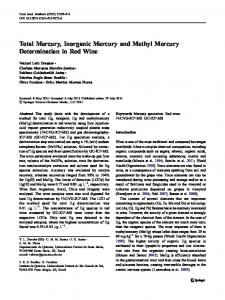
For the design of these biosensors, different techniques have been extensively used such as colorimetric, fluorescence, bioluminescence, chemiluminescence and many other methods (Shahdordizadeh et al., 2017). In this section, we highlight recent optical FNA-based biosensors for simultaneous detection of Hg2+ and Pb2+ ions. 3.1. Colorimetric biosensors As the most accepted types of optical sensors, the colorimetric biosensors have attracted remarkable attention due to their features, including rapid response, low cost, and suitable for on-site analyses without the demand of trained technician (Feng et al., 2014; Mao et al., 2017). Colorimetric biosensors utilize the color reporting groups with a high extinction coefficient to achieve the efficient detection of different analytes (Huang et al., 2018). Therefore, in colorimetric assays, target recognition event could be distinguished only by the naked eye; and, users are able to accurately detect targets without any requirement of precise equipment (Bostan et al., 2017). Nowadays, different types of nanoparticles (NPs), quantum dots (QDs), and carbon nanotubes (CNTs) are used in biosensor designing, due to their exceptional advantages including regular structure, high surface reaction activity, great catalytic efficiency, large surface-tovolume ratio, intense adsorption ability, and significant chemical and thermal stability (Chen et al., 2017; Cui et al., 2015; Hashkavayi and Raoof, 2017; Martín-Yerga et al., 2017; Shawky et al., 2017). Colorimetric biosensors especially utilize gold nanoparticles (AuNPs) due to unique properties, including ease of synthesis, large surface area, high chemical stability, feasible surface modifications, and well biocompatibility (Borghei et al., 2016; Chen et al., 2015; Liu et al., 2014). The tremendous potential of AuNPs in colorimetric FNA-based biosensors is based on size-dependent optical properties. The variation in their particle size induces changing in surface resonance frequency, followed by rapidly color change in the colloid form of AuNPs (Bostan et al., 2017; Robati et al., 2016; Shahdordizadeh et al., 2017). Consequently, dispersion/aggregation behavior of AuNPs could be considered as the fundamental strategy to analyze monitoring in the sample under analysis. Chen et al. (2015) developed a label free colorimetric assay for detection of Hg2+ and Pb2+ ions using AuNPs combined with graphene oxide (GO), which enhances the stability of AuNPs against salt-induced aggregation. GO-AuNP nanohybrids demonstrate significant peroxidase-like activity, which can catalyze the oxidation of the peroxidase substrate 3,3,5,5-tetramethylbenzidine (TMB) by H2O2. In the absence of Hg2+ and Pb2+ ions, the aptamer-GO-AuNP collections formed, which would be able to stay suspended in the solution, even after the addition of salt. Subsequently, the catalytic oxidation of TMB in the presence of H2O2 led to a color change from red to light blue, clearly distinguishable by the naked eye. In the presence of Hg2+ or Pb2+ ions, the aptamers switched from random coil structure into a relatively rigid conformations, therefore left the GO-AuNP nanohybrids. GO-AuNP nanohybrids are no longer protected by the aptamers, and high-salt conditions inhibited the peroxidase-like activity of GO-AuNP nanohybrids by producing salt-induced aggregated nanohybrids. Thus, the presence of Hg2+ or Pb2+ ions has been confirmed by a change in the solution color from blue to colorless. Originally, an efficient binary DNA-based colorimetric biochip was constructed by Shi et al. (2015) for the on-site and parallel recognition of Hg2+ and Pb2+ ions. As shown in Fig. 1, both the guanine and the thymine rich DNA hairpin probes, modified by amino and biotin groups, have been immobilized on a plastic chip in their folded forms. Binding with Hg2+ and Pb2+ compelled the DNA sequences to change their folded arrangements to the unfolded ones. Such an event allowed AuNPs-streptavidin conjugate (or streptavidin-horseradish peroxidase conjugate) to join the biotin groups and therefore, the visual signals were obviously detected by a dual-color staining protocol. In comparison with conventional organic dyes, the silica NPs (SiO2-
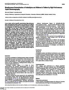
3.2. Fluorescent biosensors Fluorescence, as one of the most sensitive detection techniques, is widely used to determine very low amounts of analyte. So, fluorescence is applied as a powerful technique to construct the wide range of biosensors (Rubab et al., 2018). Because of low cost, wide response range, rapid detection and easy operation, fluorescent biosensors have received significant attention in the heavy metal ion monitoring (Dolati et al., 2017; Farzin et al., 2017; Xu et al., 2010). Among various fluorescence biosensing systems, special attention has been paid to the biosensors based on Förster Resonance Energy Transfer (FRET), as a nonradiative process whereby energy transfer occurs from an excited state donor to a proximal ground state acceptor through long-range dipole-dipole interactions (Chen et al., 2013; Tan et al., 2013). Auto fluorescent proteins, organic dyes and inorganic nanostructures could be used as the energy donor and acceptor fluorophores in the FRETbased biosensors. However, inorganic quenchers including GO, CNTs, and AuNPs have been extremely used in the construction of these biosensors, because of their special fluorescent properties such as high chemical stability, great spectral resolution, and extremely sharp emission bands (Dolati et al., 2017; Li et al., 2011). Through functionalizing the 5′ and 3′ termini of thrombin-binding aptamer (TBA), with the FAM fluorophore and the DABCYL quencher, respectively, Liu et al. (2009a) developed a FRET-based biosensor for the highly selective and sensitive detection of Hg2+ and Pb2+ ions. The random coil structure of TBA strand changed into the G-quadruplex and hairpin-like structures, upon Pb2+ and Hg2+ binding, respectively. Subsequently, the different degrees of fluorescence decrease at 518 nm (excitation at 475 nm) have been produced through the FRET process, which confirmed the presence of both ions. In this paper, the effect of buffer composition has been examined on the detection response of the designed biosensor, which showed a sensitivity decrease upon increasing the stability of metal-anion complexes. The investigation of the aptasensor selectivity illustrated that the concentrations more than 1.0 μM of some ions, unfortunately, decreased the FAM fluorescence, leading to false positive signals and therefore, the biosensor inefficiency. To overcome the problem, several masking reagents including phytic acid, NaCN, and a random DNA strand have been applied in the sensing system. As shown in Fig. 2, Chung’s group (Chung et al., 2013) reported a novel turn-on biosensing strategy for simultaneous detection of Hg2+ and Pb2+ ions sensitively and selectively, by applying the superior fluorescence-quenching property of AuNPs. The sensing method is based on the displacement of fluorophore-labeled oligonucleotides from AuNPs upon the binding of the target ions with the aptamers. The TexasRed® with excitation at 585 nm and emission at 615 nm and also, the Cy5.5™ with excitation at 680 nm and emission at 706 nm were chosen as the fluorophores to label the aptamers. In the absence of Hg2+ and Pb2+ ions, the fluorescence transfer from the TexasRed® and the Cy5.5™ fluorophores to the AuNPs happened, because of their close proximity to the NPs' surface. However, the formation of G-quadruplex 132
Biosensors and Bioelectronics 116 (2018) 130–147
Z. Khoshbin et al.
Fig. 1. Representation of the principle of the DNA hairpin-based colorimetric biochip for simultaneous detection of Hg2+ and Pb2+ ions. Two DNA hairpin probes containing thymine-rich and guanine-rich fragments, modified by amino and biotin groups at the two termini, were immobilized on a plastic chip in their folded forms. In the presence of Hg2+ and Pb2+, the DNA sequences change their folded arrangements to the unfolded ones, allowing nanogold-streptavidin conjugate (or streptavidin-horseradish peroxidase conjugate) to join the biotin groups and therefore, the visual signals were obviously detected by a dual-color staining protocol. (a) Single color staining protocol and (b) Dual-color staining protocol. Reproduced from Ref. Shi et al. (2015).
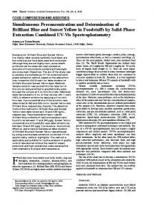
dimensional (3D) sensing surface was exquisitely designed by Qu et al. (2017) for sensitive detection of Hg2+ and Pb2+ ions. The DNMs were fabricated through the incorporation of the self-assembled target-specific DNA tetrahedral structured probes (TSPs) into a droplet array, and subsequently, transferring them to the designated locations for immobilization and sensing-interface engineering. The constructed DNMs were spaced along the microchannel with highly ordered probe orientations and circular arrangement, giving a microarray biosensor with advanced sensitivity and selectivity. A bubble-mediated shuttle reaction was used inside the DNM functionalized microchannel by using air bubbles to enhance probe-target interactions, and therefore, improve the target-capturing efficiency. With applying bubble-mediated technique, there is no need to design and construct a complex microchannel, and also immense user intervention. As shown in Fig. 3, a sample plug array containing an aqueous solution plug of metal ions and Cy3-labeled reporter, an air bubble plug, and a solution plug of washing buffer was driven to the microchannel from the inlet using a constant flow rate. The sample plug array moved successively through the DNM sensing interface. The syringe pump was paused when the sample plugs array approached the outlet of the capillary. The sample plugs array then flowed backwards through the DNM sensing interface. Based on Fig. 3, the binding between the Hg2+ specific oligonucleotide and its Cy3-labeled reporter strand was induced by Hg2+ ions through the formation of T-Hg2+-T configuration that resulted a fluorescence signal at 580 nm (excitation at 532 nm) as a criterion of Hg2+ presence. A Pb2+ specific oligonucleotide, its substrate strand, and a Cy3-labled
structures caused the displacement of the Cy5.5™-labeled complementary strands from the surface of the AuNPs; therefore, the recovered fluorescence could be seen as a confirmation of Pb2+ ion presence. Such behavior was also observed for the TexasRed®-labeled complementary strands by the formation of the hairpin-like structures of the thymine-rich aptamers in the presence of Hg2+ ion. The presence of approximately 500-fold higher amounts of the other metal ions with respect to Hg2+ and Pb2+ amounts had no significant effect on the fluorescence enhancements, which confirmed the excellent specificity of the designed aptasensor. Originally, Wang and Si (2013) developed a novel aptasensor based on multi-walled carbon nanotube (MWCNT) long-range energy transfer for multicolor fluorescent detection of Hg2+, Pb2+, and Ag+ ions. FAM, ROX, and Cy5 fluorophores with excitation (emission) wavelengths of 480 (520), 565 (604), and 630 (663) nm, respectively, were chosen to modify the Hg2+, Pb2+ and Ag+ aptamers. In the absence of ions, the fluorophore-labeled aptamers with random coil structures wrapped onto the MWCNTs' surface via strong π-π stacking interactions that quenched the fluorescence. In the presence of Hg2+ and Pb2+ ions, the structural changes of the aptamers were achieved through the formation of the hairpin-like and the G-quadruplex structures, respectively. The fluorophore-labeled aptamers were subsequently divorced from the MWCNTs' surface, demonstrating an increment of fluorescence emission. Lately, an efficient microchannel functionalized with the DNA nanostructured microarrays (DNMs) accompanied by tubular three133
Biosensors and Bioelectronics 116 (2018) 130–147
Z. Khoshbin et al.
Fig. 2. Schematic outline of the “turn-on” fluorescent based biosensor for simultaneous detection of Hg2+ and Pb2+ ions using assembled DNA probe on the surface of AuNPs. Formation of hairpin-like structures of the thymine-rich aptamers in the presence of Hg2+ ion caused the displacement of the TexasRed®-labeled complementary strands from the AuNPs; and fluorescence was generated as a confirmation of Hg2+ presence. The displacement of the Cy5.5™-labeled complementary strands from the AuNPs was induced by the formation of the G-quadruplex structures of the guanine-rich aptamers in the presence of Pb2+ ion; and fluorescence was recovered as a result of Pb2+ presence. Reproduced from Ref. Chung et al. (2013).
reporter were intelligently used for Pb2+ detection. The presence of Pb2+ induced cleavation of the substrate strand, and subsequently, the Pb2+ specific oligonucleotide was hybridized with the Cy3-labled reporter strand, producing a fluorescence signal as a confirmation of Pb2+ existence. The sensing of Ag+ ion was also carried out besides the detection of the others. Upconversion nanoparticles (UCNPs) are considered as a newly emerged class of imaging agents with the ability to convert the low excitation energy photons into visible emissions (Dehghani et al., 2018). In recent years, the UCNPs have attracted growing interest due to their potential applications in biosensing and bioimaging (Han et al., 2016; Huang et al., 2015; Jin et al., 2017; Tan et al., 2016b). The UCNPs have proven to exhibit unique properties, including long
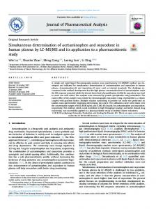
lifetime, no autofluorescence, high chemical stability and photo-stability, low toxicity, narrow emission spectrum and low background fluorescence (Chen et al., 2018; He et al., 2017; Kim et al., 2018; Liu et al., 2017b; Wang et al., 2010, 2009; Zhu et al., 2018). Therefore, the UCNPs have great potential for fabrication of different fluorescent biosensors as a novel approach. Wu and colleagues (Wu et al., 2014) presented a sensitive FRET-based aptasensor for the simultaneous detection of Hg2+ and Pb2+ ions, using the UCNPs conjugated to the ionresponsive aptamers as donors and the AuNPs attached on the complementary DNAs as acceptors. As illustrated in Fig. 4, the distance between the donor and acceptor was decreased in the absence of Hg2+ and Pb2+ ions, due to hybridizing the ion-responsive aptamers by their corresponding complementary DNA strands. Because of a good overlap 134
Biosensors and Bioelectronics 116 (2018) 130–147
Z. Khoshbin et al.
Fig. 3. Construction of 3D tubular DNM sensor inside microchannels for the detection of Hg2+, Pb2+, and Ag+ ions based on bubble-mediated shuttle reaction. A series of metal-ion specific sequences were adapted to DNA TSPs. DNMs were immobilized on an aldehyde-functionalized microchannel through amine groups of three vertices of TSPs. The DNM functionalized microchannel was connected to the as-prepared sample plugs array via the inlet, and subsequently connected to a syringe pump via the outlet. The sample plug array containing an aqueous solution plug of metal ions and Cy3-labeled reporter, an air bubble plug, and a solution plug of washing buffer were drived to the microchannel from the inlet using a constant flow rate. The sample plug array moved successively through the DNM sensing interface. The syringe pump was paused when the sample plugs array approached the outlet of the capillary. The sample plugs array then flowed backwards through the DNM sensing interface. Reproduced from Ref. Qu et al. (2017).
between the UCNPs’ fluorescence emission and the AuNPs absorption spectrum, the red and green upconversion fluorescence at 660 and 542 nm, respectively, were quenched in the absence of Hg2+ and Pb2+ ions. By preferring to bind to their corresponding targets, the aptamers formed the specific arrangements in the presence of Pb2+ and Hg2+ ions. Therefore, the dual FRET was disrupted, and the red and green upconversion fluorescence was restored as a criterion for verifying Hg2+ and Pb2+ presence, when excited with a 980 nm laser. The mentioned fluorescent biosensors usually require the chemical modification of the aptamer with either a fluorophore or a quencher. The labeling of aptamer with fluorescent reporters for target monitoring increases the fabrication costs (Feng et al., 2014). Against labeledfluorescent biosensors, the label-free ones are affordable and easy to use (Wang et al., 2015b); so label-free fluorescent biosensors have attracted growing interest for Hg2+ and Pb2+ detection. DNA intercalating dyes as one of the most common signal reporters are extremely used for fabrication of label-free fluorescence aptasensors. The free dye molecules represent negligible fluorescence intensity, while strong fluorescence signal could be observed when they intercalate into DNA strand. Lin et al. (2011a) developed a label-free FRET-based biosensor for detection of Hg2+ and Pb2+ ions, using two non-fluorescent DNA sequences, polythymine (T33) and polyguanine (G33), and two reporters, benzothiazolium-4-quinolinium dimer derivative (TOTO-3) and terbium ions (Tb3+). Upon interaction with Hg2+ ions, the T33 random-
coil structure changed to a hairpin-like structure that facilitated the intercalating of the greater amounts of TOTO-3. Consequently, the DNA/TOTO-3 complexes with Hg2+ ions resulted more strongly fluorescence at 660 nm in comparison with what that was for free TOTO-3, when excited at 620 nm. The G33 strand could able to form a G-quartet structure in the presence of Tb3+ ions through the cooperative coordination of nitrogen and oxygen atoms of the base and phosphate groups. The efficient energy transfer from nucleobases to Tb3+ ions increased the fluorescence intensity of the G33/Tb3+ conjugates. As Pb2+ ions wined in competition with Tb3+ ions to interact with the G33 strand, the extent of the G33-Tb3+ complexes decreased upon increasing the Pb2+ concentration. Therefore, a decrease in fluorescence at 545 nm was induced as a criterion of Pb2+ presence, when excited at 290 nm. No covalent linkage of any dyes to the DNA strands made this sensing approach a relatively cost-effective sensing technique with great practical potential for the detection of Hg2+ and Pb2+ ions in real samples. Zhan’s group (Zhan et al., 2015) achieved the quantitative detection of Hg2+ and Pb2+ ions using GeneFinder™ (GF) with excitation wavelength of 490 nm as a DNA intercalator. The FNA-based recognition probe has been designed by adding six thymidines (T) to both ends of T30695 strand with sequence of 5′-GGGTGGGTGGGTGGGT-3′. Fig. 5 shows that the modified T30695 remained in random-coil state in the absence of two investigated ions. Next, the added intercalator GFs could
135
Biosensors and Bioelectronics 116 (2018) 130–147
Z. Khoshbin et al.
Fig. 4. Schematic illustration of dual FRET between the upconversion nanoparticles and controlled AuNPs for the simultaneous detection of Hg2+ and Pb2+ ions. NaYF4: Yb, Ho UCNPs and Mn2+-doped NaYF4: Yb, Er UCNPs, and controlled AuNPs were employed as the donors and the acceptors, respectively, to construct the biosensor. The two donor-acceptor pairs were formed by hybridizing the aptamers and their corresponding complementary DNA, inducing the quenched green and red upconversion fluorescence. In the presence of Hg2+ and Pb2+, the aptamers preferred to bind to their corresponding analytes and formed a hairpin-like and the G-quadruplex structures for Hg2+ and Pb2+, respectively, restoring the green and red upconversion fluorescence. Reproduced from Ref. Wu et al. (2014).
4. Electrochemical FNA-based biosensors
bind to the modified T30695 by electrostatic interactions, producing moderate strong fluorescence signal. Upon addition of Hg2+ and Pb2+ ions, the special structures of the modified T30695 strand were induced, resulting the diverse number of the stacked intercalator GFs on them. Subsequently, the different extent of fluorescence intensity about 536 nm could be achieved as a criterion of Hg2+ and Pb2+ presence. Xu et al. (2015) also applied the modified T30695 as the recognition probe using SYBR Green I (SG) with excitation wavelength of 490 nm as a DNA intercalator. The intercalation of different SG concentration could be induced by the formation of T-Hg2+-T hairpin conformation and G-quadruplex structure in the presence of Hg2+ and Pb2+ ions, respectively. Therefore, a label-free fluorescent FNA-based biosensor for Hg2+ and Pb2+ detection was successfully constructed by monitoring of the SG fluorescence changes about 535 nm upon addition of these two ions. Besides, the detection limits of Hg2+ and Pb2+ ions were reformed to a rate of 10.45 nM and 2.65 nM, respectively, compared to Zhan’s group research (Zhan et al., 2015).
Electrochemical sensors are made of electrodes and an electrochemical segment, which measure the changes in current, impedance, conductance, voltage or potential difference signals, created by the biochemical interactions between the biorecognition element and target analyte. Based on the type of the measured signal, electrochemical biosensors are further classified into amperometric, impedimetric, conductometric, voltammetric and potentiometric categories (Ragavan et al., 2018; Rubab et al., 2018; Song et al., 2012). Electrochemical FNA-based biosensors are among the widely used biosensors, which are composed of a FNA layer (the biorecognition element) immobilized on the electrode surface (the electrochemical transducer), to detect the target analyte that interact with FNA unit (Chiorcea-Paquim et al., 2018; Huang et al., 2018; Ragavan et al., 2018). Due to their inherent advantages including small size, rapid response, low cost, superior stability, and high sensitivity, the electrochemical biosensors have attracted great interest in the detection of heavy metal ions (Cui et al., 2015; Li et al., 2016b; Olaoye and Manderville, 2017; Tang et al., 2014; Wang et al., 2015a, 2016b; Zhang 136
Biosensors and Bioelectronics 116 (2018) 130–147
Z. Khoshbin et al.
Fig. 5. Scheme of depicting the label free fluorescent assay for detection of Hg2+ and Pb2+ ions using GeneFinder™ as a DNA intercalating dye and the modified T30695 as an integrated FNA. In the absence of Hg2+ and Pb2+, the modified T30695 remained in random-coil status. Then, the added GF intercalated with the modified T30695, releasing moderate strong fluorescence. While in the presence of Hg2+ and Pb2+, the modified T30695 formed T-Hg2+-T or G-quadruplex structure, and subsequently, interacted with diverse number of GFs. Finally, extremely strong or very weak fluorescence was produced as a criterion of Hg2+ and Pb2+ presence. Reproduced from Ref. Zhan et al. (2015).
simultaneous detection of Hg2+ and Pb2+ ions. For example, Lin et al. (2011b) designed a novel label-free DNA-based biosensor for simultaneous detection of Hg2+ and Pb2+ ions using the difference in chargetransfer resistance (ΔRCT) before and after DNA-ion interactions, monitored by electrochemical impedance spectroscopy with [Fe(CN)6]4-/3as redox probe. As illustrated in Fig. 6, the sensing strategy toward the target ions is based on the formation of the G-quadruplex structure and the T-Hg2+-T complex in the presence of Pb2+ and Hg2+ ions, resulting the decreased RCT values. Taking advantage of the ΔRCT values, Hg2+ and Pb2+ ions could be selectively detected with high precision. The designed biosensor has been successfully evaluated by Hg2+ and Pb2+ detection in newborn calf serum and lake water samples. In this paper, the sensing of Ag+ ion was also carried out besides the simultaneous determination of Hg2+ and Pb2+ ions. In order to simultaneous detection of the three coexist metal ions in a sample, the use of masking agents is necessary; EDTA to mask Pb2+ and Hg2+ for Ag+ detection, cysteine to mask Ag+ and Hg2+ for Pb2+ detection, and the mixture of G-rich and C-rich DNA strands to mask Pb2+ and Ag+ for Hg2+ detection. An electrochemical assay based on DNA conformational changes has been established for sensitive detection and quantification of Hg2+ and Pb2+ ions, simultaneously, by Shi et al. (2012). The conformational changes of the Hg2+-specific oligonucleotide and Pb2+-specific DNAzyme were induced during the interaction with Pb2+ and Hg2+ ions
et al., 2017). For example, Zhou et al. (2016b) developed a sensitive electrochemical biosensor to detect Pb2+ ion based on synergetic catalysis of hemin/G-quadruplex DNAzyme and Au-Pd hybrid nanostructures. In the presence of Pb2+, the substrate strand was cleaved, which resulted the releasing of the enzyme strand. Then, G-rich DNAlabeled Au-Pd nanostructures was linked with the released enzyme strand with assistance of a helper DNA. A large number of hemin/Gquadruplex-based DNAzyme complexes were formed on the electrode with an addition of hemin to be applied as nicotinamide adenine dinucleotide hydrogen (NADH) oxidase and peroxidase mimics. Finally, hemin/G-quadruplex-based DNAzyme complexes, attached to the porous Au-Pd nanoparticles, could catalyze the reduction of H2O2 generated from NADH in the presence of O2, which produced an electronic signal as a criterion of Pb2+ presence. Among different electrochemical biosensing methods, impedimetric ones are considered as a potentially powerful technique with ultra-high sensitivity for label-free detection of very small quantities of different analytes (Rushworth et al., 2014). An impedimetric biosensing technique relies on the electron transfer kinetics between the redox probe and the electrode, which produces the electrochemical impedance signal significantly affected by molecular weight and/or high charge density of the target (Du and Dong, 2016). In recent years, several electrochemical biosensors have emerged using electrochemical impedance spectroscopy (EIS) technique as an efficient sensing tool for the
137
Biosensors and Bioelectronics 116 (2018) 130–147
Z. Khoshbin et al.
Fig. 6. Schematic illustration of the immobilized DNA-based biosensor for simultaneous detection of Hg2+, Pb2+, and Ag+ ions. The sensing strategy was based on the formation of the G-quadruplex structure and the THg2+-T and C-Ag+-C complexes in the presence of Pb2+, Hg2+ and Ag+ ions, respectively, resulting the decreased RCT values. Reproduced from Ref. Lin et al. (2011).
(Fig. 7), that could be tracked by EIS. The Hg2+ and Pb2+ detection could be obtained by applying ΔRCT parameter that was greater for Pb2+ in comparison with what caused by Hg2+, presumably due to the difference in DNA conformational changes. In recent years, carbon-based nanomaterials, such as graphene and carbon nanotubes, have received remarkable interest to be used in the biosensor designing field. Their special features including environmental security, speedy electron transfer rate, unrivaled structural
characteristics, and physicochemical proficiencies make carbon-based nanomaterials efficient to construct the electrochemical biosensors for detection and quantification of heavy metal ions. Graphene oxide (GO), as one of the carbon-based nanomaterials, has been extensively applied to develop the electrochemical FNA-based biosensors, because of its superior thermal, mechanical, and also photophysical properties (Liu et al., 2012; Toda et al., 2015). Wang et al. (2015c) developed a DNAzyme-based impedimetric biosensor for Hg2+ and Pb2+ detection. Fig. 7. Schematic illustration of the DNAzymebased impedimetric biosensor for simultaneous detection of Hg2+ and Pb2+ ions. The induced DNA conformational changes in the presence of Hg2+ and Pb2+ led to decrease in chargetransfer resistance of DNA films, which permitted selective detection of Hg2+ and Pb2+ ions. Reproduced from Ref. Shi et al. (2012).
138
Biosensors and Bioelectronics 116 (2018) 130–147
Z. Khoshbin et al.
Fig. 8. Schematic of Hg2+ and Pb2+ detection using the electrochemical biosensor based on NH2-rGO. The gold electrode was modified by a multilayer rGO functionalized with amino groups by using N2 plasma enhanced chemical vapor deposition (PECVD). Three kinds of DNA strands were immobilized on the amino-rGO placed on a gold electrode. In the presence of Hg2+ and Pb2+, conformational changes of DNA strands were achieved that induced the differences in RCT values before and after DNA interactions with the ions. Reproduced from Ref. Wang et al. (2015).
into the G-quadruplex structure in the presence of Pb2+ ion. Then, the structure-switching of electrically charged aptamers disrupted the charge distribution in the vicinity of graphene surface, induced the changes of electric properties in comparison with what that was for the absence of Pb2+. Zhou et al. (2016a) developed DNA-functionalized molybdenum disulfide (MoS2) nanosheet/gold nanoparticle hybrid FET device for the ultrasensitive detection of Hg2+ in an aqueous environment. Frist, Au electrodes were placed onto a silicon substrate, followed by attachment of MoS2 film on it. Then, the Hg2+ specific aptamers were efficiently decorated onto AuNPs, which formed T-Hg2+-T hairpin conformation in the presence of Hg2+ ion. Subsequently, the change in the MoS2 electrical conductivity was induced as the Hg2+ sensor signal. FET technique is widely applied in the biosensor designing for simultaneous detection of different metal ions (Bi et al., 2009; Luo et al., 2009). However, there is still no report about the application of FET method along with FNAs to construct the FET-FNA based biosensors for detection of Hg2+ and Pb2+, simultaneously. Therefore, it is a novel direction to expand the FET technique for fabrication of the FET-FNA based biosensors, enormously, to attain the simultaneous detection of Hg2+ and Pb2+ ions. A summary of the available reports on the FNA-based biosensors for simultaneous Hg2+ and Pb2+ determination is provided in Table 2. Based on Table 2, the EIS assays possess lower limit of detection values in comparison with the others. Considering the detection limit as a criterion of biosensor diagnostic power, the EIS method can be extremely efficient to design FNA-based biosensors for simultaneous detection of Hg2+ and Pb2+ ions. The linear range of detection approaches, given in Table 2, clarify that the ESI fabricated FNA-based biosensors are able to detect and determine Hg2+ and Pb2+ in the greater widespread concentration range. It can be considered as another superiority of the ESI fabricated FNA-based biosensors over the others. According to Table 2, the developed FNA-based biosensors by using the optical and electrochemical techniques are capable to be applied for simultaneous detection of Hg2+ and Pb2+ in real samples. Some advantages and disadvantages of the FNA-based biosensors are summarized in Table 3. According to Table 3, the colorimetric FNA-based biosensors are simple and cost-effective, because of the distinction of Hg2+ and Pb2+ only by the naked eye and also, no need for fluorescent
They improved the biosensor functionality with placing the aminomodified reduced graphene oxide (NH2-rGO) nanofilm on a gold electrode, as a highly favorable anchorage for both the DNAzyme and the DNA strands (Fig. 8). Indeed, the DNA strands immobilization on the electrode surface could be facilitated in the presence of amino groups due to the strong electrostatic adsorption between the positive charged ammonium of the NH2-rGO and the negative charged phosphates groups of the oligonucleotides. The presence of target ions could be recognized through the evaluation of the RCT values before and after DNA interactions with Hg2+ and Pb2+ ions. The designed biosensor has been successfully evaluated through Hg2+ and Pb2+ detection in the serum and tomato juice samples. Field effect transistor (FET) method, as one of the most efficient potentiometric biosensing techniques, has attracted remarkable attention, due to its capability for portable instrumentation, sensitive measurements, simultaneous and parallel analyte detection, rapid response time, easy operation, etc. (Kaisti, 2017; Park et al., 2014). In a FET, the electric current crosses along a semiconductor path (the channel), which is connected to two electrodes, (the source and the drain). A third electrode (gate) that is coupled to the device through a narrow dielectric layer (typically SiO2), modulates the conductance between these two electrodes. The FET characteristics can be determined by a plot of the current across drain electrodes as a function of the gate voltage at a constant drain voltage (de Moraes and Kubota, 2016). Recent years have witnessed of the extensive application of FET technique to fabricate FET-based biosensors (Afsharimani et al., 2017; Bansod et al., 2017; Cao et al., 2014; Maity et al., 2017; Wang et al., 2016b). Also, there are many researches that clarify the widespread application of FET technique to develop FET-FNA based biosensors for detection of heavy metal ions (Knopfmacher et al., 2014; Tan et al., 2016a; Wang et al., 2016a, 2018a). For example, Li et al. (2017) developed a label-free graphene FET aptasensor for Pb2+ detection by using the G-quadruplex structure-switching signaling principle for the first time. Graphene was intelligently chosen to the fabrication of the FET-based aptasensor, because of its unique advantages including extremely high charge mobility and capacity, facile functionalization, high sensitivity to its local environment, excellent thermal and electrical conductivity, and biocompatibility. The G-rich aptamers were attached on the graphene surface, which switched from random coil 139
5.0 nM (0.3 nM) 121 pM (128 pM)
FRET from the TexasRed® and the Cy5.5TM fluorophores to the AuNPs
Long-range energy transfer from dyes to MWCNTs
Target interaction with the DNA nanostructured microarrays inside the microchannel Dual FRET between the upconversion nanoparticles and controlled gold nanoparticles Intercalating of TOTO-3 dye into T33 strand/Competition with Tb3+ to form G-quartet structure Different stacking of intercalator GF on the modified T30695
Fluorescence
Fluorescence
Fluorescence
Fluorescence
140
The parameters in the parenthesis are related to Pb2+ ion.
EIS
EIS
Calf serum and lake water Buffer solution, human serum and river water Serum and tomato juice
0.1 nM− 10 μM (10 pM−10 μM) 1 pM− 10 μM (1 pM−10 μM) 0.01–100 nM (0.01–100 nM)
0.1 nM (10.0 pM) 1.0 pM (0.1 pM) 5.4 pM (7.8 pM)
Difference in charge-transfer resistance (ΔRCT) before and after DNAion interactions Difference in charge-transfer resistance (ΔRCT) before and after DNAion interactions Difference in charge-transfer resistance (ΔRCT) before and after DNAion interactions
EIS
Water
Water
soil and water
Sea food and human serum
River water
Not reported
Serum
Soil and pond water
0–300 nM (0–382 nM)
5.7 nM (10 nM)
Different stacking of intercalator SG on the modified T30695
0–200 nM (0–386 nM)
25–500 nM (3–50 nM)
10 nM (1.0 nM) 16.15 nM (12.65 nM)
0.5–500 nM (0.1–100 nM)
Not reported
20–150 nM (20–150 nM)
10 pM− 1 µM (10 pM−1 µM)
10–200 nM (0.5–30 nM)
Tap and lake water
Human serum and human plasma
0.05–10 μM (0.025–10 μM) Not reported
River water
Practical samples
0–50 µM (0–50 µM)
Linear range
150 pM (50 pM)
10 nM (20 nM)
15 nM (20 nM)
Down to 5 nM (Down to 5 nM) Not reported
Fluorescence
Fluorescence
Fluorescence
Fluorescence
Colorimetry
Binding-induced exposure of cryptic site, accompanied by GNP/ enzyme-catalyzed signals Blue shift in the Bragg diffraction peak position of the CPCH films, induced by hydrogel shrinking FRET between FAM and DABCYL
Colorimetry
0.3 µM (0.5 µM)
Peroxidase-like activity of AuNPs
Colorimetry
Limit of detection
Strategy
Detection method
Table 2 Available FNA-based biosensors for simultaneous detection of Hg2+ and Pb2+ ions.
Turn-off (Turnoff) Turn-on (Turnon) Turn-on (Turnon) Turn-off (Turnoff) Turn-on (Turnon) Turn-on (Turnon) Turn-on (Turnon) Turn-on (Turnon) Turn-on (Turnoff) Turn-on (Turnoff) Turn-on (Turnoff) Turn-on (Turnon) Turn-on (Turnon) Turn-on (Turnon)
Sensor mode
Wang et al. (2015)
Shi et al. (2012)
Lin et al. (2011)
Xu et al. (2015)
Zhan et al. (2015)
Lin et al. (2011)
Wu et al. (2014)
Qu et al. (2017)
Wang et al. (2013)
Chung et al. (2013)
Liu et al. (2009)
Ye et al. (2012)
Shi et al. (2015)
Chen et al. (2015)
Researches
Z. Khoshbin et al.
Biosensors and Bioelectronics 116 (2018) 130–147
Biosensors and Bioelectronics 116 (2018) 130–147
Z. Khoshbin et al.
Table 3 Some advantages and disadvantages of the FNA-based biosensors for simultaneous detection of Hg2+ and Pb2+ ions. Detection method
Advantages
Disadvantages
Researches
Colorimetry
Simple, label-free, biocompatible, cost effective and suitable approach without any requirement of sophisticated instruments Easy-to-use applications with good recovery yield The simple, rapid, sensitive and on-site detection method
Time-consuming of GO preparation and its purification (about one week)
Chen et al. (2015)
Influence of the assay conditions (such as target binding time, blocking protocol and staining time) on the analytical results of biochip Need to keep all the experimental conditions the same during performing quantitative analysis Limited access to the quantitation in the low nM range by the nature of logarithmic calibration curve Incubation times of 30 and 35 min for Hg2+ and Pb2+ detection, respectively Need to hydrolysis time of 1 h to achieve an obvious color response
Shi et al. (2015)
Colorimetry
Applicable for multiplex analyses of heavy metal ions in real-world samples
Colorimetry
Fluorescence
Fluorescence
Fluorescence
Fluorescence Fluorescence
Fluorescence
Screening a wide concentration range of heavy metal ions with high selectivity and reversibility Ability of rehydration of the aptamers from dried gels to store and protect them for further usage Using only a single DNA-based sensor to detect both Hg2+ and Pb2+ ions
Superior fluorescence-quenching property of AuNPs a high resistance of the AuNP-conjugated aptamers to enzymatic degradation of nucleases Quick response of biosensor within 10 min Being efficient in environmental applications where several metal ions simultaneously co-exist Simple method with low sample and reagent consumption and short analytical response time Rapid response achieved within 5 min Applicable for in vitro bioassays and clinical diagnostics Able to detect other metal ions or contaminants in food safety analysis and environment monitoring Employing the UCNPs with unique properties, including long lifetime, and low toxicity Simple detection process due to no use of masking agents
A rapid and relatively cost-effective sensing technique
Fluorescence
Fluorescence
EIS EIS
EIS
Simple and the feasible assay to detect Hg2+ and Pb2+ in real matrices Great potential for the development of a cost effective tool for environmental monitoring Easy, reliable and convenient biosensor for detection of Pb2+ and Hg2+ in aqueous solution No interference of other organic compounds, even with high concentration A label free detection method for simultaneous detection of Pb2+, Ag+, and Hg2+ ions Inexpensive instrumentation, simple sample preparation process, rapid, and on-site monitoring suitable for parallel analysis Hg2+ and Pb2+ in medical diagnosis and environmental monitoring Improvement of the biosensor efficiency by using the NH2rGO
Ye et al. (2012)
The biosensor inefficiency induced by a decrease of the FAM fluorescence in the presence of some ions with the concentrations more than 1.0 μM Need to use the masking reagents such as phytic acid, NaCN, and a random DNA Adverse effects of buffer solutions to the biosensor efficiency Time-consuming of AuNP synthesis process
Liu et al. (2009)
Time-consuming of pretreatment process of MWCNTs
Wang et al. (2013)
Chung et al. (2013)
Qu et al. (2017) Time-consuming of synthesis and surface modification of UCNPs Time-consuming of the conjugation process of the AuNPs to the aptamers Adverse effects of buffer solutions (Tris-acetate, sodium acetate, sodium phosphate and sodium tetraborate) and some nonspecific metal ions with high concentration Adverse effects of pH of buffer solution more than 7.4 to the biosensor functionality Decrease of biosensor functionality upon increasing the temperature from 20 to 70 °C Competition between Hg2+ concentrations of 100 nM or greater and Tb3+ to interactions with G33
Wu et al. (2014)
Lin et al. (2011)
Zhan et al. (2015)
Xu et al. (2015)
Need to use the masking reagents to mask the interfering metal ions
Lin et al. (2011)
Need to use the masking reagents to mask the interfering metal ions
Shi et al. (2012)
Time-consuming of preparation process of DNA-gold electrode films Time-consuming of preparation of the Au electrode modified with NH2rGO
Wang et al. (2015)
of the fluorescent FNA-based biosensors is affected by the type of the buffer solution. The temperature and the pH of the buffer solution can also affect the sensing efficiency of the constructed biosensor. As a consequence, it is necessary to optimize some factors including the buffer type, the pH of the buffer, temperature, and to minimize the need for the usage of masking reagents for improvement the functionality of the FNA-based biosensors.
labels and intercalating dyes. Besides, the need for a specialist has been successfully eliminated in these biosensors. Tables 2 and 3 illustrate that the application of the nanomaterials such as CNTs, GO, AuNPs, and UCNPs increases the biosensor sensitivity to the targets and improves the detection limit of the fabricated biosensor. But, the preparation processes of these nanomaterials are time-consuming, which, as a defect, enhances the biosensor fabrication time. Table 3 shows that there is no need to apply masking reagents for eliminating the adverse effects of the interfering metal ions in the constructed colorimetric FNA-based biosensors, which is considered as another advantages of these biosensors. Based on Table 3, the response
5. Other biosensors Due to their ability to recognize various biologically relevant 141
Biosensors and Bioelectronics 116 (2018) 130–147
Z. Khoshbin et al.
Fig. 9. Schematic illustration of the papain-based biosensor for detection of Hg2+, Pb2+, and Cu2+ ions. Papain was directly adsorbed on the surface of AuNPs, forming papain-functionalized gold nanoparticles (P-AuNPs). With adding Pb2+ or Hg2+ (or a combination of them), the binding between the ions and papain induced aggregation of AuNPs that changed the color of the PAuNPs solution from red to purple or blue. Reproduced from Ref. Guo et al. (2011).
Fig. 10. Schematic of preparation of the novel ELISA probe for Hg2+ and Pb2+ detection. The sensing of Cd2+ ion was also carried out. (A) Goat anti-mice IgG and HRP were immobilized on AuNPs. Goat anti-mice IgG with the ability of binding to any murine IgG McAb made the probe versatile to bind with murine IgG McAb. (B) The analytical technique steps: (1) Antigens of Pb(II)-ICTBE-BSA, Hg(II)-ICTBE-BSA and Cd(II)-ICTBE-BSA was coated on the microplate. (2) The mixture solution of McAbs and heavy metal ions was added to the system. The metal ions competed with coating antigens to bind with McAbs. (3) The McAb-metal ion complex was removed with washing buffer; subsequently, the probes were captured by McAbs bound with coating antigens. (4) TMB solution was added and the colorful products were produced by the catalyzing oxidation of HRP and TMB. Reproduced from Ref. Zhou et al. (2011).
cysteine residues could directly adsorbed on the surface of AuNPs to form papain-functionalized gold nanoparticles (P-AuNPs). With adding Pb2+ or Hg2+ (or a combination of them), the color change of the PAuNPs solution was achieved as a result of the aggregation of AuNPsinduced by the binding between each ion and papain (Fig. 9). Because of the simplicity and rapidity of the P-AuNPs detection system, this sensing approach demonstrated the potential application for
molecules with high specificity, proteins have received a great potential for construction of robust biosensors (Tamura and Hamachi, 2014). The recent decade has been witnessed the application of proteins in heavy metal ion sensing (Gu et al., 2011; Jiang et al., 2015; Lei et al., 2015; Ravikumar et al., 2015). A simple, cost-effective and rapid proteinbased biosensor for Hg2+, Pb2+, and Cu2+ detection was designed by Guo and colleagues (Guo et al., 2011). Papain as a protein with seven 142
Biosensors and Bioelectronics 116 (2018) 130–147
Z. Khoshbin et al.
Fig. 11. Schematic illustration of immunochromatographic strip for simultaneous determination of Hg2+, Pb2+, and Cd2+ ions. Hg(II)-ITCBE-BSA, Cd(II)-ITCBE-BSA, Pb(II)ITCBE-BSA and goat anti-mouse IgG antibody were coated on the NC membrane as TLs and CL, respectively. Anti-Hg(II)-ITCBE McAb-particle, anti-Cd(II)-ITCBE McAb-particle and anti-Pb(II)-ITCBE McAb-particle were applied to the conjugate pad. The flow of the McAbparticle conjugates along the strip resulted in characteristic TL inversely proportional to the concentration of metal ion, enabling visual and simultaneous determination of Hg(II), Cd(II) and Pb(II) in a single strip. Reproduced from Ref. Zhou et al. (2013).
and Pb(II)-ITCBE-BSA, respectively. Anti-Hg(II)-ITCBE McAb-AuNPs and anti-Pb(II)-ITCBE McAb-AuNPs were also applied to the conjugate pad. In the absence of Hg2+ and Pb2+ ions, the McAb-AuNPs conjugates would freely migrate into the nitrocellulose (NC) membrane and be captured by coating antigens and goat anti-mouse IgG coated on the membrane, appearing the lines CL and TLs. Comparatively, in the presence of Hg2+ and Pb2+, the McAb-AuNPs conjugates would be bound firstly by the metal ions, leading fewer or no McAb-AuNPs conjugates being captured by immobilized Hg(II)-ITCBE-BSA and Pb (II)-ITCBE-BSA. Consequently, the color density of the TLs was decreased by increasing the concentration of the metal ions. Silwana et al. (2014) constructed a novel polymer immobilized HRP-based biosensor for an amperometric determination of Hg2+ and Pb2+ ions. Conducting polymers play a significant role in biomolecule adsorption for biosensor construction. For the first-time, Poly(anilineco-2,2′-dithiodianiline) [PANI-co-PDTDA)] conducting co-polymer was used as a polymer matrix containing HRP to successful immobilization of HRP on the platinum (Pt) electrode to enhance the electrochemistry of HRP. The Pt/PANI-co-PDTDA/HRP biosensor functionality was evaluated through the measuring of HRP inhibition induced by the ions. Resulting biosensor could analysis Hg2+ and Pb2+ ions in tap and river water samples.
monitoring of water quality. Enzyme-linked immunosorbent assay (ELISA), as an immunoassay method, applies the specificity of antibodies with the effective catalytic features of enzymes to detect some analytes such as antibodies, antigens, proteins, glycoproteins, and small molecular compounds in biological samples with high specificity (Gonzalez et al., 2018; Pang et al., 2017; Preechakasedkit et al., 2018; Zhang et al., 2018a). As a result of its significant advantages such as no need for sophisticated instruments, easy operation, and high sensitivity and selectivity, ELISA has been applied successfully to the detection of the heavy metal ions (He et al., 2011; Xiang et al., 2010; Zhao et al., 2011; Zou et al., 2013). Zhou et al. (2011) established a versatile and highly sensitive ELISA detection method for Hg2+, Pb2+, and Cd2+ determination by immobilizing goat anti-mice IgG and horseradish peroxidase (HRP) on the AuNPs (Fig. 10). The specificity of goat anti-mice IgG to bind with murine monoclonal antibodies (McAb) made the probe to be versatile for murine McAb binding. The high surface-to-volume ratio of AuNPs induced loading of a large amount of HRP enzyme, which improved the probe sensibility. The HRP molecules attached on AuNPs could catalytically oxidize the substrate, which produced the visible product for signal amplification proportional to the concentration of metal ions. The antigens of Hg(II)-ITCBE-BSA and Pb(II)-ITCBE-BSA were coated on the microplate. There was a competition between the ions and the coating antigens to bind with the McAb. After removing the McAbmetal ion complex with washing buffer, the probes were captured by McAbs bound with coating antigens. Consequently, the colorful products were produced by the catalyzing oxidation of HRP and TMB. In another work, Zhou et al. (2013) designed an immunochromatographic strip with the aim of rapid, on-set, and simultaneous determination of Hg2+, Pb2+, and Cd2+ ions in water samples. As depicted in Fig. 11, the control line (CL) and test lines (TLs) were separately coated with goat anti-mouse IgG, Hg(II)-ITCBE-BSA
6. Conclusions and future perspectives Mercury and lead as two the most toxic heavy metal ions have been converted to the global concern; because of their serious threats on human. Also, their non-biodegradability in the environment makes it essential to establish an efficient and reliable method for quantifying of these ions. Especially, the simultaneous detection of different heavy metal ions in a single sample still remains a great challenge that has received an immense importance from the environmental and 143
Biosensors and Bioelectronics 116 (2018) 130–147
Z. Khoshbin et al.
biological point of view. Therefore, the development of simple and sensitive sensors with the capability of simultaneous evaluation should be a prospective direction for functional nucleic acid-based biosensors. In this work, we make mainly a comprehensive review about the recent development of functional nucleic acid-based biosensors for simultaneous detection of Hg2+ and Pb2+ ions. Optical biosensors have witnessed immense progress because of their remarkable advantages including rapid response, and high sensitivity without the need for the precise instrument. Colorimetric and fluorescent biosensors have opened promising optical approaches with good biocompatibility and excellent efficiency for simultaneous determination of Hg2+ and Pb2+ ions. With their potential in naked-eye analysis and on-site detection, the colorimetric biosensors are successfully designed for Hg2+ and Pb2+ detection, mainly involving AuNPs and SiO2-NPs. Notably, fluorescence assays are the most common sensing techniques for simultaneous Hg2+ and Pb2+ detection, including the labeled and labelfree fluorescent biosensors. The gathered information in this review shows that the response of the fluorescent FNA-based biosensors is affected by the type of the buffer solution, its pH value, and temperature. As a consequence, it is necessary to optimize these factors to improve the functionality of the fluorescent FNA-based biosensors for simultaneous detection of Hg2+ and Pb2+ ions. Recent progress in the electrochemical aptasensors has represented their ability to Hg2+ and Pb2+ monitoring by high sensitivity as low as picomole level. Among the electrochemical techniques, EIS has a great potential to fabricate the FNA-based biosensors for simultaneous detection of Hg2+ and Pb2+ ions. A summary of the collected researches in the review shows that EIS assays, applied to detect Hg2+ and Pb2+ ions simultaneously, possess lower limit of detection values in comparison with the others. The ESI fabricated FNA-based biosensors are also able to detect and determine Hg2+ and Pb2+ in the greater widespread concentration range. It can be considered as the superiority of the ESI fabricated FNA-based biosensors over the others. FET, as one of the most efficient potentiometric biosensing techniques, has been widely used in the biosensor designing for simultaneous detection of different metal ions. But, there is still no report about the application of FET method along with FNAs to construct the FET-FNA based biosensors for detection of Hg2+ and Pb2+, simultaneously. So, it is a future perspective to apply the FET technique for fabrication of the FETFNA based biosensors for the simultaneous detection of Hg2+ and Pb2+ ions. Despite all developments in Hg2+ and Pb2+ monitoring approaches, very few commercial products are available for their simultaneous detection in the complex matrix of the biological samples such as blood, urine, etc. Therefore, future research efforts should be focused on the construction of the commercial aptasensors with simultaneous detection capability in the real biological samples. Moreover, the discovery of new DNAzymes and aptamers towards Hg2+ and Pb2+ ions should be an important future direction to increase accuracy and sensitivity of FNA-based biosensors. In addition, the exploitation of commercially portable devices for these metal ion assays may become another potential direction due to their simplicity and popularity. It is hoped that the recent biosensors, presented in this review, be promising as commercial diagnostic tools.
Alsaafin, A., McKeague, M., 2017. Functional nucleic acids as in vivo metabolite and ion biosensors. Biosens. Bioelectron. 94, 94–106. Bansod, B., Kumar, T., Thakur, R., Rana, S., Singh, I., 2017. A review on various electrochemical techniques for heavy metal ions detection with different sensing platforms. Biosens. Bioelectron. 94, 443–455. Bao, T., Wen, W., Zhang, X., Xia, Q., Wang, S., 2015. An exonuclease-assisted amplification electrochemical aptasensor for Hg2+ detection based on hybridization chain reaction. Biosens. Bioelectron. 70, 318–323. Bi, X., Agarwal, A., Yang, K.-L., 2009. Oligopeptide-modified silicon nanowire arrays as multichannel metal ion sensors. Biosens. Bioelectron. 24 (11), 3248–3251. Borghei, Y.-S., Hosseini, M., Dadmehr, M., Hosseinkhani, S., Ganjali, M.R., Sheikhnejad, R., 2016. Visual detection of cancer cells by colorimetric aptasensor based on aggregation of gold nanoparticles induced by DNA hybridization. Anal. Chim. Acta 904, 92–97. Bostan, H.B., Danesh, N.M., Karimi, G., Ramezani, M., Shaegh, S.A.M., Youssefi, K., Charbgoo, F., Abnous, K., Taghdisi, S.M., 2017. Ultrasensitive detection of ochratoxin A using aptasensors. Biosens. Bioelectron. 98, 168–179. Cao, A., Mescher, M., Bosma, D., Klootwijk, J.H., Sudhölter, E.J., Smet, L.Cd, 2014. Ionophore-containing siloprene membranes: direct comparison between conventional ion-selective electrodes and silicon nanowire-based field-effect transistors. Anal. Chem. 87 (2), 1173–1179. Chen, G.-H., Chen, W.-Y., Yen, Y.-C., Wang, C.-W., Chang, H.-T., Chen, C.-F., 2014. Detection of mercury (II) ions using colorimetric gold nanoparticles on paper-based analytical devices. Anal. Chem. 86 (14), 6843–6849. Chen, G., Song, F., Xiong, X., Peng, X., 2013. Fluorescent nanosensors based on fluorescence resonance energy transfer (FRET). Ind. Eng. Chem. Res. 52 (33), 11228–11245. Chen, X., Lan, J., Liu, Y., Li, L., Yan, L., Xia, Y., Wu, F., Li, C., Li, S., Chen, J., 2018. A paper-supported aptasensor based on upconversion luminescence resonance energy transfer for the accessible determination of exosomes. Biosens. Bioelectron. 102, 582–588. Chen, X., Zhai, N., Snyder, J.H., Chen, Q., Liu, P., Jin, L., Zheng, Q., Lin, F., Hu, J., Zhou, H., 2015. Colorimetric detection of Hg 2+ and Pb 2+ based on peroxidase-like activity of graphene oxide–gold nanohybrids. Anal. Methods 7 (5), 1951–1957. Chen, Z., Li, H., Jia, W., Liu, X., Li, Z., Wen, F., Zheng, N., Jiang, J., Xu, D., 2017. Bivalent aptasensor based on silver-enhanced fluorescence polarization for rapid detection of lactoferrin in milk. Anal. Chem. Chiorcea-Paquim, A.-M., Eritja, R., Oliveira-Brett, A.M., 2018. Electrochemical and AFM characterization of G-quadruplex electrochemical biosensors and applications. J. Nucleic Acids 2018. Chung, C.H., Kim, J.H., Jung, J., Chung, B.H., 2013. Nuclease-resistant DNA aptamer on gold nanoparticles for the simultaneous detection of Pb 2+ and Hg 2+ in human serum. Biosens. Bioelectron. 41, 827–832. Cui, L., Wu, J., Ju, H., 2015. Electrochemical sensing of heavy metal ions with inorganic, organic and bio-materials. Biosens. Bioelectron. 63, 276–286. Damborský, P., Švitel, J., Katrlík, J., 2016. Optical biosensors. Essays Biochem. 60 (1), 91–100. Danesh, N.M., Ramezani, M., Emrani, A.S., Abnous, K., Taghdisi, S.M., 2016. A novel electrochemical aptasensor based on arch-shape structure of aptamer-complimentary strand conjugate and exonuclease I for sensitive detection of streptomycin. Biosens. Bioelectron. 75, 123–128. de Moraes, A.C.M., Kubota, L.T., 2016. Recent trends in field-effect transistors-based immunosensors. Chemosensors 4 (4), 20. Dehghani, S., Nosrati, R., Yousefi, M., Nezami, A., Soltani, F., Taghdisi, S.M., Abnous, K., Alibolandi, M., Ramezani, M., 2018. Aptamer-based biosensors and nanosensors for the detection of Vascular Endothelial Growth Factor (VEGF): a review. Biosens. Bioelectron. Deng, W., Hong, L.-R., Zhao, M., Zhuo, Y., Gao, M., 2015. Electrochemiluminescencebased detection method of lead (II) ion via dual enhancement of intermolecular and intramolecular co-reaction. Analyst 140 (12), 4206–4211. Dey, D., Goswami, T., 2011. Optical biosensors: a revolution towards quantum nanoscale electronics device fabrication. BioMed. Res. Int. 2011. Dolati, S., Ramezani, M., Abnous, K., Taghdisi, S.M., 2017. Recent nucleic acid based biosensors for Pb 2+ detection. Sens. Actuators B: Chem. Du, Y., Dong, S., 2016. Nucleic acid biosensors: recent advances and perspectives. Anal. Chem. 89 (1), 189–215. Emrani, A.S., Danesh, N.M., Ramezani, M., Taghdisi, S.M., Abnous, K., 2016. A novel fluorescent aptasensor based on hairpin structure of complementary strand of aptamer and nanoparticles as a signal amplification approach for ultrasensitive detection of cocaine. Biosens. Bioelectron. 79, 288–293. Erdmann, V.A., Barciszewski, J., 2012. From Nucleic Acids Sequences to Molecular Medicine. Springer Science & Business Media. Faccio, G., Bannwarth, M., Schulenburg, C., Steffen, V., Jankowska, D., Pohl, M., Rossi, R., Maniura-Weber, K., Boesel, L., Richter, M., 2016. Encapsulation of FRET-based glucose and maltose biosensors to develop functionalized silica nanoparticles. Analyst 141 (13), 3982–3984. Farzin, L., Shamsipur, M., Sheibani, S., 2017. A review: aptamer-based analytical strategies using the nanomaterials for environmental and human monitoring of toxic heavy metals. Talanta 174, 619–627. Feng, C., Dai, S., Wang, L., 2014. Optical aptasensors for quantitative detection of small biomolecules: a review. Biosens. Bioelectron. 59, 64–74. Gao, F., Gao, C., He, S., Wang, Q., Wu, A., 2016. Label-free electrochemical lead (II) aptasensor using thionine as the signaling molecule and graphene as signal-enhancing platform. Biosens. Bioelectron. 81, 15–22. Ge, S., Wu, K., Zhang, Y., Yan, M., Yu, J., 2016. based biosensor relying on flower-like reduced graphene guided enzymatically deposition of polyaniline for Pb 2+ detection. Biosens. Bioelectron. 80, 215–221.
Acknowledgement We greatly appreciate the supports of this work by Research Council of Ferdowsi University of Mashhad (Grant No. 3/44446). References Abduljalil, J.M., 2018. Bacterial Riboswitches and RNA Thermometers: Nature and Contributions to Pathogenesis. Non-coding RNA Research. Afsharimani, N., Uluutku, B., Saygin, V., Baykara, M.Z., 2017. Self-assembled molecular films of alkanethiols on graphene for heavy metal sensing. J. Phys. Chem. C.
144
Biosensors and Bioelectronics 116 (2018) 130–147
Z. Khoshbin et al.
hydrogels for bioanalytical and biomedical applications. Chem. Soc. Rev. 45 (5), 1410–1431. Li, L., Wen, Y., Xu, L., Xu, Q., Song, S., Zuo, X., Yan, J., Zhang, W., Liu, G., 2016b. Development of mercury (II) ion biosensors based on mercury-specific oligonucleotide probes. Biosens. Bioelectron. 75, 433–445. Li, Y., Lu, Y., 2009. Functional Nucleic Acids for Analytical Applications. Springer. Li, Y., Wang, C., Zhu, Y., Zhou, X., Xiang, Y., He, M., Zeng, S., 2017. Fully integrated graphene electronic biosensor for label-free detection of lead (II) ion based on Gquadruplex structure-switching. Biosens. Bioelectron. 89, 758–763. Liebelt, B.D., Finocchiaro, G., Heimberger, A.B., 2016. Principles of Immunotherapy. In: Handbook of Clinical Neurology. Elsevier, pp. 163–181. Lin, Y.-W., Liu, C.-W., Chang, H.-T., 2011a. Fluorescence detection of mercury (II) and lead (II) ions using aptamer/reporter conjugates. Talanta 84 (2), 324–329. Lin, Z., Li, X., Kraatz, H.-B., 2011b. Impedimetric immobilized DNA-based sensor for simultaneous detection of Pb2+, Ag+, and Hg2+. Anal. Chem. 83 (17), 6896–6901. Liu, C.-W., Huang, C.-C., Chang, H.-T., 2009a. Highly selective DNA-based sensor for lead (II) and mercury (II) ions. Anal. Chem. 81 (6), 2383–2387. Liu, G., Li, Z., Zhu, J., Liu, Y., Zhou, Y., He, J., 2015. Studies on the thymine–mercury–thymine base pairing in parallel and anti-parallel DNA duplexes. N. J. Chem. 39 (11), 8752–8762. Liu, J., Cao, Z., Lu, Y., 2009b. Functional nucleic acid sensors. Chem. Rev. 109 (5), 1948–1998. Liu, J., Guan, Z., Lv, Z., Jiang, X., Yang, S., Chen, A., 2014. Improving sensitivity of gold nanoparticle based fluorescence quenching and colorimetric aptasensor by using water resuspended gold nanoparticle. Biosens. Bioelectron. 52, 265–270. Liu, J., Tang, J., Gooding, J.J., 2012. Strategies for chemical modification of graphene and applications of chemically modified graphene. J. Mater. Chem. 22 (25), 12435–12452. Liu, L., Zhou, X., Lu, Y., Shan, D., Xu, B., He, M., Shi, H., Qian, Y., 2017a. Facile screening of potential xenoestrogens by an estrogen receptor-based reusable optical biosensor. Biosens. Bioelectron. 97, 16–20. Liu, X., Ren, J., Su, L., Gao, X., Tang, Y., Ma, T., Zhu, L., Li, J., 2017b. Novel hybrid probe based on double recognition of aptamer-molecularly imprinted polymer grafted on upconversion nanoparticles for enrofloxacin sensing. Biosens. Bioelectron. 87, 203–208. Lu, Y., Li, X., Wang, G., Tang, W., 2013. A highly sensitive and selective optical sensor for Pb 2+ by using conjugated polymers and label-free oligonucleotides. Biosens. Bioelectron. 39 (1), 231–235. Luo, L., Jie, J., Zhang, W., He, Z., Wang, J., Yuan, G., Zhang, W., Wu, L.C.M., Lee, S.-T., 2009. Silicon nanowire sensors for Hg 2+ and Cd 2+ ions. Appl. Phys. Lett. 94 (19), 193101. Maity, A., Sui, X., Tarman, C.R., Pu, H., Chang, J., Zhou, G., Ren, R., Mao, S., Chen, J., 2017. Pulse-driven capacitive lead ion detection with reduced graphene oxide fieldeffect transistor integrated with an analyzing device for rapid water quality monitoring. ACS Sens. 2, 1653–1661. Mao, Y., Fan, T., Gysbers, R., Tan, Y., Liu, F., Lin, S., Jiang, Y., 2017. A simple and sensitive aptasensor for colorimetric detection of adenosine triphosphate based on unmodified gold nanoparticles. Talanta 168, 279–285. Martín-Yerga, D., Fanjul-Bolado, P., Hernández-Santos, D., Costa-García, A., 2017. Enhanced detection of quantum dots by the magnetohydrodynamic effect for electrochemical biosensing. Analyst 142 (9), 1591–1600. Mehta, J., Bhardwaj, S.K., Bhardwaj, N., Paul, A., Kumar, P., Kim, K.-H., Deep, A., 2016. Progress in the biosensing techniques for trace-level heavy metals. Biotechnol. Adv. 34 (1), 47–60. Olaoye, O., Manderville, R., 2017. Aptamer utility in sensor platforms for the detection of toxins and heavy metals. J. Toxins 4, 12–23. Pang, B., Zhao, C., Li, L., Song, X., Xu, K., Wang, J., Liu, Y., Fu, K., Bao, H., Song, D., 2017. Development of a low-cost paper-based ELISA method for rapid Escherichia coli O157: H7 detection. Anal. Biochem. Pang, S., Liu, S., Su, X., 2015. An ultrasensitive sensing strategy for the detection of lead (II) ions based on the intermolecular G-quadruplex and graphene oxide. Sens. Actuators B: Chem. 208, 415–420. Park, J., Nguyen, H.H., Woubit, A., Kim, M., 2014. Applications of field-effect transistor (FET)-type biosensors. Appl. Sci. Converg. Technol. 23 (2), 61–71. Preechakasedkit, P., Siangproh, W., Khongchareonporn, N., Ngamrojanavanich, N., Chailapakul, O., 2018. Development of an automated wax-printed paper-based lateral flow device for alpha-fetoprotein enzyme-linked immunosorbent assay. Biosens. Bioelectron. 102, 27–32. Qian, Z.S., Shan, X.Y., Chai, L.J., Chen, J.R., Feng, H., 2015. A fluorescent nanosensor based on graphene quantum dots–aptamer probe and graphene oxide platform for detection of lead (II) ion. Biosens. Bioelectron. 68, 225–231. Qu, X., Yang, F., Chen, H., Li, J., Zhang, H., Zhang, G., Li, L., Wang, L., Song, S., Tian, Y., 2017. Bubble-mediated ultrasensitive multiplex detection of metal ions in three-dimensional DNA nanostructure-encoded microchannels. ACS Appl. Mater. Interfaces 9 (19), 16026–16034. Ragavan, K., Kumar, S., Swaraj, S., Neethirajan, S., 2018. Advances in biosensors and optical assays for diagnosis and detection of malaria. Biosens. Bioelectron. 105, 188–210. Ravikumar, Y., Nadarajan, S.P., Lee, C.-S., Rhee, J.-K., Yun, H., 2015. A new generation fluorescent based metal sensor-iLOV protein. J. Microbiol. Biotechnol. 25, 503–510. Robati, R.Y., Arab, A., Ramezani, M., Langroodi, F.A., Abnous, K., Taghdisi, S.M., 2016. Aptasensors for quantitative detection of kanamycin. Biosens. Bioelectron. 82, 162–172. Rubab, M., Shahbaz, H.M., Olaimat, A.N., Oh, D.-H., 2018. Biosensors for rapid and sensitive detection of Staphylococcus aureus in food. Biosens. Bioelectron. Rushworth, J.V., Ahmed, A., Griffiths, H.H., Pollock, N.M., Hooper, N.M., Millner, P.A.,
Gonzalez, A., Gaines, M., Gallegos, L.Y., Guevara, R., Gomez, F.A., 2018. Thread-paper, and fabric enzyme-linked immunosorbent assays (ELISA). Methods. Gu, Z., Zhao, M., Sheng, Y., Bentolila, L.A., Tang, Y., 2011. Detection of mercury ion by infrared fluorescent protein and its hydrogel-based paper assay. Anal. Chem. 83 (6), 2324–2329. Guo, Y., Li, J., Zhang, X., Tang, Y., 2015. A sensitive biosensor with a DNAzyme for lead (II) detection based on fluorescence turn-on. Analyst 140 (13), 4642–4647. Guo, Y., Wang, Z., Qu, W., Shao, H., Jiang, X., 2011. Colorimetric detection of mercury, lead and copper ions simultaneously using protein-functionalized gold nanoparticles. Biosens. Bioelectron. 26 (10), 4064–4069. Hai, H., Yang, F., Li, J., 2013. Electrochemiluminescence sensor using quantum dots based on a G-quadruplex aptamer for the detection of Pb 2+. RSC Adv. 3 (32), 13144–13148. Han, G.-M., Li, H., Huang, X.-X., Kong, D.-M., 2016. Simple synthesis of carboxyl-functionalized upconversion nanoparticles for biosensing and bioimaging applications. Talanta 147, 207–212. Hao, C., Xua, L., Xing, C., Kuang, H., Wang, L., Xu, C., 2012. Oligonucleotide-based fluorogenic sensor for simultaneous detection of heavy metal ions. Biosens. Bioelectron. 36 (1), 174–178. Hashkavayi, A.B., Raoof, J.B., 2017. Design an aptasensor based on structure-switching aptamer on dendritic gold nanostructures/Fe 3 O 4@ SiO 2/DABCO modified screen printed electrode for highly selective detection of epirubicin. Biosens. Bioelectron. 91, 650–657. He, H., Wu, F., Xu, M., Yang, S., Sun, C., Yang, Y., 2011. Development and validation of a competitive indirect enzyme-linked immunosorbent assay for the determination of mercury in aqueous solution. Anal. Methods 3 (8), 1859–1864. He, L., Yang, L., Zhu, H., Dong, W., Ding, Y., Zhu, J.-J., 2017. A highly sensitive biosensing platform based on upconversion nanoparticles and graphene quantum dots for the detection of Ag+. Methods Appl. Fluoresc. 5 (2), 024010. Helmling, C., Klötzner, D.-P., Sochor, F., Mooney, R.A., Wacker, A., Landick, R., Fürtig, B., Heckel, A., Schwalbe, H., 2018. Life times of metastable states guide regulatory signaling in transcriptional riboswitches. Nat. Commun. 9 (1), 944. Hidding, J., A therapeutic battle: Antibodies vs. Aptamers. Huang, J., Su, X., Li, Z., 2017. Metal Ion Detection Using Functional Nucleic Acids and Nanomaterials. Biosens. Bioelectron. Huang, K.-W., Yu, C.-J., Tseng, W.-L., 2010. Sensitivity enhancement in the colorimetric detection of lead (II) ion using gallic acid–capped gold nanoparticles: improving size distribution and minimizing interparticle repulsion. Biosens. Bioelectron. 25 (5), 984–989. Huang, L.-J., Yu, R.-Q., Chu, X., 2015. DNA-functionalized upconversion nanoparticles as biosensors for rapid, sensitive, and selective detection of Hg 2+ in complex matrices. Analyst 140 (15), 4987–4990. Huang, P.-J.J., Liu, J., 2015. Rational evolution of Cd2+-specific DNAzymes with phosphorothioate modified cleavage junction and Cd2+ sensing. Nucleic Acids Res. 43 (12), 6125–6133. Huang, R., He, N., Li, Z., 2018. Recent progresses in DNA nanostructure-based biosensors for detection of tumor markers. Biosens. Bioelectron. Huang, Y., Yan, J., Fang, Z., Zhang, C., Bai, W., Yan, M., Zhu, C., Gao, C., Chen, A., 2016. Highly sensitive and selective optical detection of lead (II) using a label-free fluorescent aptasensor. Rsc Adv. 6 (93), 90300–90304. Jiang, T., Guo, D., Wang, Q., Wu, X., Li, Z., Zheng, Z., Yin, B., Xia, L., Tang, J., Luo, W., 2015. Developing a genetically encoded green fluorescent protein mutant for sensitive light-up fluorescent sensing and cellular imaging of Hg (II). Anal. Chim. Acta 876, 77–82. Jin, B., Wang, S., Lin, M., Jin, Y., Zhang, S., Cui, X., Gong, Y., Li, A., Xu, F., Lu, T.J., 2017. Upconversion nanoparticles based FRET aptasensor for rapid and ultrasenstive bacteria detection. Biosens. Bioelectron. 90, 525–533. Kaiser, L., Weisser, J., Kohl, M., Deigner, H.-P., 2018. Small molecule detection with aptamer based lateral flow assays: applying aptamer-C-reactive protein cross-recognition for ampicillin detection. Sci. Rep. 8 (1), 5628. Kaisti, M., 2017. Detection principles of biological and chemical FET sensors. Biosens. Bioelectron. 98, 437–448. Kasprowicz, A., Stokowa-Sołtys, K., Wrzesiński, J., Jeżowska-Bojczuk, M., Ciesiołka, J., 2015. In vitro selection of deoxyribozymes active with Cd 2+ ions resulting in variants of DNAzyme 8–17. Dalton Trans. 44 (17), 8138–8149. Kharma, N., Varin, L., Abu-Baker, A., Ouellet, J., Najeh, S., Ehdaeivand, M.-R., Belmonte, G., Ambri, A., Rouleau, G., Perreault, J., 2015. Automated design of hammerhead ribozymes and validation by targeting the PABPN1 gene transcript. Nucleic Acids Res. 44 (4) (e39-e39). Kim, J., Kwon, J.H., Jang, J., Lee, H., Kim, S., Hahn, Y.K., Kim, S.K., Lee, K.H., Lee, S., Pyo, H., 2018. Rapid and background-free detection of avian influenza virus in opaque sample using NIR-to-NIR upconversion nanoparticle-based lateral flow immunoassay platform. Biosens. Bioelectron. Knopfmacher, O., Hammock, M.L., Appleton, A.L., Schwartz, G., Mei, J., Lei, T., Pei, J., Bao, Z., 2014. Highly stable organic polymer field-effect transistor sensor for selective detection in the marine environment. Nat. Commun. 5, 2954. Lee, J.H., Wang, Z., Lu, Y., 2012. DNAzyme-Based Sensing for Metal Ions in Ocean Platform. In: Tiquia-Arashiro, S.M. (Ed.), Molecular Biological Technologies for Ocean Sensing. Springer, pp. 103–116. Lei, C., Wang, Z., Nie, Z., Deng, H., Hu, H., Huang, Y., Yao, S., 2015. Resurfaced fluorescent protein as a sensing platform for label-free detection of copper (II) ion and acetylcholinesterase activity. Anal. Chem. 87 (3), 1974–1980. Li, H., Tian, J., Wang, L., Zhang, Y., Sun, X., 2011. Multi-walled carbon nanotubes as an effective fluorescent sensing platform for nucleic acid detection. J. Mater. Chem. 21 (3), 824–828. Li, J., Mo, L., Lu, C.-H., Fu, T., Yang, H.-H., Tan, W., 2016a. Functional nucleic acid-based
145
Biosensors and Bioelectronics 116 (2018) 130–147
Z. Khoshbin et al.
Wang, L., Wen, Y., Li, L., Yang, X., Jia, N., Li, W., Meng, J., Duan, M., Sun, X., Liu, G., 2018c. Sensitive and Label-free Electrochemical Lead Ion Biosensor based on a DNAzyme triggered G-quadruplex/hemin conformation. Biosens. Bioelectron. Wang, M., Mi, C.-C., Wang, W.-X., Liu, C.-H., Wu, Y.-F., Xu, Z.-R., Mao, C.-B., Xu, S.-K., 2009. Immunolabeling and NIR-excited fluorescent imaging of HeLa cells by using NaYF4: Yb, Er upconversion nanoparticles. ACS Nano 3 (6), 1580–1586. Wang, M., Zhang, S., Ye, Z., Peng, D., He, L., Yan, F., Yang, Y., Zhang, H., Zhang, Z., 2015c. A gold electrode modified with amino-modified reduced graphene oxide, ion specific DNA and DNAzyme for dual electrochemical determination of Pb (II) and Hg (II). Microchim. Acta 182 (13–14), 2251–2258. Wang, R., Zhou, X., Shi, H., 2015d. Triple functional DNA–protein conjugates: signal probes for Pb 2+ using evanescent wave-induced emission. Biosens. Bioelectron. 74, 78–84. Wang, S.-E., Si, S., 2013. Aptamer biosensing platform based on carbon nanotube longrange energy transfer for sensitive, selective and multicolor fluorescent heavy metal ion analysis. Anal. Methods 5 (12), 2947–2953. Wu, S., Duan, N., Shi, Z., Fang, C., Wang, Z., 2014. Dual fluorescence resonance energy transfer assay between tunable upconversion nanoparticles and controlled gold nanoparticles for the simultaneous detection of Pb 2+ and Hg 2+. Talanta 128, 327–336. Wu, Z.-H., Lin, J.-H., Tseng, W.-L., 2012. Oligonucleotide-functionalized silver nanoparticle extraction and laser-induced fluorescence for ultrasensitive detection of mercury (II) ion. Biosens. Bioelectron. 34 (1), 185–190. Xiang, J.-J., Zhai, Y.-F., Tang, Y., Wang, H., Liu, B., Guo, C.-W., 2010. A competitive indirect enzyme-linked immunoassay for lead ion measurement using mAbs against the lead-DTPA complex. Environ. Pollut. 158 (5), 1376–1380. Xu, H., Zhan, S., Zhang, D., Xia, B., Zhan, X., Wang, L., Zhou, P., 2015. A label-free fluorescent sensor for the detection of Pb 2+ and Hg 2+. Anal. Methods 7 (15), 6260–6265. Xu, J.-P., Song, Z.-G., Fang, Y., Mei, J., Jia, L., Qin, A.J., Sun, J.Z., Ji, J., Tang, B.Z., 2010. Label-free fluorescence detection of mercury (II) and glutathione based on Hg2+DNA complexes stimulating aggregation-induced emission of a tetraphenylethene derivative. Analyst 135 (11), 3002–3007. Yang, D., Liu, X., Zhou, Y., Luo, L., Zhang, J., Huang, A., Mao, Q., Chen, X., Tang, L., 2017. Aptamer-based biosensors for detection of lead (ii) ion: a review. Anal. Methods 9 (13), 1976–1990. Ye, B.-F., Zhao, Y.-J., Cheng, Y., Li, T.-T., Xie, Z.-Y., Zhao, X.-W., Gu, Z.-Z., 2012. Colorimetric photonic hydrogel aptasensor for the screening of heavy metal ions. Nanoscale 4 (19), 5998–6003. Yun, W., Cai, D., Jiang, J., Zhao, P., Huang, Y., Sang, G., 2016. Enzyme-free and label-free ultra-sensitive colorimetric detection of Pb 2+ using molecular beacon and DNAzyme based amplification strategy. Biosens. Bioelectron. 80, 187–193. Zhan, S., Wu, Y., Wang, L., Zhan, X., Zhou, P., 2016. A mini-review on functional nucleic acids-based heavy metal ion detection. Biosens. Bioelectron. 86, 353–368. Zhan, S., Xu, H., Zhang, D., Xia, B., Zhan, X., Wang, L., Lv, J., Zhou, P., 2015. Fluorescent detection of Hg 2+ and Pb 2+ using GeneFinder™ and an integrated functional nucleic acid. Biosens. Bioelectron. 72, 95–99. Zhang, C., Cui, H., Han, Y., Yu, F., Shi, X., 2018a. Development of a biomimetic enzymelinked immunosorbent assay based on molecularly imprinted polymers on paper for the detection of carbaryl. Food Chem. 240, 893–897. Zhang, C., Lai, C., Zeng, G., Huang, D., Tang, L., Yang, C., Zhou, Y., Qin, L., Cheng, M., 2016. Nanoporous Au-based chronocoulometric aptasensor for amplified detection of Pb 2+ using DNAzyme modified with Au nanoparticles. Biosens. Bioelectron. 81, 61–67. Zhang, D., Yin, L., Meng, Z., Yu, A., Guo, L., Wang, H., 2014a. A sensitive fluorescence anisotropy method for detection of lead (II) ion by a G-quadruplex-inducible DNA aptamer. Anal. Chim. Acta 812, 161–167. Zhang, J., Tang, Y., Teng, L., Lu, M., Tang, D., 2015. Low-cost and highly efficient DNA biosensor for heavy metal ion using specific DNAzyme-modified microplate and portable glucometer-based detection mode. Biosens. Bioelectron. 68, 232–238. Zhang, L., Mi, N., Zhang, Y., Wei, M., Li, H., Yao, S., 2013a. Label-free DNA sensor for Pb 2+ based on a duplex–quadruplex exchange. Anal. Methods 5 (21), 6100–6105. Zhang, M., Ge, L., Ge, S., Yan, M., Yu, J., Huang, J., Liu, S., 2013b. Three-dimensional paper-based electrochemiluminescence device for simultaneous detection of Pb 2+ and Hg 2+ based on potential-control technique. Biosens. Bioelectron. 41, 544–550. Zhang, W., Liu, Q.X., Guo, Z.H., Lin, J.S., 2018b. Practical application of aptamer-based biosensors in detection of low molecular weight pollutants in water sources. Molecules 23 (2), 344. Zhang, Y., Wang, L.-J., Zhang, C.-Y., 2014b. Highly sensitive detection of telomerase using a telomere-triggered isothermal exponential amplification-based DNAzyme biosensor. Chem. Commun. 50 (15), 1909–1911. Zhang, Y., Zhang, C., Ma, R., Du, X., Dong, W., Chen, Y., Chen, Q., 2017. An ultra-sensitive Au nanoparticles functionalized DNA biosensor for electrochemical sensing of mercury ions. Mater. Sci. Eng.: C 75, 175–181. Zhao, H., Nan, T., Tan, G., Gao, W., Cao, Z., Sun, S., Li, Z., Li, Q.X., Wang, B., 2011. Development of two highly sensitive immunoassays for detection of copper ions and a suite of relevant immunochemicals. Anal. Chim. Acta 702 (1), 102–108. Zhou, G., Chang, J., Pu, H., Shi, K., Mao, S., Sui, X., Ren, R., Cui, S., Chen, J., 2016a. Ultrasensitive mercury ion detection using dna-functionalized molybdenum disulfide nanosheet/gold nanoparticle hybrid field-effect transistor device. ACS Sens. 1, 295–302. Zhou, Q., Lin, Y., Lin, Y., Wei, Q., Chen, G., Tang, D., 2016b. Highly sensitive electrochemical sensing platform for lead ion based on synergetic catalysis of DNAzyme and Au–Pd porous bimetallic nanostructures. Biosens. Bioelectron. 78, 236–243. Zhou, Q., Xing, A., Zhao, K., 2014. Simultaneous determination of nickel, cobalt and mercury ions in water samples by solid phase extraction using multiwalled carbon
2014. A label-free electrical impedimetric biosensor for the specific detection of Alzheimer's amyloid-beta oligomers. Biosens. Bioelectron. 56, 83–90. Scognamiglio, V., Pezzotti, G., Pezzotti, I., Cano, J., Buonasera, K., Giannini, D., Giardi, M.T., 2010. Biosensors for effective environmental and agrifood protection and commercialization: from research to market. Microchim. Acta 170 (3–4), 215–225. Shahdordizadeh, M., Taghdisi, S.M., Ansari, N., Langroodi, F.A., Abnous, K., Ramezani, M., 2017. Aptamer based biosensors for detection of Staphylococcus aureus. Sens. Actuators B: Chem. 241, 619–635. Sharma, R., Ragavan, K., Thakur, M., Raghavarao, K., 2015. Recent advances in nanoparticle based aptasensors for food contaminants. Biosens. Bioelectron. 74, 612–627. Shawky, S.M., Awad, A.M., Allam, W., Alkordi, M.H., El-Khamisy, S.F., 2017. Gold aggregating gold: a novel nanoparticle biosensor approach for the direct quantification of hepatitis C virus RNA in clinical samples. Biosens. Bioelectron. 92, 349–356. Shen, T., Yue, Q., Jiang, X., Wang, L., Xu, S., Li, H., Gu, X., Zhang, S., Liu, J., 2013. A reusable and sensitive biosensor for total mercury in canned fish based on fluorescence polarization. Talanta 117, 81–86. Shi, L., Liang, G., Li, X., Liu, X., 2012. Impedimetric DNA sensor for detection of Hg 2+ and Pb 2+. Anal. Methods 4 (4), 1036–1040. Shi, X., Gao, X., Zhang, L., Li, Y., Fan, L., Yu, H.-Z., 2015. Binary DNA hairpin-based colorimetric biochip for simultaneous detection of Pb 2+ and Hg 2+ in real-world samples. Analyst 140 (8), 2608–2612. Silwana, B., Van Der Horst, C., Iwuoha, E., Somerset, V., 2014. Amperometric determination of cadmium, lead, and mercury metal ions using a novel polymer immobilised horseradish peroxidase biosensor system. J. Environ. Sci. Health Part A 49 (13), 1501–1511. Song, K.-M., Lee, S., Ban, C., 2012. Aptamers and their biological applications. Sensors 12 (1), 612–631. Su, H., Wang, Z., Liu, G., 2014. Near-Infrared Fluorescence Imaging Probes for Cancer Diagnosis and Treatment. In: Cancer Theranostics. Elsevier, pp. 55–67. Taghdisi, S.M., Danesh, N.M., Beheshti, H.R., Ramezani, M., Abnous, K., 2016. A novel fluorescent aptasensor based on gold and silica nanoparticles for the ultrasensitive detection of ochratoxin A. Nanoscale 8 (6), 3439–3446. Tamura, T., Hamachi, I., 2014. Recent progress in design of protein-based fluorescent biosensors and their cellular applications. ACS Chem. Biol. 9 (12), 2708–2717. Tan, D., He, Y., Xing, X., Zhao, Y., Tang, H., Pang, D., 2013. Aptamer functionalized gold nanoparticles based fluorescent probe for the detection of mercury (II) ion in aqueous solution. Talanta 113, 26–30. Tan, F., Cong, L., Saucedo, N.M., Gao, J., Li, X., Mulchandani, A., 2016a. An electrochemically reduced graphene oxide chemiresistive sensor for sensitive detection of Hg 2+ ion in water samples. J. Hazard. Mater. 320, 226–233. Tan, G.R., Wang, M., Hsu, C.Y., Chen, N., Zhang, Y., 2016b. Small upconverting fluorescent nanoparticles for biosensing and bioimaging. Adv. Opt. Mater. 4 (7), 984–997. Tang, J., Lu, M., Tang, D., 2014. Target-initiated impedimetric proximity ligation assay with DNAzyme design for in situ amplified biocatalytic precipitation. Analyst 139 (12), 2998–3001. Tchounwou, P.B., Yedjou, C.G., Patlolla, A.K., Sutton, D.J., 2012. Heavy metal toxicity and the environment. In: Molecular, Clinical and Environmental Toxicology. Springer, pp. 133–164. Toda, K., Furue, R., Hayami, S., 2015. Recent progress in applications of graphene oxide for gas sensing: a review. Anal. Chim. Acta 878, 43–53. Verdian-Doghaei, A., Housaindokht, M.R., 2015. Spectroscopic study of the interaction of insulin and its aptamer–sensitive optical detection of insulin. J. Lumin. 159, 1–8. Verdian-Doghaei, A., Housaindokht, M.R., Abnous, K., 2014. A fluorescent aptasensor for potassium ion detection-based triple-helix molecular switch. Anal. Biochem. 466, 72–75. Verdian-Doghaei, A., Housaindokht, M.R., Bozorgmehr, M., Abnous, K., 2015. Conformational switch of insulin-binding aptamer into G-quadruplex induced by K+ and Na+: an experimental and theoretical approach. J. Biomol. Struct. Dyn. 33 (6), 1153–1163. Verdian, A., 2018. Apta-nanosensors for detection and quantitative determination of acetamiprid–A pesticide residue in food and environment. Talanta 176, 456–464. Wang, C., Cui, X., Li, Y., Li, H., Huang, L., Bi, J., Luo, J., Ma, L.Q., Zhou, W., Cao, Y., 2016a. A label-free and portable graphene FET aptasensor for children blood lead detection. Sci. Rep. 6, 21711. Wang, D., Noël, V., Piro, B., 2016b. Electrolytic gated organic field-effect transistors for application in biosensors—A Review. Electronics 5 (1), 9. Wang, F., Banerjee, D., Liu, Y., Chen, X., Liu, X., 2010. Upconversion nanoparticles in biological labeling, imaging, and therapy. Analyst 135 (8), 1839–1854. Wang, G.-L., Gu, T.-T., Dong, Y.-M., Wu, X.-M., Li, Z.-J., 2015a. Photoelectrochemical aptasensing of lead (II) ion based on the in situ generation of photosensitizer of a selfoperating photocathode. Electrochem. Commun. 61, 117–120. Wang, H., Liu, Y., Liu, G., 2018a. Reusable resistive aptasensor for Pb (II) based on the Pb (II)-induced despiralization of a DNA duplex and formation of a G-quadruplex. Microchim. Acta 185 (2), 142. Wang, H., Zhang, Y., Ma, H., Ren, X., Wang, Y., Zhang, Y., Wei, Q., 2016a. Electrochemical DNA probe for Hg2+ detection based on a triple-helix DNA and Multistage Signal Amplification Strategy. Biosens. Bioelectron. 86, 907–912. Wang, H., Zhang, Y., Ma, H., Ren, X., Wang, Y., Zhang, Y., Wei, Q., 2016b. Electrochemical DNA probe for Hg 2+ detection based on a triple-helix DNA and Multistage Signal Amplification Strategy. Biosens. Bioelectron. 86, 907–912. Wang, J., Cheng, W., Meng, F., Yang, M., Pan, Y., Miao, P., 2018b. Hand-in-hand RNA nanowire-based aptasensor for the detection of theophylline. Biosens. Bioelectron. 101, 153–158. Wang, K., Liao, J., Yang, X., Zhao, M., Chen, M., Yao, W., Tan, W., Lan, X., 2015b. A labelfree aptasensor for highly sensitive detection of ATP and thrombin based on metalenhanced PicoGreen fluorescence. Biosens. Bioelectron. 63, 172–177.
146
Biosensors and Bioelectronics 116 (2018) 130–147
Z. Khoshbin et al.
resonance energy transfer (LRET) using upconversion nanoparticles. Biosens. Bioelectron. 100, 475–481. Zhu, G., Zhang, C.-Y., 2014. Functional nucleic acid-based sensors for heavy metal ion assays. Analyst 139 (24), 6326–6342. Zhu, Y., Deng, D., Xu, L., Zhu, Y., Wang, L., Qi, B., Xu, C., 2015. Ultrasensitive detection of lead ions based on a DNA-labelled DNAzyme sensor. Anal. Methods 7 (2), 662–666. Zhu, Y., Zeng, G.-M., Zhang, Y., Tang, L., Chen, J., Cheng, M., Zhang, L.-H., He, L., Guo, Y., He, X.-X., 2014. Highly sensitive electrochemical sensor using a MWCNTs/GNPsmodified electrode for lead (II) detection based on Pb 2+-induced G-rich DNA conformation. Analyst 139 (19), 5014–5020. Zou, J., Tang, Y., Zhai, Y., Zhong, H., Song, J., 2013. A competitive immunoassay based on gold nanoparticles for the detection of chromium in water samples. Anal. Methods 5 (11), 2720–2726.
nanotubes as adsorbent after chelating with sodium diethyldithiocarbamate prior to high performance liquid chromatography. J. Chromatogr. A 1360, 76–81. Zhou, W., Ding, J., Liu, J., 2017. 2-aminopurine-modified DNA homopolymers for robust and sensitive detection of mercury and silver. Biosens. Bioelectron. 87, 171–177. Zhou, Y., Li, Y.-S., Meng, X.-Y., Zhang, Y.-Y., Yang, L., Zhang, J.-H., Wang, X.-R., Lu, S.-Y., Ren, H.-L., Liu, Z.-S., 2013. Development of an immunochromatographic strip and its application in the simultaneous determination of Hg (II), Cd (II) and Pb (II). Sens. Actuators B: Chem. 183, 303–309. Zhou, Y., Tian, X.-L., Li, Y.-S., Zhang, Y.-Y., Yang, L., Zhang, J.-H., Wang, X.-R., Lu, S.-Y., Ren, H.-L., Liu, Z.-S., 2011. A versatile and highly sensitive probe for Hg (II), Pb (II) and Cd (II) detection individually and totally in water samples. Biosens. Bioelectron. 30 (1), 310–314. Zhu, D., Miao, Z.Y., Hu, Y., Zhang, X.J., 2018. Single-step, homogeneous and sensitive detection for microRNAs with dual-recognition steps based on luminescence
147