JOURNAL OF CLINICAL MICROBIOLOGY, Dec. 1995, p. 3091–3095 0095-1137/95/$04.0010 Copyright q 1995, American Society for Microbiology
Vol. 33, No. 12
Simultaneous Detection and Genotyping of Three Genomic Groups of Borrelia burgdorferi Sensu Lato in Dutch Ixodes ricinus Ticks by Characterization of the Amplified Intergenic Spacer Region between 5S and 23S rRNA Genes SJOERD G. T. RIJPKEMA,1* MARC J. C. H. MOLKENBOER,1 LEO M. SCHOULS,2 FRANS JONGEJAN,3 AND JOOP F. P. SCHELLEKENS1 Laboratory of Bacteriology and Antimicrobial Agents1 and Molecular Microbiology Unit,2 National Institute of Public Health and the Environment, Bilthoven, and Tick Research Unit, Department of Parasitology and Tropical Veterinary Medicine, Faculty of Veterinary Medicine, Utrecht University, 3508 TD Utrecht,3 The Netherlands Received 7 June 1995/Returned for modification 2 August 1995/Accepted 24 August 1995
We developed a rapid and reliable method for the identification Borrelia burgdorferi sensu lato species in ticks. We used the DNA sequence polymorphism of the spacer region between 5S and 23S rRNA genes, which has been shown to be able to discriminate between eight genomic groups of B. burgdorferi sensu lato (D. Postic, M. Assous, P. A. D. Grimont, and G. Baranton, Int. J. Syst. Bacteriol. 44:743–752, 1994). Spacer DNA was amplified by PCR and was then hybridized to five membrane-bound oligonucleotides. The oligonucleotides were specific for B. burgdorferi sensu stricto, Borrelia garinii, Borrelia afzelii, and group VS116. A probe which reacted with all genomic groups of B. burgdorferi sensu lato was also used. Ninety-six ticks collected in the field were destructed by bead beating, and the supernatant was used directly in a PCR. B. burgdorferi sensu lato DNA was detected in 6 of 57 adult ticks (11%) and 9 of 39 nymphs (23%). B. garinii was found in three nymphs and four adults, three nymphs carried B. afzelii, and one adult and one nymph carried group VS116. Double infections with B. afzelii and group VS116 were found in two nymphs and one adult. Thus, our method can simultaneously identify three genomic groups of B. burgdorferi sensu lato in ticks collected in the field. This technique provides new ways to study the association of genomic groups present in ticks and the risk of Lyme borreliosis.
PotiB2, comprise European isolates, whereas groups DN127 and 21123 include North American isolates. Finally, there is Borrelia japonica, which is isolated from Japanese Ixodes ovatus ticks (22). There is strong evidence that the division in genomic groups has clinical relevance for European LB. Studies in Belgium, France, and Germany have presented indirect evidence of the association of B. garinii with neurological symptoms, the association of B. burgdorferi sensu stricto with arthritis, and the association of B. afzelii with acrodermatitis chronica atrophicans (1, 3, 11). In a study of patients with LB in The Netherlands, Van Dam et al. (32) identified 56 of 57 spirochetal isolates from patients with erythema migrans or acrodermatitis chronica atrophicans as B. afzelii and 9 of 10 Borrelia isolates from patients with disseminated LB as B. garinii (32). Because of this apparent association between genomic groups and the clinical symptoms of LB, knowledge of the presence of B. afzelii, B. garinii, and B. burgdorferi sensu stricto in ticks may give important information on the risk of LB in tick-inhabited areas. Identification of the genomic group by serotyping with monoclonal antibodies or RFLP analysis of rRNA genes requires isolation of the spirochete, a cumbersome and timeconsuming procedure (4, 35). Typing methods which use PCR to amplify species-specific polymorphic DNA sequences are advantageous in this respect, because isolation of the organism can be omitted. Recently, three methods of differentiating B. burgdorferi sensu lato into genomic groups have been described: by PCR with genomic group-specific primers, analysis of the PCR product by DNA sequencing, or RFLP analysis (10, 12, 19). In the study described here, we used the spacer region between the 5S and 23S rRNA genes (rDNA) of B. burgdorferi
Borrelia burgdorferi sensu lato, the causative agent of the zoonosis Lyme borreliosis (LB), is transmitted by ticks of the genus Ixodes (7). The onset of LB is often marked by the development of an erythema migrans rash at the site of the tick bite. The infection can persist for years and may affect the nervous system, cause arthritis, or result in a chronic cutaneous manifestation, acrodermatitis chronica atrophicans (30). B. burgdorferi sensu lato has been divided into three groups on the basis of DNA relatedness: B. burgdorferi sensu stricto, Borrelia garinii, and Borrelia afzelii (4, 8). This division corresponds to the specific rRNA gene restriction patterns, protein electrophoresis patterns, multilocus enzyme electrophoresis patterns, and reactivities of monoclonal antibodies with outer surface protein A (Osp) or the reactivities of polyclonal antisera with OspC (4, 6, 31, 35). The genetic relatedness of B. burgdorferi sensu lato isolates has also been linked to the restriction fragment length polymorphism (RFLP) of the spacer region between the 5S (rrf) and 23S (rrl) rRNA genes (29). The 5S and 23S rRNA genes are tandemly duplicated in B. burgdorferi sensu lato, and this constellation has not been found in other members of the genus Borrelia or in other eubacteria (14, 29). Recently, the number of B. burgdorferi sensu lato genomic groups has been extended to eight by MseI RFLP analysis of the intergenic spacer region between the 5S and 23S rRNA genes (22). Two of the newly identified groups, VS116 and * Corresponding author. Mailing address: Laboratory of Bacteriology and Antimicrobial Agents, National Institute of Public Health and the Environment, P.O. Box 1, 3720 BA Bilthoven, The Netherlands. Phone: 31-30-2742107. Fax: 31-30-2744418. Electronic mail address:
[email protected]. 3091
3092
RIJPKEMA ET AL.
J. CLIN. MICROBIOL.
TABLE 1. B. burgdorferi sensu lato isolates used in PCR and RLB
Isolate
20047 A39S ACA-1 AR-1 AR-2 B31 BO23 DK-1 ECM1 G1 HB-4 M19 N34 PKo
Genomic group of B. burgdorferi sensu lato by RLB
B. garinii B. afzelii B. afzelii B. garinii Group VS116 B. burgdorferi sensu stricto B. afzelii B. afzelii B. afzelii B. garinii B. burgdorferi sensu stricto Group VS116 B. garinii B. afzelii
Source
Origin
Supplier
Tick Skin Skin Tick Tick Tick
France The Netherlands Sweden The Netherlands The Netherlands New York State
D. Postic A. Van Dam E. Åsbrink RIVMa RIVM ATCC 35210b
Skin Skin Skin CSFc Blood
Germany Denmark Germany Germany New York State
U. Go ¨bel K. Hansen U. Go ¨bel R. Marconi S. Cutler
Tick Tick Skin
The Netherlands Germany Germany
M. Nohlmans R. Ackermann V. Preac-Mursic
a RIVM, National Institute of Public Health and the Environment, Bilthoven, The Netherlands. b ATCC, American Type Culture Collection. c CSF, cerebrospinal fluid.
sensu lato as the target for a nested PCR and determined the genomic group by hybridization of the PCR product to multiple genomic group-specific oligonucleotide probes immobilized on a membrane. This system, designated reverse line blotting (RLB), has other additional advantages: DNA sequencing of the PCR product is not required, small amounts of PCR product can be typed, different genomic groups can be detected concurrently, and coinfections of different genomic groups of B. burgdorferi sensu lato can be distinguished. Coinfections with two or more genomic groups of B. burgdorferi sensu lato have been found in ticks and patients with LB (10, 12, 18, 33). A previous survey showed that between 13 and 43% of ticks collected on the Dutch island of Ameland contained Borrelia spp., and we isolated representatives of B. garinii and group VS116 from these ticks (24, 27). In the study described here,
we used PCR and RLB to identify the presence of genomic groups of B. burgdorferi sensu lato in ticks collected in the field. We show that three genomic groups are present in 96 Ixodes ricinus ticks collected on the Dutch island of Ameland and that double infections with strains from two genomic groups occur among these ticks. MATERIALS AND METHODS Study area and tick collections. In May 1994, ticks were collected by blanketdragging in the woodlands on the island of Ameland in The Netherlands (538279N, 58459E). I. ricinus ticks collected from Ameland were maintained in the laboratory for four generations at 188C with 100% relative humidity and a photoperiod of 14 h of light and 10 h of darkness. Lysates of laboratory-reared ticks were negative for Borrelia DNA by PCR. Ticks that were used for PCR were stored in 70% ethanol at 2208C. Although the ticks investigated in the present study originated from the same batch collected in May 1994, different individual ticks were used either for indirect fluorescence antibody assay (IFA) or for PCR. Borrelia strains. The genomic groups and the biological and geographical origins of the B. burgdorferi sensu lato strains used in the study are given in Table 1.. The nomenclature for the B. burgdorferi sensu lato species was adopted from that of Postic et al. (22). For use in PCR and RLB, B. burgdorferi sensu lato strains were cultured in BSKII medium at 348C, and DNA was isolated as described previously (5, 36). DNA prepared from Borrelia hermsii and Borrelia anserina was a gift from A. P. van Dam (Academic Medical Center, Amsterdam, The Netherlands), and Treponema pallidum subsp. pallidum cells originated from our own bacterial strain collection. Detection of Borrelia spp. in ticks by immunofluorescence. Ticks were screened for the presence of B. burgdorferi sensu lato by using an IFA as described previously (24). Preparation of tick samples for PCR. Ticks were processed for PCR by mechanical destruction (9). Fifty-seven adult and 39 nymphal I. ricinus ticks were air dried on paper and cut in half lengthwise. The two halves were transported to a reaction vessel (1.7 ml; Multi Technology Inc., Salt Lake City, Utah) which contained 200 ml of Saiki buffer (Perkin-Elmer, Gouda, The Netherlands [26]) supplemented with 2.5 mM MgCl2 and 50 ml of zirconium beads of 0.5 and 0.1 mm, respectively, for the two halves (Biospecs, Bartelsvilles, Okla.). The suspension was vortexed on a Mini bead beater (Biospecs) for 3 min at a speed of 3,000 rpm and chilled on ice. After 2 min of centrifugation, the supernatant was stored at 2208C until it was used for PCR. Detection of B. burgdorferi sensu lato DNA by PCR. All tick samples and DNAs of B. burgdorferi sensu lato isolates were tested in duplicate by nested PCR. Taq DNA polymerase (Amplitaq), primers (Table 2), and buffers were obtained from Perkin-Elmer. The first PCR was performed in a reaction volume of 25 ml containing 0.625 U of Taq DNA polymerase, Saiki buffer supplemented with 2.5 mM MgCl2, 200 mM (each) deoxynucleoside triphosphate (dNTP), 5 pmol each of primers 23SN1 and 23SC (Table 2), and 5 ml of the supernatant of the processed tick sample. Finally, the mixture was sealed with 50 ml of paraffin oil. PCR tubes were briefly vortexed and centrifuged and were then transferred to a thermal cycler (Hybaid Omnigene TR3 SM2; Biozym, Landgraaf, The Nether-
TABLE 2. Oligonucleotide sequences of primers and probes used in PCR and RLB for detection and identification of B. burgdorferi sensu lato genomic groups Designation
Target specificity
Nucleotide sequence
Position on 5S-23S intergenic spacer region
Primers for first PCRa 23SN1 23SC1
B. burgdorferi sensu lato B. burgdorferi sensu lato
59-ACCATAGACTCTTATTACTTTGAC 59-TAAGCTGACTAATACTAATTACCC
469–446 92–115
Primers for second PCRa 23SN2 5SCB
B. burgdorferi sensu lato B. burgdorferi sensu lato
59-ACCATAGACTCTTATTACTTTGACCA 59-biotin-GAGAGTAGGTTATTGCCAGGG
469–444 243–263
Probes for RLBb S1 Ss Ga Af VS116
B. burgdorferi sensu lato B. burgdorferi sensu stricto B. garinii B. afzelii VS116
59-a-CTTTGACCATATTTTTATCTTCCAc 59-a-AACACCAATATTTAAAAAACATAA 59-a-AACATGAACATCTAAAAACATAAA 59-a-AACATTTAAAAAATAAATTCAAGG 59-a-CATTAAAAAAATATAAAAAATAAATTTAAGG
453–430 322–299 322–298 305–278 303–278
a
The positions and sequences of the primers are derived from Schwartz et al. (29). Probe positions are derived from Schwartz et al. (29), and sequences were obtained from Postic et al. (22). The nucleotide sequence and orientation of the probe are complementary and inverse to those for the 5SCB primer. c a, aminolink spacer. b
VOL. 33, 1995
IDENTIFICATION OF B. BURGDORFERI SENSU LATO IN TICKS
3093
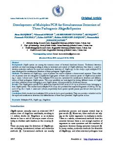
FIG. 1. Genotyping of Borrelia isolates and simultaneous detection and genotyping of genomic groups of B. burgdorferi sensu lato in tick samples. The indicated Borrelia isolates are in the following lanes: 1, A39S; 2, B31; 3, AR-2; 4, AR-1; 22, B. hermsii; 23, B. anserina; 24, group M19. The indicated ticks are in the following lanes: 5, 6, 8, and 10, adults infected with B. garinii; 7, adult infected with group VS116; 9, adult infected with group VS116 and B. afzelii; 11, 17, and 20, nymphs infected with B. afzelii; 12, 19, and 21, nymphs infected with B. garinii; 14 and 15, nymphs infected with B. afzelii and group VS116; 18, nymph infected with group VS116; 13 and 16, nymphs negative by PCR.
lands). The program for the first PCR consisted of an initial denaturation (1 min 948C) and then 25 rounds of temperature cycling (948C for 30 s, 528C for 30 s, and 728C for 1 min). For the second PCR, the tubes were transported to a safety cabinet. A 5-ml reaction mixture containing 0.625 U of Taq DNA polymerase, Saiki buffer supplemented with 2.5 mM MgCl2, 1 mM (each) dNTP, and 5 pmol of biotin-labeled primer 5SCB and of primer 23SN2 was added to each tube. PCR tubes were briefly vortexed and centrifuged and were then transferred to a thermal cycler, and after a denaturation step (1 min at 948C), 40 rounds of temperature cycling (948C for 30 s, 558C for 30 s, and 728C for 1 min) were performed. Contamination of tick samples with B. burgdorferi sensu lato DNA was monitored by using negative control samples (without tick sample) which had been subjected to identical procedures. The product of the first PCR had a size of approximately 380 bp, and DNA amplification by the second PCR resulted in a DNA molecule of 225 bp. To ascertain that amplification had taken place, 5 ml of the PCR mixture was analyzed on ethidium bromide-stained 2% agarose gels in a Tris-borate-EDTA (pH 8.2) buffer. All PCR samples which showed bands, albeit faint, bands of an erroneous size, or a smear were tested by RLB. Samples from ticks which were negative by PCR were spiked with 100 fg of B31 DNA (approximately 20 genome equivalents) to determine whether PCR was inhibited by tick components. The specificity of the nested PCR was determined with boiled T. pallidum (5 3 103 cells) and 25 ng of B. hermsii or B. anserina DNA. The sensitivity of the nested PCR was determined with DNAs from strains B31 (B. burgdorferi sensu stricto), A39S (B. afzelii), AR-1 (B. garinii), and AR-2 (group VS116) at concentrations of 0.1 to 100 fg of DNA per PCR sample. Identification of genomic groups of B. burgdorferi sensu lato by RLB. RLB is a modification of the reverse dot blot, and probes are applied to the membrane as lines instead of dots (25). In one assay, the reactivity of 45 PCR products with up to 45 different probes can be determined (16). After amplification of DNA by PCR, the product was hybridized to four genomic group-specific oligonucleotides (Table 2) (B. burgdorferi sensu stricto, B. afzelii, B. garinii, and group VS116) and one probe which reacts with all genomic groups of B. burgdorferi sensu lato (Table 2); the latter probe served as a control on hybridization procedures. The probes were covalently linked to an activated Biodyne C membrane (Pall Europe Ltd., Portsmouth, United Kingdom) by the 59 aminolink group. For this purpose, the membrane was activated by incubation for 15 min in 16% 1-ethyl-3-(3dimethyl-aminopropyl)carbodiimide (Merck), rinsed with water, and placed in a miniblotter system (Immunetics, Cambridge, Mass.). Five slots were filled with 150 ml of an oligonucleotide suspension, which consisted of 12.5 to 100 pmol of probe (Perkin-Elmer) in 500 mM NaHCO3 (pH 8.4). After 1 min of incubation, excess solution was aspirated, and the blot was removed from the miniblotter and inactivated by incubation with 100 mM NaOH for 10 min. After a rinse with water, the blot was incubated at 568C in 23 SSPE (360 mM NaCl, 20 mM NaH2PO4, 2 mM EDTA [pH 7.2])–0.1% sodium dodecyl sulfate (SDS) for 10 min, and after a rinse in 23 SSPE, the blot was stored at 48C or was used immediately. The filter was again placed in the miniblotter, but the orientation was rotated 908 to the previous position, resulting in a perpendicular position of the slots on the lines which contained the oligonucleotides. The slots were filled with 150 ml of heat-denatured PCR products (10 ml of the PCR mixture suspended in 140 ml of 23 SSPE–0.1% SDS, boiled for 10 min, and chilled on ice) and incubated for 60 min at 458C. The slots were aspirated, and the blot was removed from the miniblotter and washed twice for 10 min at 408C with 23 SSPE–0.5% SDS. Ten milliliters of streptavidin-peroxidase conjugate (Boehringer Mannheim GmbH, Mannheim, Germany) diluted 1:4,000 in 23 SSPE– 0.5% SDS was added to the blot; this was followed by an incubation at 408C for 30 min. Subsequently, the blot was rinsed twice with 23 SSPE–0.5% SDS at 408C. Finally, the blot was briefly rinsed twice with 23 SSPE. Bound streptavidin-
labeled PCR product was detected by chemiluminescence, which was performed with the ECL detection system (Amersham International plc, Den Bosch, The Netherlands), and visualized by exposure of the blot to an X-ray film (Hyperfilm; Amersham). PCR-amplified DNAs from B. hermsii, B. anserina, or B. burgdorferi sensu lato isolates were used to determine the specificity of the RLB. To determine the ability of the RLB to detect a mixture of two genomic groups simultaneously, different amounts of B. afzelii A39S DNA (500, 100, 50, 10, and 5 fg) were mixed with 500 fg of B. garinii AR-1 DNA. The DNA in the mixture was amplified by nested PCR and was hybridized to genomic group-specific probes in RLB. Statistical analysis. The chi-square test of the package GraphPad Instat (version 1.14; GraphPad Software, San Diego, Calif.) was used to compare B. burgdorferi sensu lato infection rates in ticks obtained by IFA and PCR. A P value of #0.05 was considered significant.
RESULTS Specificity and sensitivity of PCR and RLB. The sensitivity of the nested PCR was tested with a range of B. burgdorferi sensu lato DNA concentrations diluted in water and pooled tick lysate. I. ricinus ticks, which were maintained in the laboratory and which were free of Borrelia spp., were used as the source for a pool of tick lysates. In water, 5 fg of Borrelia DNA (from strains B31, AR-1, or A39S) was detected, and in tick lysates the detection limit of the nested PCR was 10 fg of DNA. The DNAs of T. pallidum and B. anserina were not amplified in the nested PCR, and amplification of DNA from B. hermsii yielded a fragment with a size of 500 bp (data not shown). Fourteen B. burgdorferi sensu lato strains were tested by PCR and RLB, and the correct genomic group of all strains was identified from amplified 5S-23S rDNA sequences (Table 1). The reactivities of PCR samples from strains B31, A39S, AR-1, AR-2, and M19 in RLB are shown in Fig. 1. The PCR product of isolate M19 reacted with the probe for group VS116 (Fig. 1, lane 24). PCR products from B. hermsii or B. anserina were negative in the hybridization assay (Fig. 1, lanes 22 and 23). RLB could distinguish between the presence of B. afzelii DNA and B. garinii DNA in mixtures, which had been amplified by PCR, when 50 to 500 fg of B. garinii DNA was added to 500 fg of B. afzelii DNA (data not shown). Therefore, the presence of two genomic groups in a sample can be detected by PCR and RLB if the ratio between genomic groups does not exceed 1:10. Detection and identification of B. burgdorferi sensu lato in ticks by PCR and RLB. A total of 96 ticks were investigated by PCR for the presence of B. burgdorferi sensu lato. Six of 57 (11%) adult ticks and 9 of 39 nymphs (23%) were positive for
3094
RIJPKEMA ET AL.
J. CLIN. MICROBIOL.
TABLE 3. B. burgdorferi sensu lato infection rate in I. ricinus ticks collected on the Dutch North Sea island of Ameland as determined by IFA, PCR, and RLB Infection rate by: Tick stage
Nymphs Adults a
IFA
Genomic group by RLBa PCR
No. tested
No. (%) positive
No. tested
No. (%) positive
Ss
Ga
Af
VS116
Af 1 VS116
120 90
23 (19) 18 (20)
39 57
9 (23) 6 (11)
0 0
3 4
3 0
1 1
2 1
Ss, B. burgdorferi sensu stricto; Ga, B. garinii; Af, B. afzelii; VS116, group VS116; Af 1 VS116, infection of B. afzelii and group VS116.
B. burgdorferi sensu lato (Table 3). The discrepancy in the Borrelia infection rate for nymphs and adults was not statistically significant (P 5 0.96). Tick lysates, which were negative for B. burgdorferi sensu lato by PCR (n 5 81), were spiked with strain B31 DNA, and subsequently, Borrelia DNA was detected in all samples (data not shown). This finding indicated that PCR was not strongly inhibited by tick components because approximately 20 copies of the Borrelia genome could be detected in these tick samples. The analysis of 15 positive PCR samples in RLB is shown in Fig. 1. Among the samples of adult ticks, B. garinii was present in four samples (lanes 5, 6, 8, and 10), group VS116 was present in one sample (lane 7), and a double infection of group VS116 and B. afzelii was found in one sample (lane 9). Among the samples of nymphs, three samples contained B. garinii (lanes 12, 19, and 21), three samples contained B. afzelii (lanes 11, 17, and 20), and one sample reacted with the VS116 probe (lane 18). The two remaining samples contained B. afzelii and group VS116 (lanes 14 and 15). The overall results are summarized in Table 3. Detection of B. burgdorferi sensu lato in ticks by IFA. B. burgdorferi sensu lato infection rates in adult and nymphal I. ricinus ticks collected on Ameland in 1994 were 20 and 19%, respectively (Table 3). Of the adult ticks, 9 of 42 (21%) females and 9 of 48 (19%) males contained Borrelia spirochetes; this difference was not statistically significant (P 5 0.96). The observed discrepancies between the B. burgdorferi sensu lato infection rates determined by IFA or PCR for adult ticks (P 5 0.20) or nymphs (P 5 0.76) were not statistically significant. Infection rates for nymphs (20%; P 5 0.74) or adults (24%; P 5 0.69) were also not significantly different from those found in the survey performed in 1993 (24). DISCUSSION Our aim was to develop a method for the detection and identification of genomic groups of B. burgdorferi sensu lato isolates in ticks which does not require the prior isolation of Borrelia isolates. This would allow a more accurate description of the predominance of various genomic groups of B. burgdorferi sensu lato in their natural habitat. Such information is needed to more precisely define areas where individuals would be at high risk of contracting systemic LB such as neuroborreliosis or Lyme arthritis. We used the 5S-23S rDNA intergenic spacer region as the target for nested PCR and hybridization. Previously, the 16S23S rDNA region and the 23S-23S rDNA region have been used for a PCR to detect B. burgdorferi sensu stricto DNA in ticks and patient biopsy specimens, respectively (19, 28). By this PCR it was possible to detect 10 fg of B. burgdorferi sensu lato DNA diluted in tick lysate, which corresponds to approximately two organisms per sample. The PCR was shown to be species specific, and in RLB, the anticipated genomic group was identified from the PCR product of each B. burgdorferi sensu lato isolate (n 5 14).
B. burgdorferi sensu lato DNA was amplified by PCR in 16% of ticks collected on Ameland in 1994. Complete inhibition of PCR by tick components has been found for Amblyomma americanum ticks (13). Therefore, PCR-negative ticks were spiked, and we established that approximately 20 copies of the Borrelia genome could be detected in these ticks; however, this does not rule out the possibility that these ticks contained lower numbers of Borrelia spirochetes. In a different group of ticks from the same batch, the presence of Borrelia spirochetes was analyzed by IFA. Infection rates for male and female ticks differed only marginally. A slightly higher percentage of infected nymphs was detected by PCR than by IFA, whereas the infection rate in adults was higher by IFA than by PCR. These differences, however, were not statistically significant and may have been due to a selection of individuals for each group. Hybridization of 15 PCR-positive samples from ticks by RLB revealed three genomic groups of B. burgdorferi sensu lato: B. afzelii, B. garinii, and group VS116. These findings are in accordance with those from other studies performed in The Netherlands and Europe. B. garinii and B. afzelii are found in ticks and patients with LB throughout Europe, and both Borrelia species are also present in Dutch ticks (2, 20, 22, 32, 34, 35). Until now, only four Borrelia strains isolated from ticks collected in England, Switzerland, The Netherlands, and Japan have been classified as genomic group VS116 (2, 21, 22, 23, 27). Borrelia strains which belong to group M19, which is closely related to B. garinii, have been isolated from ticks collected throughout The Netherlands (20). In the present study, we show that group VS116 is found among ticks on Ameland and that strain M19 belongs to genomic group VS116 (Fig. 1). Thus, B. burgdorferi group VS116 is well established among Dutch ticks. The relevance of this genomic group for clinical LB remains to be investigated. Until now, patient isolates belonging to group VS116 have not been described. Kuiper et al. (17) reported a seropositivity rate of 20% among healthy Dutch forestry workers. Possibly, the majority of infections with B. burgdorferi group VS116 result in asymptomatic seropositivity. To examine this hypothesis, urine samples from asymptomatic seropositive individuals, which may contain B. burgdorferi sensu lato DNA, could be investigated by PCR (15). Double infections with B. afzelii and group VS116 were found in two nymphs and one adult tick (20% of PCR-positive ticks). Cross-reactivity between PCR products is unlikely because the probes for VS116 and B. afzelii differ in 8 of 22 nucleotides, and the reference strains used in the present study did not display such cross-hybridization (Table 2 and Fig. 1). Thus, two different genomic groups were present in these ticks. Previous studies showed that double infections occur in European ticks. B. burgdorferi sensu lato strains with two different OspA genotypes have been isolated from 36% of infected ticks in Switzerland (18). Recently, sequencing of OspA DNA amplified from ticks collected in the field revealed one double infection among 40 PCR-positive ticks, which is considerably
IDENTIFICATION OF B. BURGDORFERI SENSU LATO IN TICKS
VOL. 33, 1995
lower than our rate of double infection (12). This variation might be explained by differences in the tick populations studied or differences in study methodology. The typing method described in the latter study could not detect both genomic groups of B. burgdorferi sensu lato if the ratio of a mixture exceeded 1:5, whereas our method could distinguish two genomic groups at a ratio of 1:10. In one study (10), the simultaneous presence of B. burgdorferi sensu stricto, B. afzelii, or B. garinii was found in 8 of 18 patients with neuroborreliosis, and B. garinii was detected in all of these samples, consistent with the association of B. garinii with neuroborreliosis. Only these three genomic groups could be identified; therefore, further investigation of coinfections, including the involvement of newly recognized genomic groups such as VS116, is needed. In conclusion, amplification and hybridization of the 5S-23S rDNA intergenic spacer region to multiple genomic groupspecific oligonucleotides provide an accurate and rapid method of determining the presence of all genomic groups of B. burgdorferi sensu lato in ticks collected in the field.
14.
15.
16.
17.
18.
19.
20.
ACKNOWLEDGMENTS We are grateful to D. Postic and I. Saint Girons for sharing the 5S-23S intergenic spacer sequence data with us prior to publication. We thank D. Postic, E. Åsbrink, U. Go ¨bel, K. Hansen, R. Marconi, S. Cutler, M. Nohlmans, R. Ackermann, V. Preac-Mursic, and A. van Dam for the gift of Borrelia isolates and B. A. M. van der Zeijst and J. D. A. van Embden for critical reading of the manuscript.
21.
22.
23. REFERENCES 1. Anthonissen, F. M., M. Dekesel, P. P. Hoet, and G. H. Bigaignon. 1994. Evidence for the involvement of different genospecies of Borrelia in the clinical outcome of Lyme disease in Belgium. Res. Microbiol. 145:327–331. 2. Assous, M. V., D. Postic, and G. Baranton. 1994. Clinical and epidemiological implications of Borrelia burgdorferi sensu lato taxonomy, p. 148–162. In Y. Yanaghari and T. Masuzawa (ed.), Present status of Lyme disease and biology of Lyme Borrelia. Proceedings of the International Symposium on Lyme Disease in Japan. Kanzanji, Hamamatsu, Shizuoka, Japan. 3. Assous, M. V., D. Postic, G. Paul, P. Nevot, and G. Baranton. 1993. Western blot analysis of sera from Lyme borreliosis patients according to the genomic species of the Borrelia strains used as antigens. Eur. J. Clin. Microbiol. Infect. Dis. 12:261–268. 4. Baranton, G., D. Postic, I. Saint Girons, P. Boerlin, J.-C. Piffaretti, M. Assous, and P. A. D. Grimont. 1992. Delineation of Borrelia burgdorferi sensu stricto, Borrelia garinii sp. nov., and group VS461 associated with Lyme borreliosis. Int. J. Syst. Bacteriol. 42:378–383. 5. Barbour, A. G. 1984. Isolation and cultivation of Lyme disease spirochetes. Yale J. Biol. Med. 57:521–525. 6. Boerlin, P., O. Pe´ter, A.-G. Bretz, D. Postic, G. Baranton, and J.-C. Piffaretti. 1992. Population genetic analysis of Borrelia burgdorferi isolates by multilocus enzyme electrophoresis. Infect. Immun. 60:1677–1683. 7. Burgdorfer, W., A. G. Barbour, S. F. Hayes, J. L. Benach, E. Grunwaldt, and J. P. Davies. 1982. Lyme disease—a tick-borne spirochetosis? Science 216: 1317–1319. 8. Canica, M. M., F. Nato, L. du Merle, J. C. Mazie, G. Baranton, and D. Postic. 1993. Monoclonal antibodies for the identification of Borrelia afzelii sp. nov. associated with late cutaneous manifestations of Lyme borreliosis. Scand. J. Infect. Dis. 25:441–448. 9. De Kok, J. B., C. d’Oliveira, and F. Jongejan. 1993. Detection of the protozoan parasite Theileria annulata in Hyalomma ticks by the polymerase chain reaction. Exp. Appl. Acarol. 17:839–846. 10. Demaerschalck, I., A. Ben Massoud, M. De Kesel, B. Hoyois, Y. Lobet, P. Hoet, G. Bigaignon, A. Bollen, and E. Godfroid. 1995. Simultaneous presence of different Borrelia burgdorferi sensu lato genospecies in biological fluids of Lyme disease patients. J. Clin. Microbiol. 33:602–608. 11. Dressler, F., R. Ackermann, and A. C. Steere. 1994. Antibody responses to three genomic subgroups of Borrelia burgdorferi in European Lyme borreliosis. J. Infect. Dis. 169:313–318. 12. Eiffert, H., A. Ohlenbusch, H.-J. Christen, R. Thomssen, A. Spielman, and F.-R. Mathuschka. 1995. Nondifferentiation between Lyme disease spirochetes from vector ticks and human cerebrospinal fluid. J. Infect. Dis. 171: 476–479. 13. Ewing, S. A., J. E. Dawson, A. A. Kocan, R. W. Barker, C. K. Warner, R. J. Panciera, J. C. Fox, K. M. Kocan, and E. F. Blouin. 1995. Experimental transmission of Ehrlichia chaffeensis (Rickettsiales: Ehrlichieae) among
24.
25.
26.
27. 28.
29.
30. 31.
32.
33. 34.
35.
36.
3095
white-tailed deer by Amblyomma americanum (Acari: Ixodidae). J. Med. Entomol. 32:368–374. Fukunaga, M., and M. Sohnaka. 1992. Tandem repeat of the 23S and 5S ribosomal RNA genes in Borrelia burgdorferi, the etiological agent of Lyme disease. Biochem. Biophys. Res. Commun. 183:952–957. Karch, H., H.-I. Huppertz, M. Bo¨hme, H. Schmidt, D. Wiebecke, and A. Schwarzkopf. 1994. Demonstration of Borrelia burgdorferi DNA in urine samples from healthy humans whose sera contain B. burgdorferi-specific antibodies. J. Clin. Microbiol. 32:2312–2314. Kaufhold, A., A. Podbielski, G. Baumgarten, M. Blokpoel, J. Top, and L. Schouls. 1994. Rapid typing of group A streptococci by the use of DNA amplification and nonradioactive allele-specific oligonucleotide probes. FEMS Microbiol. Lett. 119:19–26. Kuiper, H., B. M. de Jongh, A. P. Nauta, H. Houweling, L. G. Wiessing, A. W. Moll van Charant, and L. Spanjaard. 1991. Lyme borreliosis in Dutch forestry workers. J. Infect. 23:279–286. Leuba-Garcia, S., M. D. Kramer, R. Wallich, and L. Gern. 1994. Characterization of Borrelia burgdorferi isolated from different organs of Ixodes ricinus ticks collected in Nature. Zentralbl. Bakteriol. Parasitenko. Infektionskr. Hyg. Abt. 1 Orig. 280:468–475. Liveris, D., A. Gazumyan, and I. Schwartz. 1995. Molecular typing of Borrelia burgdorferi sensu lato by PCR-restriction fragment length polymorphism analysis. J. Clin. Microbiol. 33:589–595. Nohlmans, L. M. K. E., R. de Boer, A. E. J. M. van den Bogaard, and C. P. A. van Boven. 1995. Genotypic and phenotypic analysis of Borrelia burgdorferi isolates from The Netherlands. J. Clin. Microbiol. 33:119–125. Pe´ter, O., and A. G. Bretz. 1992. Polymorphism of outer surface proteins of Borrelia burgdorferi as a tool for classification. Zentralbl. Bakteriol. Parasitenko. Infektionskr. Hyg. Abt. 1 Orig. 277:28–33. Postic, D., M. Assous, P. A. D. Grimont, and G. Baranton. 1994. Diversity of Borrelia burgdorferi sensu lato evidenced by restriction fragment length polymorphism of rrf (5S)-rrl(23S) intergenic spacer amplicons. Int. J. Syst. Bacteriol. 44:743–752. Postic, D., and G. Baranton. 1994. Molecular fingerprinting and phylogeny of Borrelia burgdorferi sensu lato, p. 133–147. In Y. Yanaghari and T. Masuzawa (ed.), Present status of Lyme disease and biology of Lyme Borrelia. Proceedings of the International Symposium on Lyme Disease in Japan. Kanzanji, Hamamatsu, Shizuoka, Japan. Rijpkema, S., J. Nieuwenhuijs, F. F. J. Franssen, and F. Jongejan. 1994. Infection rates of Borrelia burgdorferi in different instars of Ixodes ricinus ticks from the Dutch North Sea island of Ameland. Exp. Appl. Acarol. 18:531– 542. Saiki, R. K., C.-A. Chang, C. H. Levenson, T. C. Warren, C. D. Boehm, H. H. Kazazian, and H. A. Erlich. 1988. Diagnosis of sickle cell anemia and b-thalassemia with enzymatically amplified DNA and nonradioactive allelespecific oligonucleotide probes. N. Engl. J. Med. 319:537–541. Saiki, R. K., D. H. Gelfland, S. Stoffer, S. J. Scharf, R. Higuchi, G. T. Horn, K. B. Mullis, and H. A. Erlich. 1988. Primer-directed enzymatic amplification of DNA with a thermostable DNA polymerase. Science 239:487–491. Saint Girons, I. Personal communication. Schwartz, I., G. P. Wormser, J. J. Schwartz, D. Cooper, P. Weissensee, A. Gazumyan, E. Zimmerman, N. S. Goldberg, S. Bittker, G. L. Campbell, and C. S. Pavia. 1992. Diagnosis of early Lyme disease by polymerase chain reaction amplification and culture of skin biopsies from erythema migrans lesions. J. Clin. Microbiol. 30:3082–3088. Schwartz, J. J., A. Gazumyan, and I. Schwartz. 1992. rRNA gene organization in the Lyme disease spirochete, Borrelia burgdorferi. J. Bacteriol. 174: 3757–3765. Steere, A. C. 1989. Lyme disease. N. Engl. J. Med. 321:586–596. Theisen, M., B. Fredriksen, A.-M. Lebech, J. Vuust, and K. Hansen. 1993. Polymorphism in ospC gene of Borrelia burgdorferi and immunoreactivity of OspC proteins: implication for taxonomy and diagnosis for use of OspC protein as a diagnostic antigen. J. Clin. Microbiol. 31:2570–2576. Van Dam, A. P., H. Kuiper, K. Vos, A. Widjojokusumo, B. M. de Jongh, L. Spanjaard, A. C. P. Ramselaar, M. D. Kramer, and J. Dankert. 1993. Different genospecies of Borrelia burgdorferi are associated with distinct clinical manifestations of Lyme borreliosis. Clin. Infect. Dis. 17:708–717. Wienecke, R., U. Neubert, and M. Volkenandt. 1993. Cross immunity among types of Borrelia burgdorferi. Lancet 342:435. Wienecke, R., N. Zo ¨chling, U. Neubert, E.-M. Schlu ¨pen, M. Meurer, and M. Volkenandt. 1994. Molecular subtyping of Borrelia burgdorferi in erythema migrans and acrodermatitis chronica atrophicans. J. Invest. Dermatol. 103: 19–22. Wilske, B. A., V. Preac-Mursic, U. B. Go ¨bel, B. Graf, S. Jauris, E. Soutschek, E. Schwab, and G. Zumstein. 1993. An OspA serotyping system for Borrelia burgdorferi based on the reactivity with monoclonal antibodies and OspA sequence analysis. J. Clin. Microbiol. 31:340–350. Wilson, K. 1987. Preparation of genomic DNA of bacteria, p. 241–245. In F. M. Ausubel, R. Brent, R. E. Kingston, et al. (ed.), Current protocols in molecular biology. Greene Publishing Associates, Brooklyn, N.Y.