Stefan Cular1, Venkat R. Bhethanabotla1, Darren W. Branch2. 1Sensors ..... [17] K. Lange, F. Bender, A. Voigt, H. Gao, and M. Rapp, "A Surface Acoustic Wave.
Simultaneous Surface Manipulation and Sensing in a Biosensor Using a Hexagonal SAW Device Stefan Cular1, Venkat R. Bhethanabotla1, Darren W. Branch2 1 Sensors Research Laboratory, Department of Chemical Engineering, University of South Florida, Tampa, FL, USA 33620 2 Biosensors and Nanomaterials Deaprtment, Sandia National Laboratories, Albuquerque, NM, USA 87185 Abstract We present the development of a hexagonal surface acoustic wave (SAW) device to simultaneously sense and manipulate the biological sensing film in biosensor applications. The primary objective is to improve sensitivity and selectivity. A secondary objective is to regenerate the sensor for reuse. A hexagonal device fabricated in 36° lithium tantalate allows for propagation of both Rayleigh and shear horizontal (SH) wave modes simultaneously. The high electro-acoustic coupling in this piezoelectric material allows for efficient transfer of energy from electrical to mechanical form. In this device, the Rayleigh acoustic waves stress the bonds between the sensing film and analyte forcing only the analyte with higher affinity for the sensing film to stay bound, while the SH SAWs are used for sensing. Additionally, the acoustic waves work to efficiently mix the liquid samples flowing through the micro-channels of the micro-sensor system, reducing the effects of diffusion-limited processes. Results from using a sensing film of anti-mouse IgG covalently bound to the sensorsurface and mouse IgG as the analyte in buffer solution have shown improved sensor response, determined using fluorescent microscopy. Manipulation of liquid samples was achieved by strongly exciting the piezoelectric substrate with power levels of ~12 mW which is significantly greater than the 1 mW used for sensing. The larger electrical power creates an acoustic wave via piezoelectric coupling that can physically force loosely bound species from binding sites, reducing noise that can lead to inaccurate measurements. Introduction SAW devices have been used in many sensor applications in both gaseous [1-3] and liquid environments [4-6]. Each application has its own requirements. For example, the use of a SAW device as a biosensor implies the device must not inherently be attenuated by the environment it is supposed to operate in. This implication restricts the SAW biosensors to SH– SAWs and a specialized SH-SAW device that creates a Love mode wave from a thin film deposited on its surface [7, 8]. SAW sensors have been shown to work well as high sensitivity biosensors; however, as with all other biosensors non-specifically bound (NSB) protein interactions can cause a less than ideal sensor response and concentration determination [9, 10]. Some possible responses seen as a result of NSB proteins include: exaggerated response due to multi-layer formation, false responses due to miscellaneous proteins covering the surface, and no response due to poor alignment of the functional groups. Minor improvements to biosensor responses can be achieved from thorough rinsing, use of ultrasonic baths to remove NSB proteins, and pre-
treatment of the analyte containing fluids. Each one of these processes adds to the complexity and decreases the functionality of a biosensor to be operated without specialized training in everyday environments. Developments in acoustic wave applications have demonstrated NSB protein removal with relatively low power consumption thus significantly decreasing the uncertainty of the sensors response [9, 10].
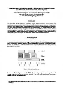
Figure 1. Hexagonal SAW device schematic
Materials and Methods A. Sensor Device Design The hexagonal SAW is a composite of three traditional delay line SAW devices rotated about the center of the die, which is approximately 20 x 20 mm. The individual delay line devices are comprised of identical bi-directional interdigital transducers (IDTs) with an aperture of 47 , delay length of 197 , and feature size of 4 m. The delay path is shorted to eliminate unwanted waves and eliminate the electrical effect [11]. A standard metallization procedure of 100 nm titanium adhesion layer followed by 700 nm gold layer was used. Figure 1 is an illustration of the hexagonal SAW layout. The IDT designs tested included design considerations to improve the phase linearity and decrease the phase noise [11, 12]. B. Micro-fluidic Sensor Fixture and Test-bed Sensitivity required for detection of many biological markers is on the order of a few nano-grams per milliliter, which is obtainable by many types of sensors; however, SAW sensors are some of the most sensitive and easy to implement devices [13-15]. Challenges associated with this order of mass sensitivity become largely a fixture and test parameter issue since any variation in fluid flow or pressure will cause a significant sensor response [16]. To address this issue, we have chosen to use a precision syringe pump (Harvard Apparatus PHD 2000) which unlike peristaltic pumps provides a smooth continuous flow with no pulses. The equipment shown in the illustration, Figure 2, is highly adaptable for all of our required fluid requirements from nano-liters to milli-liters per minute [17]. Additionally, making the configurability and operation of the test bed simple is a LabView® virtual instrument interface that controls and records all electronic operations including flow rate, flow direction, and valve position. Due to the design of the hexagonal SAW a typical micro-fluidic fixture is not feasible, so an in-house design has been designed and fabricated. The test fixture is constructed of polycarbonate, which has low moisture absorption, high strength, no centerline porosity, easy machineablity, and can be polished to be optically clear. The micro-fluidic fixture has a printed circuit board with SMA connectors to connect to electronic test equipment. C. Experimental Procedures 1. Sensor Preparation The sensors were designed for biosensor applications; however, additional preparations of the sensor-surfaces were necessary. Primary to the preparations was to insulate the IDTs from the liquid environment meeting three conditions 1) the insulating
Figure 2. Schematic of microfluidic test bed for liquid phase sampling of biologic samples.
material must not attenuate the SAWs excessively, 2) the material must not be highly permeable to water, and 3) the material must permit attachment of antibodies. High molecular weight polystyrene (Sigma Aldrich) was chosen as a solution. Coating of the sensor was achieved by dissolving polystyrene in 2-butoxyethyl acetate (Sigma Aldrich) to 4 weight percent then spin coating the sensors. The sensors were then annealed at 120°C for 1 hour. Following the annealing, the sensors where mounted in the micro-fluidic fixture and characterized using a vector network analyzer (Agilent 8753ES). 2. Sensing From a calculated exposed area of the sensor surface and cross-sectional areas of the antibodies used, a concentration of proteins was specified to ensure 100% coverage of the sensor surface without excessive multi-layer formation of non-specifically bound proteins. The calculated protein concentration was applied to the surface of the sensor through the microfluidic fixture for 1 hour to allow adequate surface adsorption to the polystyrene. Having the sensor functionalized with an antibody, goat anti-mouse IgG (Pierce), varying concentrations of antigen, mouse anti-rabbit IgG (Pierce) were flowed across the sensor at a constant 0.15 ml/min. The antigen concentrations were calculated and made to ensure less than 25% surface coverage. To further insure a good response of the sensor, a pH of 7.4 was maintained by using of phosphate buffered saline (PBS) solution. 3. Surface Manipulation The second task of this project was the use of high amplitude waves to remove loosely bound materials from the sensors surface. The first tests to show the surface was being manipulated were conducted using a Leica DMI4000 fluorescent microscope. With the prepared sensors, fluorescently labeled proteins were adsorbed onto the surface of the device for 1 hour to achieve saturation. Sensors where then flushed with 3 ml of PBS. Following the flushing the devices were subjected to high amplitude waves using a 4 watt RF power amplifier (Mini-Circuits TIA-1000-1R8). Calculations showed that the actual power delivered to the IDT was on the order of milli-watts due to insertion loss of the device and attenuation by the films on the surface. 4. Simultaneous Surface Manipulation and Sensing The final task of these experiments was the combination of the high amplitude waves manipulating the surface and the use of low amplitude waves for sensing changes in the sensing film simultaneously. The sensors were first prepared with an antibody film as described above, and used to sense a known concentration of antigen in solution. After the sensing test, the sensors were coated with known concentrations of a NSB protein, Bovine Serum Albumin (BSA). Following the BSA application, the sensors were flushed with 3 ml of PBS then subjected to high amplitude waves while monitoring the changes with a different delay path. Results and Discussion The sensors used in this project are theoretically capable of detecting nano-grams of mass change on their surfaces. The first experiments showed that this was the case with the detection of low levels of mouse IgG binding with a functionalized surface. A representative data set of the sensing capability is shown in Figure 3. N The first drop in the graph is the result of flushing away the excess antibody from the 1 hour adsorption process. Following the
Anti-mouse IgG on surface of device
9
0.0004
Phase Normalized
0.0006
0.0002
Flush with PBS to remove excess anti-mouse IgG
Flush with PBS to remove excess BSA
Flush with PBS to remove excess mouse IgG
0 Inject 324 ng mouse IgG
-0.0002
Inject 500 ng BSA
-0.0004 -0.0006 -0.0008 0
500
1000
1500
2000
2500
3000
3500
Time (s)
Figure 3. Normalized phase response for the coating of a sensor with anibodies (138 ng/ml anti-mouse IgG in PBS) followed by the detection of of the antigen (324 ng mouse IgG), and the coating of the sensor with non-specifically binding BSA (500 ng). first large drop, the antigen is injected and flushed away. In this event, there are actually two measurements that are significant. First, the phase change upon injecting a known concentration sample, and second, the non-returning of the phase change value to the baseline (residual difference in phase change) even after extensive flushing with PBS. The large step up seen upon flushing the system with PBS to remove the excess BSA may appear to be removal of the BSA, but attenuation measurements show that this is not the case. In fact, it is a complex non-linear response upon the adhesion of a large amount of BSA. In the next step, BSA is injected to completely coat the surface in preparation for testing the non-specifically bound protein removal function of the sensor. Upon first inspection one will note the magnitude of this response is significantly different from the response of the specific antigen. This is the result of leaving the linear response regime of the surface acoustic wave (SAW) sensor by applying too much material onto the surface. For many biosensors this would result in having to discard the sensor and starting over with a new one. However, this situation allows us to clearly demonstrate the simultaneous manipulation and sensing functionality of our sensor system. Coating of the sensor-surface beyond its functioning point is often encountered. Another commonly encountered situation is not having a pure sample of the one analyte protein of interest. Such a sample requires extensive filtering and/or processing before a concentration determination with the sensor. The results in Figure 4 address this issue. More specifically, they demonstrate the feasibility of simultaneous sensing and manipulation of the sensing film is in one device using two types of SAWs propagating in different directions. The
Figure 4. Normalized phase angle response of sensor to the removal of excess BSA with just PBS followed by the removal of NSB BSA using high amplitude waves. data show the addition of a high concentration of BSA to the surface of the SAW sensor followed by the removal of some of the BSA remaining on the surface upon extensive flushing with PBS. Further removal is achieved through the application of the power amplified signal for 50 seconds on a different SAW delay path than that used for sensing. Due to the limitations of the currently used, first design of the hexagonal SAW device, more of the sensing delay line is exposed to the sample solutions than what the second and third delay paths are capable of manipulating. Conclusion We have shown the development of a hexagonal SAW device to simultaneously manipulate biological films and sense changes in analyte concentration using different acoustic waves across the same sensing film. The results have shown low-level sensing of proteins in solution is possible with this new SAW sensor. Additionally, the sensor has the functionality to reduce error in responses by removing NSB proteins and other loosely bound material that are common in complex samples. With further design improvements and studies, we believe this device has wide application to bio-sensing especially where non-purified samples containing many different proteins need to be analyzed without the use of technicians and laboratory equipment. Acknowledgments Support for this work has been provided by NSF grant number DGE-0221681, University of South Florida IDRG, and DoD Grant W81XWH-05-1-0585.
References [1] J. W. Grate, "Acoustic wave microsensor arrays for vapor sensing," Chem.Rev., vol. 100, pp. 2627-2648, 2000. [2] D. S. Ballantine, Acoustic wave sensors: theory, design, and physico-chemical applications. San Diego: Academic Press, 1997. [3] A. Chaudhari, D. Srinivasagupta, S. Cular, V. R. Bhethanabotla, and Joseph, "Surface acoustic wave sensors using nanocrystalline palladium for hydrogen gas detection.," presented at AIChE Annual Meeting, Austin, Texas., 2004. [4] M. P. d. Cunha, D. C. Malocha, D. W. Puccio, J. Thiele, and T. B. Pollard, "LGX pure shear horizontal SAW for liquid sensor applications.," Sensors J. IEEE, vol. 3(5), pp. 554 – 561, 2003. [5] F. Josse, F. Bender, and R. Cernosek, "Guided shear horizontal surface acoustic wave sensors for chemical and biochemical detection in liquids," Analytical Chemistry, vol. 73, pp. 5937-5944, 2001. [6] F. Martin, M. I. Newton, G. McHale, K. A. Melzak, and E. Gizeli, "Pulse Mode Shear Horizontal-surface Acoustic Wave (SH-SAW) System for Liquid Based Sensing Applications," Biosensors & Bioelectronics, vol. 19, pp. 627-632, 2004. [7] D. W. Branch and S. M. Borzik, "Low-level Detection of a Bacillus anthracis Simulant Using Love-wave Biosensors on 36 deg YX LiTaO3," Biosensors & Bioelectronics, vol. 19, pp. 849-859, 2004. [8] N. Barie, T. Wessa, M. Bruns, and M. Rapp, "Love waves in SiO2 layers on STWresonators based on LiTaO3," Talanta, vol. 62, pp. 71-79, 2004. [9] S. Cular, D. W. Branch, V. R. Bhethanabotla, G. D. Meyer, and H. G. Craighead, "Removal of Nonspecific Binding on Microsensors Using Surface Acoustic Waves," presented at AIChE Annual Meeting, Cincinnati, OH, USA, 2005. [10] G. D. Meyer, J. M. Moran-Mirabal, D. W. Branch, and H. G. Craighead, "Nonspecific Binding Removal from Protein Microassays using Thickness Shear Mode Resonators," Accepted IEEE Sensors, 2004. [11] D. P. Morgan, Surface-Wave Devices for Signal Processing. New York: Elsevier, 1991. [12] C. Campbell, Surface acoustic wave devices and their signal processing applications. Boston: Academic Press, 1989. [13] M. Rapp, T. Wessa, and H. J. Ache, "Modification of Commercially Available Low-Loss SAW Devices Towards an Immunosensor for in-situ Measurements in Water," presented at IEEE Ultrasonics Symposium, 1995. [14] F. Bender, K. Lange, N. Barie, J. Kondoh, and M. Rapp, "On-line monitoring of Polymer depostion for tailoring the waveguide characteristics of love-wave biosensors," Langmuir, vol. 20, pp. 2315-2319, 2004. [15] T. Wessa, M. Rapp, and H. Sigrist, "Immunosensing of photoimmobilized proteins on surface acoustic wave sensors," Colloids and Surfaces, vol. 15, pp. 139-146, 1999. [16] N. Barie, H. Sigrist, and M. Rapp, "Development of Immunosenors Based on Commercially Available Surface Acoustic Wave (SAW) Devices," Analusis, vol. 27, pp. 622-629, 1999. [17] K. Lange, F. Bender, A. Voigt, H. Gao, and M. Rapp, "A Surface Acoustic Wave Biosensor Concept with Low Flow Cell Volumes for Label-Free Detection," Analytical Chemistry, vol. 75, pp. 5561-5566, 2003.