Article
Single-Cell Profiling Identifies Key Pathways Expressed by iPSCs Cultured in Different Commercial Media Maciej Daniszewski, Quan Nguyen, Hun S. Chy, ..., Alex W. Hewitt, Joseph E. Powell, Alice Pe´bay
[email protected]
HIGHLIGHTS HiPSCs maintained in StemFlex and E8 were compared by single-cell transcriptomics Data revealed similar expression of core pluripotency genes Data revealed a shared three subpopulation structure different in pluripotency states
Daniszewski et al., iScience 7, 30–39 September 28, 2018 ª 2018 The Author(s). https://doi.org/10.1016/ j.isci.2018.08.016
Article
Single-Cell Profiling Identifies Key Pathways Expressed by iPSCs Cultured in Different Commercial Media Maciej Daniszewski,1,2,11 Quan Nguyen,3,11 Hun S. Chy,4,5 Vikrant Singh,6 Duncan E. Crombie,1,2 Tejal Kulkarni,1,2 Helena H. Liang,1,2 Priyadharshini Sivakumaran,1,2 Grace E. Lidgerwood,1,2 Damia´n Herna´ndez,1,2 Alison Conquest,1,2 Louise A. Rooney,1,2 Sophie Chevalier,1,2 Stacey B. Andersen,3 Anne Senabouth,3 James C. Vickers,7 David A. Mackey,8 Jamie E. Craig,9 Andrew L. Laslett,4,5 Alex W. Hewitt,1,2,6,11 Joseph E. Powell,3,10,11 and Alice Pe´bay1,2,11,12,* SUMMARY We assessed the pluripotency of human induced pluripotent stem cells (iPSCs) maintained on an automated platform using StemFlex and TeSR-E8 media. Analysis of transcriptome of single cells revealed similar expression of core pluripotency genes, as well as genes associated with naive and primed states of pluripotency. Analysis of individual cells from four samples consisting of two different iPSC lines each grown in the two culture media revealed a shared subpopulation structure with three main subpopulations different in pluripotency states. By implementing a machine learning approach, we estimated that most cells within each subpopulation are very similar between all four samples. The single-cell RNA sequencing analysis of iPSC lines grown in both media reports the molecular signature in StemFlex medium and how it compares to that observed in the TeSR-E8 medium.
INTRODUCTION Somatic cells can be reprogrammed into induced pluripotent stem cells (iPSCs) using a combination of transcription factors involved in the maintenance of pluripotency (Park et al., 2007; Takahashi and Yamanaka, 2006; Takahashi et al., 2007; Yu et al., 2007). Like human embryonic stem cells (hESCs), human iPSCs provide a powerful means by which to investigate the pathogenesis of disease because these cells can be differentiated into relevant cell types of interest for disease modeling, drug screening, and delving into the fundamental aspects of disease pathology, as well as for regenerative medicine. Numerous protocols have been described for the maintenance of human pluripotent stem cells, using different matrices and culture media (International Stem Cell Initiative Consortium et al., 2010). The original method of maintenance of hESCs was based on a feeder layer of embryonic fibroblasts and a serum-based medium (Reubinoff et al., 2000; Thomson, 1998). It evolved to more defined conditions using serum-free media with key signaling molecules (Furue et al., 2008; Pe´bay et al., 2005; Vallier et al., 2005; Xu et al., 2005) and/or feeder-free conditions (including the use of Matrigel, collagen, vitronectin, or laminin) (Klimanskaya et al., 2005; Ludwig et al., 2006). Various commercialized serum-free media have now become available with an ongoing evolution to fully defined media, from KnockOut Serum Replacement medium supplemented with basic fibroblast growth factor to the TeSR family media (mTeSR1, TeSR2, and TeSR-E8) (Chen et al., 2011; Ludwig et al., 2006; Sun et al., 2009), and the newly released StemFlex, a medium with proprietary compounds, and hence of unknown formulation and with robustness efficiency remaining to be characterized. Although there have been clear advances in the maintenance, standardization, and upscaling of pluripotent stem cell culture, some limitations remain and need to be addressed to realize the translational potential of iPSCs (Kim and Kino-oka, 2018). Indeed, human variability is a main source of differences observed between cell lines generated and maintained in various laboratories (Allegrucci and Young, 2006; Allegrucci et al., 2007), with genetic background and methodologies also having a significant contribution (Kilpinen et al., 2016). The automation of pluripotent stem cell culture provides an efficient way to improve these aspects, increasing throughput and standardizing many aspects of cell culture, including in iPSC generation, maintenance, passaging, and differentiation into progeny cells of interest (Crombie et al., 2017; Konagaya et al., 2015; Paull et al., 2015). These automated steps allow minimal variation in each procedure,
30
iScience 7, 30–39, September 28, 2018 ª 2018 The Author(s). This is an open access article under the CC BY license (http://creativecommons.org/licenses/by/4.0/).
1Centre
for Eye Research Australia, Royal Victorian Eye and Ear Hospital, University of Melbourne, 32 Gisborne Street, East Melbourne, VIC 3002, Australia
2Ophthalmology,
Department of Surgery, the University of Melbourne, Melbourne, VIC 3002, Australia 3Institute
for Molecular Bioscience, University of Queensland, Brisbane, QLD 4072, Australia
4Commonwealth
Scientific and Industrial Research Organisation (CSIRO) Manufacturing, Clayton, VIC 3168, Australia
5Australian
Regenerative Medicine Institute, Monash University, Clayton, VIC 3168, Australia
6School
of Medicine, Menzies Institute for Medical Research, University of Tasmania, Hobart, TAS 7000, Australia
7Wicking
Dementia Research and Education Centre, University of Tasmania, Hobart, TAS 7000, Australia
8Centre
for Ophthalmology and Visual Science, Lions Eye Institute, University of Western Australia, Perth, WA 6009, Australia
9Flinders
University, Adelaide, SA 5042, Australia
10Garvan Institute of Medical Research, Darlinghurst, Sydney, NSW 2010, Australia 11These authors contributed equally 12Lead
Contact
Continued
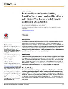
Figure 1. Quality Control of iPSCs Cultivated in StemFlex and TeSR-E8 (A) Schematic representation of the experimental plan. (B) Representative morphology of iPSCs maintained in StemFlex or TeSR-E8 post-passaging. Images of human iPSCs, at day 8 post-passaging, at different magnifications (34, 310, and 320). Cells were cultivated using an automated platform, on vitronectin, in StemFlex (IST2607, p3 1:6 ratio) or TeSR-E8 (TOB0435, p2 1:3 ratio). Images are representative of all cell lines. Scale bars: 100 mM. (C and D) Quantification of TRA-1-60-positive cells before passaging of human iPSCs maintained in StemFlex (C) or TeSRE8 (D). Scatterplot with bar of the TRA-1-60-positive (+ve) cells just before passaging, in independent iPSC lines grown in StemFlex (TOB0224 p3,5–8; TOB0431 p3,5–8; FSA0006 p3,5–8; IST2168 p3,5–8; IST2607 p3,5,6,8; TOB0474 p3,5–8; MBE2906 p3,5–8; FSA0001 p2,4–6,8; MBE1006 p2,4–6,8) or TeSR-E8 (FSA0002 p2,5,8; TOB0435 p5,8; MBE2900 p2,5,8; MBE2909 p4,5,8; TOB0199 p2,4,8; TOB0412 p3,4,8 FSA0005 p4,5,8 TOB0421 p8). The abnormal lines TOB0218 (StemFlex), MBE2899 (TeSR-E8), and MBE2901 (TeSR-E8) were not included in this analysis. Each column represents the mean G SEM of successive passages of individual lines. Two-tailed t test indicates no statistical difference in TRA-1-60 expression between the two culture conditions (p = 0.8546).
reduce inter-sample variability, and hence increase robustness of cell culture procedures (Daniszewski et al., 2017). Using an automated platform to ensure robustness of cell culture, we compared the cellular and molecular profiles of human iPSCs maintained in a standard TeSR-E8 medium and in the newly released StemFlex. Quality control was assessed by markers of pluripotency, three germ layer differentiation, and virtual karyotyping. We then compared samples grown in both media by single-cell RNA sequencing (scRNA-seq) to uncover the molecular signature underlying pluripotency, and report on whether similar pathways are modulated in both conditions.
RESULTS Donor iPSCs were derived as described in Crombie et al. (2017). Following TRA-1-60 selection, 10 lines were maintained in StemFlex medium and 10 lines in TeSR-E8 medium (Figure 1A). We had already adapted automation for the culture of pluripotent stem cells using TeSR-E8 (Crombie et al., 2017), and StemFlex also allowed maintenance on the automated platform, with colonies showing typical pluripotent stem cell morphology (Figures 1B and S1) and similar levels of expression of TRA-1-60 (Figures 1C and 1D). After 8 passages, all iPSC lines for both media expressed the pluripotency markers OCT-4 and TRA-1-60 (Figures 2A–2L). All iPSC lines in both conditions were able to differentiate to the three germ layers, as
*Correspondence:
[email protected] https://doi.org/10.1016/j.isci. 2018.08.016
iScience 7, 30–39, September 28, 2018
31
Figure 2. Expression of Pluripotency Markers of Human iPSCs Maintained in StemFlex or TeSR-E8 (A–L) Representative immunostainings for TRA-1-60 and OCT-4. Images of human iPSCs cultivated in StemFlex (TOB0474) or TeSR-E8 (MBE2901) at day 8 post-passaging (p8), for TRA-1-60 (A and G) or OCT-4 (D and J), with DAPI nuclei counterstain (B, E, H, and K) and merged images (C, F, I, and L). Images are representative of all cell lines. Scale bars: 100 mM. (M and N) Quantification of pluripotency markers. Scatterplot with bar of human iPSCs cultivated in (M) StemFlex (TOB0224; TOB0431; FSA0006; IST2168; IST2607; TOB0474; MBE2906; FSA0001; MBE1006) or (N) TeSR-E8 (TOB0421; FSA0005; TOB0199; FSA0002; MBE2909; TOB0435; MBE2900; TOB0412) at day 8 post-passaging (p16), for CD9, TRA-160, SSEA-3, OCT-4, GPR64, CDCP1, F11R, DSG2, CDH3, NLGN4X, and PCDH1. Each column represents the mean G SEM of 10 individual lines. FSA0006 and TOB0474 were analyzed twice for all markers aside from OCT-4, which was analyzed once. The abnormal lines TOB0218 (StemFlex), MBE2899 (TeSR-E8), and MBE2901 (TeSR-E8) were not included in this analysis. Two-way ANOVA followed by Sidak’s multiple comparisons test indicates no statistical difference in each pluripotency marker expression between the two culture conditions.
32
iScience 7, 30–39, September 28, 2018
assessed by embryoid body formation (Figure S2). Virtual karyotyping using SNP analysis revealed post-reprogramming anomalies in two lines grown in TeSR-E8 (MBE2899, MBE2901) and one line maintained in StemFlex (TOB0218) (Figure S3). At passage 16, iPSCs grown in both media were subjected to flow cytometry for a panel of monoclonal antibodies that detect human pluripotent stem cells (O’Brien et al., 2017), increasing the stringency of the characterization of cell pluripotency markers. Cells retained high levels of expression of the panel of pluripotent markers in both media (Figures 2M and 2N). Of note, SSEA-3 was found to be low in all samples; however, this marker is known not to be essential to pluripotency (Brimble et al., 2007). We interrogated the molecular pathways involved in StemFlex and TeSR-E8 maintenance of pluripotency. We reprogrammed two independent donor fibroblast lines, WAB0450 and WAB0069; selected iPSCs using TRA-1-60 beads; and separated each iPSC line into the two media post-passage 1 (Figure 3A). Following virtual karyotyping at passage 8 to ensure that cells did not acquire chromosomal abnormalities (Figures S4A–S4F), and for confirmation of pluripotency by embryoid body formation (Figure S2), iPSCs were harvested for scRNA-seq to identify potential variation in pluripotency, associated pathways, and potential culture medium signature. Passage 8 was chosen because the number of aberrant cells with aberrant copy number variation (CNVs) is already reduced from early passages (Hussein et al., 2011). At day 5 post-passaging, iPSCs were dissociated into single cells, and live cells were selected with propidium iodide by fluorescence-activated cell sorting (FACS). Live cells were used for library preparation and scRNA-seq, as outlined in Figure 2A. Processing of our initial analysis identified a total of 21,597 cells in four samples. We filtered a total of 635 cells, which met one or more of the following criteria: high/low mapped reads (163 cells), high mitochondrial expression (456 cells), and high ribosomal expression (57 cells), resulting in 20,962 cells remaining for subsequent analysis. We mapped reads to a reference transcriptome (GRCh38p10) containing 32,838 genes, of which we filtered 16,459 because they were detected in less than 0.1% of the total number of cells leaving 16,379 genes for the analysis. The filtered data were normalized, and cells were then divided into individual samples and clustered separately. The distribution of cells based on gene expression profile showed a clear overlap between the four culturing samples, suggesting the overall similar effects of the two media (Figure 3B). For identifying genes and pathways involved in maintaining pluripotency, we performed differential gene expression (DE) between media, assessing both individual lines and pooled lines: WAB0450_E8 was analyzed together with WAB0069_E8 and WAB0450_SF together with WAB0069_SF followed by analysis of individual lines (Table S1). Furthermore, we performed both targeted expression analysis of 53 known pluripotency markers (Figure 3C) and genome-wide expression comparisons for all 16,379 genes (Figure 3D) in the four separate samples or in the pooled media. Gene expression was compared both by expression values (Figures 3C and 3D) and by percent of expressing cells (Tables S1 and S2, Figure S5). Cell cultures retained expression of the panel of pluripotent cell surface markers (Table S2). Notably, 99.96% of all cells in TeSR-E8 and 99.97% of all cells in StemFlex expressed at least one of the three core pluripotency markers, namely, POU5F1, SOX2, and NANOG, suggesting that cells are maintained in a pluripotency state (Table S2, Figure S5). The percentage of cells expressing NANOG is at 75.5th percentile of all 16,270 reliably detected genes, higher than 13,349 genes. The distribution of number of detected cells for every gene suggests that NANOG was detected in more cells than most other genes (Figure S6). Although the total number of up- and downregulated genes in cells in the two media are similar (Figures 3C and 3D), a number of differentially regulated genes were unique for cells in each medium (Table S1). Pluripotency markers expressed higher in cells maintained in StemFlex include LEFTY1, LEFTY2, IFITM1, SFRP2, REST, OTX2, TCF3, BRIX1, KLF4, KLF5, HESX1, CRABP2, NR5A2, FOXD3, DNMR3B, UTF1, and PTEN (Figure 3C). These genes are particularly enriched for ‘‘Signaling by NODAL’’ (Reactome pathway analysis, false discovery rate [FDR] < 4 3 10 3). Gene markers expressed more in cells maintained in TeSR-E8 include TCL1B, CD7, GDF3, LIFR, GBX2, CXCL5, CDH1, FGF4, GAL, SOX2, POU5F1, DPPA2, PODXL, IFITM2, NANOG, ZFP42, TCL1A, NODAL, DPPA5, COMMD3, SEMA3A, POU3F1, PRDM14, and SALL4 (Figure 3C). These TeSR-E8-upregulated genes are enriched for ‘‘POU5F1, SOX2, NANOG repress genes related to differentiation and activate genes related to proliferation’’ (Reactome pathway analysis, FDR < 1 3 10 4). Notably, both sets of upregulated genes share the main enriched pathway ‘‘Transcriptional regulation of pluripotent stem cells’’ (Reactome pathway analysis, FDR < 4 3 10 3 and FDR 20 genes in the pathway were significantly upregulated, Table S3). For example, consistently in four samples (WAB0450_SF, WAS0450_E8, WAB0069_SF, and WAB0069_E8), the RNA levels of key pluripotency gene markers such as POU5F1, SOX2, and NANOG were higher in cells within subpopulations 1 and 2 than in cells within subpopulation 3 (Table S3). Notably, the percent of cells expressing these markers were higher in subpopulation 3 than in the remaining cells, possibly due to the relatively smaller number of cells present in this subpopulation (5%–23% of the total cells). Compared with subpopulations 1 and 2, the cells in subpopulation 3 were more primed to differentiation, with the significant upregulation (FDR