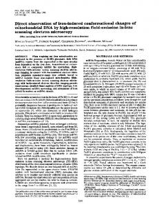
JBC Papers in Press. Published on May 10, 2017 as Manuscript M117.789495 The latest version is at http://www.jbc.org/cgi/doi/10.1074/jbc.M117.789495 Electron microscopy of UGGT
Single-particle electron microscopy structure of UDP-glucose:glycoprotein glucosyltransferase suggests a selectivity mechanism for misfolded proteins Daniel Calles-Garcia1,a , Meng Yang1,a , Naoto Soya2 , Roberto Melero3 , Marie Ménade1 , Yukishige Ito4 , Javier Vargas3,5 , Gergely L. Lukacs2 , Justin M. Kollman6 , Guennadi Kozlov1 , Kalle Gehring1 From the 1 Department of Biochemistry, McGill University, Montreal, Quebec, Canada, 2 Department of Physiology, McGill University, Montreal, Quebec, Canada, 3 Biocomputing Unit, Centro Nacional de Biotectnologíay, Madrid, Spain, 4 Synthetic Cellular Chemistry Laboratory, RIKEN, Wako, Saitama, Japan, 5 Bioengineering Lab, Escuela Politécnica Superior, Universidad San Pablo CEU, Madrid, Spain, and 6 Department of Biochemistry, University of Washington, Seattle, Washington, USA a These authors contributed equally. Running title: Electron microscopy of UGGT
Keywords: glycoprotein biosynthesis, chaperone, glycosyltransferase, ER quality control, electron microscopy, single particle analysis, small-angle X-ray scattering (SAXS) exposed hydrophobic residues and excludes folded proteins with hydrophilic surfaces. In conclusion, we have determined the UGGT structure, which enabled us to develop a plausible functional model of the mechanism for UGGT’s selectivity for misfolded glycoproteins.
ABSTRACT The enzyme UDP-glucose:glycoprotein glucosyltransferase (UGGT) mediates quality control of glycoproteins in the endoplasmic reticulum by attaching glucose to N-linked glycan of misfolded proteins. As a sensor, UGGT ensures that misfolded proteins are recognized by the lectin chaperones and do not leave the secretory pathway. The structure of UGGT and the mechanism of its selectivity for misfolded proteins have been unknown for 25 years. Here, we used negative-stain electron microscopy and small-angle X-ray scattering to determine the structure of UGGT from Drosophila melanogaster at 18 Å resolution. Three-dimensional reconstructions revealed a cage-like structure with a large central cavity. Particle classification revealed flexibility that precluded determination of a high-resolution structure. Introduction of biotinylation sites into a fungal UGGT expressed in E. coli allowed identification of the catalytic and first thioredoxin-like domain. We also used hydrogen-deuterium exchange mass spectrometry to map the binding site of an accessory protein, Sep15, to the first thioredoxin-like domain. The UGGT structural features identified suggest that the central cavity contains the catalytic site and is lined with hydrophobic surfaces. This enhances the binding of misfolded substrates with
The folding of glycoproteins that transit through the endoplasmic reticulum (ER) is reflected in the structure of their N-linked glycan. Newly synthesized proteins are modified with the intact glycan, Glc3 Man9 GlcNAc2 , which is then trimmed by glucosidases I and II to generate a monoglucosylated form. This trimmed form is recognized by the ER-specific lectin chaperones, calnexin and calreticulin, which recruit additional chaperones to fold the nascent protein (1–5). Upon release, the terminal glucose is cleaved by glucosidase II, producing a shortened glycan, Man9 GlcNAc2 , which can no longer bind the lectin chaperones. Incompletely folded or misfolded proteins after the first cycle of folding are recognized by UDPglucose:glycoprotein glucosyltransferase (UGGT), which adds back a glucose residue to regenerate the monoglucosylated form for additional rounds of lectin chaperone assisted refolding (6, 7). By glu1
Copyright 2017 by The American Society for Biochemistry and Molecular Biology, Inc.
Downloaded from http://www.jbc.org/ by guest on October 24, 2017
To whom correspondence should be addressed: Kalle Gehring, 1-514-398-7287, e-mail:
[email protected].
Electron microscopy of UGGT
RESULTS SAXS analysis–We chose to study UGGT from two sources. Drosophila melanogaster UGGT (DmUGGT) had been previously characterized biochemically and is easily purified from Sf9 insect cells due to incorporation of an N-terminal melittin signal peptide that secretes the protein into the culture media (2, 32, 33). We also studied a fungal UGGT cloned from Penicillium chrysogenum (PcUGGT), which could be expressed in E. coli and purified in high yield (Supplemental Figure S1). The two proteins are 44% and 34% identical to human UGGT, with the strongest similarity in the C-terminal catalytic domain. To characterize the proteins, we turned to SAXS and EM, two structural techniques which have been used successfully to study proteins that are difficult to crystallize. SAXS analysis of DmUGGT and PcUGGT confirmed the proteins were monomeric with no aggregation detected in Guinier plots (Figure 1A). Kratky plots of the scattering data showed an elongated bell curve that returned to zero, which is characteristic of a folded protein (Supplemental Figure S2). The radius of gyration for Dm- and PcUGGT, calculated from the slope of Guinier plots at low angle, was estimated to 4.9 nm and independent of protein concentration. The distance distribution curves for both species showed similar bellshaped curves consistent with Dmax values in the range of 100 Å to 140 Å (Figure 1B). We used the SAXS data to generate ab initio 2
Downloaded from http://www.jbc.org/ by guest on October 24, 2017
UGGT from Drosophila melanogaster and Penicillium chrysogenum. Solution analysis by small-angle X-ray and light scattering confirm that DmUGGT is monomeric and forms a complex with Sep15 in a one-to-one ratio. We identify the Sep15 binding site on UGGT using hydrogen-deuterium exchange with mass spectrometry (HDX-MS) and mutagenesis. Small-angle X-ray scattering (SAXS) and single-particle electron microscopy (EM) reveal UGGT adopts an open cage-shape with a large central cavity. We identify the domains in an EM model of the Penicillium chrysogenum protein (PcUGGT) using biotinylation sites decorated with monovalent streptavidin. The three dimensional model of UGGT allows us to propose a mechanism for substrate recognition and selection.
cosylating misfolded proteins, UGGT plays an important role in preventing misfolded proteins from exiting the ER. UGGT is conserved and essential (8, 9). It is a large monomeric protein of more than 1500 residues and found in almost all eukaryotes. The activity of UGGT has been probed using native and misfolded glycoproteins (7, 10–13), glycopeptides (14–16) and small synthetic substrates (17–22). These studies have shown a strong selectivity for glucosylation of misfolded over folded substrates. Only glycans in misfolded proteins are modified. UGGT activity requires that misfolded protein and the acceptor glycan Man9 GlcNAc2 are on the same molecule (23, 24). UGGT is known to tightly bind another ER protein, Sep15, a 15kDa selenoprotein that likely contributes to the reduction of non-native disulfide bonds (25– 27). In addition to its role as a sensor of protein folding, UGGT contributes to the maturation of the major histocompatibility complex of class 1 molecules (MHC1), ensuring that a high-affinity peptide has been loaded onto the MHC1 heavy chains (28, 29). The MHC1 is composed of MHCencoded heavy chains, which are glycosylated, and a β2 -microglobulin domain. The assembly of the complete MHC1 involves many components of the ER folding pathways. UGGT scans the peptideMHC1 interaction through glucosylating the HC and directs the MHC1 for reassociation with the calreticulin/ERp57/tapasin complex for a new peptide to be loaded onto the HC binding groove. UGGT ensures that only high-affinity peptides are loaded onto the MHC1. A mechanistic understanding of UGGT activity has been limited as the protein has proven to be a difficult crystallization target. Sequence analysis and biochemical data have suggested that it has five domains. The N-terminal portion consists of three thioredoxin-like domains followed by a β rich domain and the catalytic glucosyltransferase domain. The catalytic domain is the most strongly conserved across species (9, 30). The non-catalytic domains are less conserved with 20-40% sequence identity among species. The only crystal structure is of the third thioredoxin-like domain from Chaetomium thermophilum (31). In this report, we characterize the structure of
Electron microscopy of UGGT
3
Downloaded from http://www.jbc.org/ by guest on October 24, 2017
connected to an arm-like lobe (Figure 2B and 2D). As an additional check, we calculated the theoretical X-ray scattering curve from the Scipion DmUGGT EM model and compared it to the experimental data (Supplemental Figure S6). A good fit was observed at small angles (q