Am J Physiol Gastrointest Liver Physiol 282: G349–G358, 2002. First published October 24, 2001; 10.1152/ajpgi.00226.2001.
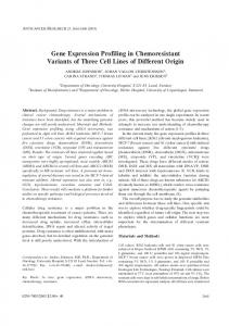
Site-specific gene expression of nNOS variants in distinct functional regions of rat gastrointestinal tract DIETER SAUR,1 WINFRIED L. NEUHUBER,2 BERND GENGENBACH,1 ANDREA HUBER,1 VOLKER SCHUSDZIARRA,1 AND HANS-DIETER ALLESCHER1 1 Department of Internal Medicine II, Technical University of Munich, 81675 Munich, and 2Department of Anatomy I, University of Erlangen, 91054 Erlangen, Germany Received 31 May 2001; accepted in final form 1 November 2001
Saur, Dieter, Winfried L. Neuhuber, Bernd Gengenbach, Andrea Huber, Volker Schusdziarra, and Hans-Dieter Allescher. Site-specific gene expression of nNOS variants in distinct functional regions of rat gastrointestinal tract. Am J Physiol Gastrointest Liver Physiol 282: G349–G358, 2002. First published October 24, 2001; 10.1152/ajpgi.00226.2001.—5⬘ mRNA variants of neuronal nitric oxide synthase (nNOS) are generated either by alternative promoter usage resulting in different mRNAs that encode for the same protein (nNOS␣) or alternative splicing encoding NH2-terminally truncated proteins (nNOS/␥) that lack the PDZ/GLGF domain for protein-protein interaction of nNOS␣. We studied the expression of 5⬘ nNOS mRNA forms and nNOS-interacting proteins (postsynaptic density protein-95; PSD-95) in the rat gastrointestinal tract and analyzed the more distinct localization of nNOS protein variants in the duodenum by immunohistochemistry with COOH- and NH2-terminal nNOS antibodies. 5⬘ nNOS mRNA variants showed a site-specific expression along the gastrointestinal tract with presence of all forms (nNOS␣-a, -b, -c; nNOS) in the muscle layer of esophagus, stomach, duodenum, longitudinal muscle layer of jejunum/ileum, proximal colon, and rectum. In contrast, a lack of nNOS␣-a and nNOS mRNA was observed in pylorus, circular muscle layer of jejunum/ ileum, and cecum. Expression of nNOS␣ and nNOS cDNAs revealed proteins of ⬃155 kDa and 135/125 kDa, respectively. Immunohistochemistry showed a differential distribution of COOH- and NH2-terminal nNOS immunoreactivity in distinct layers of rat duodenum, suggesting a cell-specific expression and distinct compartmentalization of nNOS proteins. Observed distribution of 5⬘ nNOS mRNA variants and proteins argue for a complex control of nNOS expression by usage of separate promoters, cell- and site-specific splicing mechanisms, and translational initiation. These mechanisms could be involved in gastrointestinal motor diseases and may explain the phenotype of nNOS␣ knockout mice with gastric stasis and pyloric stenosis, due to a total loss of nNOS in the pyloric sphincter region. alternative promoters; alternative splicing; transcriptional and posttranscriptional control; postsynaptic density; pyloric sphincter.
NITRIC OXIDE (NO) is an important nonadrenergic, noncholinergic mediator within the enteric nervous system (4). It is generated by enzymatic NADPH-dependent
Address for reprint requests and other correspondence: D. Saur, Dept. of Internal Medicine II, Technical Univ. of Munich, Ismaningerstr. 22, 81675 Munich, Germany (E-mail:
[email protected]). http://www.ajpgi.org
electron transfer during the conversion of L-citrulline to L-arginine by NO synthase (NOS). In addition to its action as neurotransmitter and neuromodulator (1, 4, 14, 25, 27), NO might also act as a messenger within smooth muscle or interstitial cells of Cajal (19, 24). Neuronal NOS (nNOS) (2) is the predominant isoform of NOS in the enteric nervous system (5, 12, 20, 38) besides the other constitutive and calcium-dependent endothelial NOS and the calcium-independent inducible NOS (iNOS). It has been demonstrated recently in various tissues, such as the central nervous system, skeletal muscle, testis, kidney, spleen, adrenal gland, heart, embryonic tissue, and in the rat and human gastrointestinal tract that different 5⬘ mRNA variants of nNOS are expressed (3, 9, 12, 15, 21, 26, 32, 34, 37). These variants differ in the first untranslated exon and in exon deletions or insertions within the translated region and thus in the protein structure. Differences in first exons of the 5⬘ untranslated region (UTR) are due to alternative promoter usage (26, 37), whereas exon deletions/insertions are generated by alternative splicing (9, 12, 26, 34). In rat small intestine, three different 5⬘ mRNA splice variants of nNOS have been described (12). Two (nNOS␣-a, nNOS␣-b) differ in their first untranslated exon (exon 1a and exon 1b) but encode for the same protein nNOS␣. A third variant (exon 1a/exon 3) lacks exon 2 and encodes for nNOS. Lee et al. (15) found another first exon called exon 1c in different rat tissues and demonstrated a tissue and development-specific expression of nNOS exon 1a, 1b, and 1c. In addition, one kidney-specific first exon (K1) and two alternatively spliced kidney-specific second exons (K2a and K2b) have been described (21). nNOS mutant mice, generated by targeted disruption of the nNOS gene by homologous recombination, led to a gastrointestinal phenotype closely resembling, but not identical to, hypertrophic pyloric stenosis with delayed gastric emptying of solids and fluids (10, 18). Interestingly, no other gastrointestinal abnormalities could be observed in these animals. However, this The costs of publication of this article were defrayed in part by the payment of page charges. The article must therefore be hereby marked ‘‘advertisement’’ in accordance with 18 U.S.C. Section 1734 solely to indicate this fact.
0193-1857/02 $5.00 Copyright © 2002 the American Physiological Society
G349
G350
DISTRIBUTION OF NNOS SPLICE VARIANTS
genetic model in which exon 2 and, subsequently, fulllength nNOS␣, was disrupted maintained nNOS expression due to alternative splicing, resulting in the NH2-terminally truncated proteins nNOS (135 kDa) with ⬃80% catalytic activity of nNOS␣ and nNOS␥ (125 kDa) lacking functional nNOS enzymatic activity (3). Full-length nNOS␣ but not the truncated forms (nNOS and nNOS␥) contain an NH2-terminal PDZ/ GLGF motif encoded by exon 2, enabling protein-protein interactions with various proteins such as ␣-syntrophin or proteins of the postsynaptic density (PSD) like PSD-95 or PSD-93 (3, 28). PSD-95 and PSD-93 can anchor nNOS␣ to N-methyl-D-aspartate (NMDA) receptor subunits and subsequently to the cell membrane (6). Because differences in the NH2-terminal protein structure affect functionally important parts and determines the cellular localization of nNOS, different functional roles can be attributed to membranebound nNOS␣, cytosolic nNOS, and the inactive form nNOS␥. Therefore, it is of physiological and functional interest to determine the site and organ-specific expression of nNOS variants and the presence of putative interaction partners. MATERIALS AND METHODS
Tissue preparation. Adult male Wistar rats with an individual body weight of ⬃200 g were killed by cervical dislocation. The entire gastrointestinal tract was removed and divided into the following areas: upper and lower esophagus; gastric fundus/corpus and antrum; pylorus; small intestine; proximal, medial, and distal colon; and rectum. Each area of gut was cleaned in ice-cold PBS, attached mesenterial fat was removed, and the muscle layers including the attached myenteric plexus were separated from the mucosa by scraping. In small intestine, the longitudinal muscle layer with attached myentric plexus (LMMP) was additionally peeled away from the circular muscle layer (CM). Tissues were cut into small pieces, immediately frozen in liquid nitrogen, and stored at ⫺80°C until use. RNA isolation. Total RNA was extracted from muscle layer preparations of liquid nitrogen-frozen rat gastrointestinal tissues. Specimens were homogenized with a Polytron homogenizer (Kinematica), and RNA was isolated using the guanidine isothiocynate/phenol/chloroform extraction method (Peq Lab, Schwalbach, Germany), followed by DNase I treatment (1 U/g RNA, 15 min, 25°C; GIBCO BRL, Eggenstein, Germany). RT-PCR and Southern blot analysis. RT-PCR and Southern blot analysis were performed as described previously (12). Five micrograms of total RNA were reverse transcribed at 42°C for 1 h into complementary DNA using 200 units SuperScript II RNase H⫺ reverse transcriptase (GIBCO BRL) in the presence of 100-ng random hexamer primers (Roche, Mannheim, Germany) or 25 pmol of the gene-specific antisense primer nNOS P1/ex5-AS (for all primers see Table 1) complementary to exon 5 of rat nNOS (2). As negative controls, isolated RNA amplified without reverse transcriptase or random hexamer primers were used. Site-specific expression of nNOS mRNA splice variants and PSD-95 mRNA were investigated by PCR using isoform-specific sense primers for rat nNOS exon 1a, exon 1b, and exon 1c combined with a common antisense strand primer for nNOS exon 5 (P2/ex5-AS) or sense and antisense gene-specific primers for PSD-95. One microliter of the RT-reaction mix was subjected AJP-Gastrointest Liver Physiol • VOL
Table 1. Oligonucleotides of sense- and antisensestrand primers and hybridization probes Sequence Name
5⬘
nNOS exon 1a (S) nNOS exon 1b (S) nNOS exon 1c (S) nNOS P1/ex5-AS (AS) nNOS P2/ex5-AS (AS) nNOS ex6-AS (AS) nNOS exon 3 (HP) PSD-95 (S) PSD-95 (AS) PSD-95 (HP)
3⬘
GGTTGTGCGAAGGGAAGTT GAGGGGCGACACTACCAT CACCACAGCCTCTGGAATGAAAGA GGATGAGTAGTATTGGTCGAGA TCTGGCTTCCGCGTGTGC CACACATCGAGAGGCGTT AGACCTCGATGGCAAATCG CCAAGAAATACCGCTACCAAG CACCTCCTTCGATGATCTTC AACACGGACACCCTAGAAGC
S, sense; AS, antisense; HP, hybridization probes; nNOS, neuronal nitric oxide synthase; PSD, postsynaptic density protein.
to 35 or 30 PCR cycles in a Biometra UNO I thermal cycler using 2.5 units of Taq polymerase (Sigma, Deisenhofen, Germany) with the following conditions. After a “hot start” with an initial denaturation at 95°C for 3 min, each PCR cycle involved denaturation at 94°C for 30 s, annealing at 58°C for 45 s, and extension at 72°C for 90 s. The last cycle was followed by an extension step at 72°C for 7 min. If the first round of PCR yielded no visible PCR product in the ethidium bromide-stained gel, a second round of nested PCR was performed for 20 cycles with annealing at 58°C. PCR products were size fractionated by 1.5% agarose gel electrophoresis, visualized by ethidium bromide staining, denaturated, and blotted onto Hybond N⫹ nylon membranes (AmershamPharmacia, Freiburg, Germany) by capillary transfer. Blots were hybridized overnight at 46°C with [␥-33P] ATP 5⬘ endlabeled internal probes specific for nNOS exon 3 or PSD-95 (see Table 1 for all hybridization probes). Membranes were washed twice with 2⫻ standard saline citrate (SSC) containing 0.1% SDS for 15 min at room temperature and once with 0.5⫻ SSC containing 0.1% SDS for 10 min at 44°C. Labeled products were detected by autoradiography. Finally, 20-l aliquots of the PCR products were size fractionated on a 1.5% agarose gel, excised, and purified, using a gel extraction kit (Qiagen, Hilden, Germany) and cloned into the TOPO PCRII plasmid (Invitrogen, Groningen, Netherlands) as described by the manufacturer. Nucleotide sequences were deduced by cycle sequencing of the purified plasmids (Qiagen Mini Prep Kit) with T7 sequencing primer (GATC, Konstanz, Germany). Sequences were analyzed by BLASTn homology search. Heterologous eucaryotic expression of nNOS␣ and nNOS cDNA in COS-7 cells. Eucaryotic expression vectors containing either full-length nNOS␣ or nNOS cDNAs were constructed as follows. For nNOS␣, a 5,057-bp fragment of rat nNOS cDNA, kindly provided by Dr. S. Snyder, Johns Hopkins Medical School, Baltimore, MD (2) that contained 348 bp of the 5⬘ UTR was subcloned into the mammalian expression vector pcDNA3 (Invitrogen) resulting in nNOS␣-pcDNA3. For nNOS, the 1,558-bp Xho I/Nar I 5⬘ end fragment of nNOS␣-pcDNA3 was replaced with a 792 bp Xho I/Nar I fragment representing the nNOS exon 1a/exon 3 sequence. This fragment was constructed by RT-PCR, using random hexamer-primed total RNA from LMMP of rat ileum as template. Thirty PCR cycles with annealing at 56°C for 45 s, extension at 72°C for 90 s, and denaturation at 94°C for 30 s were performed using a proofreading polymerase (Pwo, Roche) and the sense and antisense strand primers nNOS exon 1a and nNOS ex6-AS, respectively (see Table 1). The PCR product was subsequently cloned into PCRII plasmid, 282 • FEBRUARY 2002 •
www.ajpgi.org
DISTRIBUTION OF NNOS SPLICE VARIANTS
sequenced with T7 and M13 reverse primers (GATC) and subcloned in to the Xho I/Nar I site of nNOS␣-pcDNA3, resulting in nNOS-pcDNA3. COS-7 cells were grown in MEM medium supplemented with 10% fetal bovine serum, glutamate, and penicillin/streptomycin (GIBCO-BRL) and transfected with 2 g of nNOS␣pcDNA3, nNOS-pcDNA3, or an empty pcDNA3 vector by lipid-mediated transfer, using 16 l Enhancer and 10 l Effectene transfection reagent (Qiagen) as described previously (26). Protein expression and Western blot analysis. Liquid nitrogen frozen specimens from the mucosa and the longitudinal and circular muscle layer (LM/CM) with attached nerve plexus of rat duodenum were homogenized, and proteins were extracted as described before (12, 26). COS-7 cells were lysed 48 h after transfection using 1⫻ lysis buffer (50 mM Tris, pH 7.6, 0.1 mM EDTA, 0.1 mM EGTA, 3 M leupeptin, 1 M pepstatin, 1 mM phenylmethylsulfonyl fluoride, 12 mM -mercaptoethanol, 1% Triton X-100) and centrifuged at 21,000 g for 20 min. The total protein concentration was measured with protein assay kit II (Bio-Rad, Munich, Germany). Proteins were separated by 7.5% SDS-PAGE and transferred to polyvinylidene difluoride membranes (BioRad). Blots were probed with anti-rat nNOS antibodies [nNOS COOH-terminal monoclonal antibody (nNOS-C), amino acids 1095–1289 of nNOS␣, Transduction Laboratories, Heidelberg, Germany; nNOS NH2-terminal polyclonal antibody (nNOS-N), amino acids 38–57 of nNOS␣ NOS1 (K-20), Santa Cruz Biotechnologies, Heidelberg, Germany] as described previously (12, 26). Signal detection of the immunoreactive bands was facilitated by enhanced chemiluminescence (Amersham-Pharmacia). Immunohistochemistry. Detection of nNOS immunoreactivity in rat duodenum was performed as described previously (11, 23). In brief, rats were perfusion fixed with Zamboni’s solution and segments of the duodenum were excised. After cryoprotection in 20% phosphate-buffered sucrose, 12m-thick cryostat sections were mounted on poly-L-lysinecoated slides and air dried (1 h). Following a 5-min rinse in Tris-buffered saline (TBS), sections were incubated in TBS containing 1% BSA, 5% normal goat serum, and 0.5% Triton X-100. All antibodies were diluted in TBS containing 1% BSA and 0.5% Triton X-100. Immunological detection of nNOS was performed by incubating sections overnight with the COOH-terminal antibody nNOS-C and the NH2-terminal antibody nNOS-N (directed against an exon 2 encoded domain of nNOS) in a 1:100 dilution. Binding of the antibodies was visualized with goat anti-mouse IgG (nNOS-C) tagged with Alexa 488 (MoBiTec; Molecular Probes, Go¨ ttingen, Germany) or goat anti-rabbit IgG (nNOS-N) tagged with indocarbocyanin (Cy3; Dianova, Hamburg, Germany) diluted 1:800 and 1:200, respectively. Colocalization of COOH- and NH2-terminal nNOS immunoreactivity was investigated by a sequential double immunostaining protocol as described previously (11, 23). Sections were coincubated sequentially with antibody nNOS-C and nNOS-N. Binding of the antibodies was facilitated using goat anti-mouse IgG antibody tagged with Alexa 488 and goat anti-rabbit IgG antibody labeled with Cy3 in a dilution of 1:800 and 1:200, respectively. Controls included trials with omission of the primary antibodies, replacing it by buffer or normal rabbit serum (23). Preabsorption of the primary antibodies was done with the respective antigens for 3 h at room temperature. For nNOS-N antibody, a blocking peptide (amino acids 38–57 of human nNOS␣) from Santa Cruz Biotechnologies [NOS1 (K20) P] was used with a concentration of 10 g/ml. For nNOS-C antibody, expressed and purified (12) full-length AJP-Gastrointest Liver Physiol • VOL
G351
nNOS␣ proteins were used in a concentration of 5 g/ml. Sections were analyzed by confocal laser scanning microscopy (Bio-Rad MRC 1000 attached to a Nikon Diaphot 300). Fluorochromes were excited with 488 and 568 nm lines, respectively, by a Krypton-Argon laser. Single optical sections were taken with a 20⫻ objective lens (0.75 numerical aperture) and various zoom factors. When controls and “full” incubations were compared, special care was taken to keep the pinhole and gain of the photomultiplier constant. Two channel scans were coded green and red, and merged images were documented using the software package Corel PhotoPaint. RESULTS
Site-specific expression of 5’ splice variants of rat nNOS in the gastrointestinal tract. RT-PCR experiments with equal amounts of total RNA and Southern blot hybridization showed a distinct and site-specific expression of 5⬘ nNOS mRNA variants along the rat gastrointestinal tract, with expression of all four different forms (nNOS␣-a, nNOS␣-b, nNOS␣-c, nNOS; see Fig. 1 for exon structure of 5⬘ nNOS mRNA variants) in the upper and lower esophagus, gastric fundus/corpus/antrum, duodenum, LMMP of small intestine, proximal colon, and rectum (Fig. 2, Table 2). In the esophagus, nNOS␣ mRNA forms predominated and nNOS showed a low abundant expression (Fig. 2), because this form could only be detected after 35 PCR cycles of coamplification with nNOS␣-a, whereas nNOS␣-a, -b, and -c were already present after 30 cycles (data not shown). All nNOS variants were present in gastric fundus/ corpus and antrum (Fig. 2, Table 2). In contrast, in the pyloric sphincter, an almost exclusive expression of nNOS␣-b and nNOS␣-c was detected (Fig. 2, Table 2). Only one of seven preparations revealed a weak signal for nNOS␣-a and only two of seven preparations were positive for nNOS after 35 PCR cycles (Table 2). Because nNOS␣-a and nNOS are expressed in the duodenum and antrum, detection of nNOS␣-a in one and nNOS in two of seven pyloric sphincter preparations was most likely due to contaminating tissue from antrum or duodenum. The negative RT-PCR/Southern blot results for nNOS␣-a in six and nNOS in five of seven independent preparations were confirmed by a second round of nested PCR and independent RT-PCR experiments from the same RNA preparations. As positive control, we used RNA isolated from LMMP of rat small intestine. The small intestine was divided into duodenum and jejunum/ileum, which was further separated in the LMMP and the CM. In duodenum and LMMP, all four variants were found in an almost equal distribution (Fig. 2, Table 2). In contrast, in the CM of jejunum/ ileum nNOS␣-b and nNOS␣-c were the predominant forms, whereas nNOS␣-a and nNOS were not detectable in six of seven preparations, respectively (Fig. 2, Table 2). The positive result for nNOS␣-a and nNOS in one of seven preparations is most likely due to contaminating LMMP tissue. The large bowel was separated into cecum; proximal, middle, and distal colon; and rectum. We could detect 282 • FEBRUARY 2002 •
www.ajpgi.org
G352
DISTRIBUTION OF NNOS SPLICE VARIANTS
Fig. 1. Schematic exon structure of alternative spliced neuronal nitric oxide synthase (nNOS) 5⬘ mRNA transcripts extrapolated from the human gene (9). Top: three alternative transcripts (exon 1a/exon 2, exon 1b/exon 2, and exon 1c/exon 2) driven by separate promoters and spliced to the common exon 2 are shown. Different first exons are located in the 5⬘ untranslated region of nNOS mRNA and therefore encode for nNOS␣. Bottom: Exon structure of the nNOS transcript (exon 1a/exon 3) with skipping of exon 2 is shown. Arrows, positions of sense- and antisense-strand primers used in RT-PCR (Fig. 2). Translational initiation codons (AUG, CUG) are noted by a vertical arrow. Localization of the PDZ/GLGF domain, encoded by exon 2, and exon splice junctions are noted.
nNOS␣-b and nNOS␣-c in all tissue preparations of the cecum, whereas nNOS␣-a and nNOS showed a low abundant expression or were not detectable (positive results for nNOS␣-a and nNOS were only in two of five preparations) (Fig. 2, Table 2). All splice variants
were found in the proximal colon and rectum, whereas the preparations of the distal and middle colon showed varying expression patterns for nNOS␣-a and nNOS. In four and three of five preparations nNOS␣-a and nNOS mRNA were detected, respectively (Fig. 2, Ta-
Fig. 2. Site-specific expression of nNOS 5⬘ mRNA variants in the rat gastrointestinal tract as shown by Southern blot hybridization with an internal rat nNOS exon 3-specific 33P end-labeled oligonucleotide. RNA was isolated from the muscle layers of the indicated rat gastrointestinal tissues, and RT-PCR was performed using nNOS sense-specific primers for exon 1a, 1b, 1c and an antisense strand primer corresponding to the common exon 5. For primer locations see arrows in Fig. 1. A: site-specific expression of nNOS␣-a (exon 1a/exon 2) and nNOS mRNA (exon 1a/exon 3) in the rat gastrointestinal tract, whereas nNOS␣-b (exon 1b/exon 2) was present at all investigated localizations. B: ubiquitous expression of nNOS␣-c mRNA (exon 1c/exon 2) in the rat gastrointestinal tract. CM1 circular muscle layer; LM/MP1 longitudinal muscle/myenteric plexus layer.
AJP-Gastrointest Liver Physiol • VOL
282 • FEBRUARY 2002 •
www.ajpgi.org
G353
DISTRIBUTION OF NNOS SPLICE VARIANTS
Table 2. Distribution of nNOS 5⬘ end splice variants in the rat gastrointestinal tract as shown by southern blot hybridization of RT-PCR products Tissue
n
nNOS␣-a
nNOS␣-b
nNOS␣-c
nNOS
Upper esophagus Lower esophagus Fundus/corpus Antrum Pylorus Duodenum Jejunum/ileum (LMMP) Jejunum/ileum (CM) Cecum Proximal colon Middle colon Distal colon Rectum
5 5 5 5 7 5 5 7 5 5 5 5 5
⫹⫹ ⫹⫹ ⫹⫹ ⫹⫹ (⫺) ⫹⫹ ⫹⫹ (⫺) (⫺) ⫹⫹ (⫹/⫺) (⫹/⫺) ⫹⫹
⫹⫹ ⫹⫹ ⫹⫹ ⫹⫹ ⫹⫹ ⫹⫹ ⫹⫹ ⫹⫹ ⫹⫹ ⫹⫹ ⫹⫹ ⫹⫹ ⫹⫹
⫹⫹ ⫹⫹ ⫹⫹ ⫹⫹ ⫹⫹ ⫹⫹ ⫹⫹ ⫹⫹ ⫹⫹ ⫹⫹ ⫹⫹ ⫹⫹ ⫹⫹
(⫹) (⫹) ⫹⫹ ⫹⫹ (⫺) ⫹⫹ ⫹⫹ (⫺) (⫺) ⫹⫹ (⫹/⫺) (⫹/⫺) ⫹⫹
(⫺), no RT-PCR product obtained in the majority of tissue preparations; (⫹/⫺), varying expression patterns; (⫹), weak signal (only present after 35 PCR cycles); ⫹⫹, positive result in all investigated samples after 30 PCR cycles; LMMP, longitudinal muscle/myenteric plexis layer; CM, circular muscle layer.
ble 2). In contrast, nNOS␣-b and nNOS␣-c mRNAs were present at all investigated locations of the colon (Fig. 2, Table 2). The observed variance of nNOS mRNA expression patterns in the colon of different animals could be due to interindividual differences of nNOS␣-a and nNOS expression. Furthermore, the abundance of nNOS mRNA and protein expression decreases from the proximal to the distal colon (30). Expression of PSD-95, a nNOS PDZ/GLGF interacting protein, in the gastrointestinal tract. We investigated the expression of the nNOS␣ interacting PSD-95 along the rat gastrointestinal tract. By using RT-PCR and Southern blot hybridization, we could demonstrate the presence of mRNA for PSD-95 as a possible target for a membrane association of nNOS␣ at all investigated localizations (Fig. 3). Heterologous eucaryotic expression of nNOS␣ and nNOS cDNA. Expression of nNOS␣ and nNOS cDNA in COS-7 cells revealed immunoreactive bands at 155 kDa for nNOS␣ and 135 and 125 kDa for nNOS cDNA with a specific COOH-terminal monoclonal antibody (nNOS-C). A specific NH2-terminal antibody (nNOS-N) directed against an exon 2 encoded domain of nNOS revealed just a single band at 155 kDa for nNOS␣ cDNA, whereas no bands were detected for
nNOS cDNA lacking exon 2 (Fig. 4). Thus the two antibodies distinguish between full-length nNOS␣ (155 kDa; reactive with nNOS-C and nNOS-N) and the NH2-terminal truncated variants nNOS (135 kDa) and nNOS␥ (125 kDa) (both nNOS-C positive and both nNOS-N negative). nNOS and nNOS␥ proteins are generated by translation of the same cDNA (nNOScDNA) using two different start codons in exon 1a (CUG) and exon 5 (AUG) (see Fig. 1). Atypical translation initiation codons have been described in various genes, including mouse nNOS mRNA, where a CUG start codon within exon 1a similar to rat nNOS is used (3). This start codon and the surrounding sequence is highly conserved between mouse and rat with a homology of 100% between nt 395 and 431 of mouse (European Molecular Biology Laboratories accession number U50718) and nt 406 to 442 of rat nNOS exon 1a (European Molecular Biology Laboratories accession no. AF008912). Localization of COOH- and NH2-terminal nNOS immunoreactivity in the rat duodenum. To investigate a possible differential distribution of the different NH2terminal nNOS proteins (nNOS␣//␥) in the rat gastrointestinal tract, we used confocal laser scanning microscopy and double staining with nNOS antibodies
Fig. 3. Expression of the nNOS␣-PDZ/ GLGF interacting postsynaptic density protein-95 (PSD-95) in the rat gastrointestinal tract. Southern blot hybridization was performed with an internal rat PSD-95-specific 33P end-labeled oligonucleotide. RNA was isolated from the muscle layers of the indicated rat gastrointestinal tissues, and RT-PCR was performed using specific primers for PSD95.
AJP-Gastrointest Liver Physiol • VOL
282 • FEBRUARY 2002 •
www.ajpgi.org
G354
DISTRIBUTION OF NNOS SPLICE VARIANTS
Fig. 4. Heterologous eucaryotic expression of nNOS␣, nNOS, and nNOS␥. Eucaryotic expression vectors containing nNOS␣ or nNOS cDNAs were transiently transfected in to COS-7 cells. After 48 h, cells were harvested and cellular proteins were analyzed by Western blotting. Equal amounts of protein (10 g) were loaded on each lane and detected with a monoclonal antibody specific for the COOHterminal domain of nNOS (nNOS-C; left box) or an antibody specific for the exon 2 encoded NH2-terminal domain of nNOS (nNOS-N; right box). The specificity of the nNOS antibodies was verified at different dilutions and by probing with inducible NOS (iNOS) and endothelial NOS (eNOS) positive controls. Protein samples from human and rat brain or HeLa cells were used as positive or negative controls for nNOS, respectively (data not shown).
the lamina propria (Fig. 5, B and C). Therefore, the staining of these cells has to be regarded as nonspecific binding of the secondary antibody. In addition to the nonspecific epithelial reaction with nNOS-N, there was clear specific staining of submucosal nerve cell bodies and nerve fibers running within the lamina propria around crypts and in the intestinal villi with nNOS-N antibody (red). In contrast, there was no positive staining with nNOS-C antibody (green) either in submucosal neurons or in nerve fibers running to the mucosa (Fig. 5, B and C). These results indicate a differential distribution of COOH- and NH2-terminal nNOS immunoreactivity in different cell compartments, nerve fibers, and layers of the duodenum. In addition, the exclusive staining of submucosal neurons and mucosal nerve fibers with nNOS-N suggests the existence of an additional COOH-terminally truncated or extended nNOS variant. Western blot analysis of rat duodenum. NH2-terminal nNOS immunoreactivity of rat duodenum, which may represent novel COOH-terminal extended or deleted nNOS variants, was further characterized by Western blot analysis of tissue homogenates from the mucosa and the LM/CM of rat duodenum. The assay using the NH2-terminal nNOS antibody nNOS-N revealed a single band with a molecular weight of ⬃85 kDa in the mucosa and three bands with molecular weights of ⬃155, ⬃85, and ⬃30 kDa in LM/CM (Fig. 6), suggesting the possible presence of nNOS protein variants with differing COOH-terminal ends. DISCUSSION
directed against the COOH-terminal end (nNOS-C detecting nNOS␣//␥; see Fig. 4) and the NH2-terminal end (nNOS-N detecting only nNOS␣). Because it is almost impossible to characterize the whole rat gastrointestinal tract by histochemistry, we investigated rat duodenum, due to the presence of all nNOS mRNA variants in this region (see Table 2). When colocalization experiments were carried out, nNOS-C and nNOS-N antibodies resulted in a good colocalization (yellow color) in nerve cell bodies in the myenteric plexus and in some nerve fibers running within the myenteric plexus and projecting to the LM, CM, and deep muscular plexus (Fig. 5, A and B). In addition there were some cell compartments and nerve fibers in the myenteric plexus, LM, and CM that showed reactivity with either one of the antibodies, resulting in a green (nNOS-C), or red (nNOS-N) staining (Fig. 5, A and B). Neuronal staining of both antibodies could be specifically blocked with preabsorption using the respective immunogens as described in MATERIALS AND METHODS. Staining of some epithelial cells could not be abolished by preabsorption of the primary COOH-terminal antibody nNOS-C and was therefore considered to be nonspecific (Fig. 5C). Preabsorption and omission of the primary NH2-terminal antibody nNOS-N left positive some mononuclear cells, presumably macrophages, in AJP-Gastrointest Liver Physiol • VOL
We could demonstrate a distinct and site-specific expression of four different 5⬘ nNOS splice variants along the rat gastrointestinal tract. Three variants differ in their untranslated first exon (exon 1a, exon 1b, and exon 1c) resulting in nNOS␣-a, nNOS␣-b, and nNOS␣-c mRNA (Fig. 1). We have previously described exon 1a and exon 1b expression in the LMMP of rat small intestine (12), whereas the third variant (exon 1c) was found by Lee et al. (15) in rat kidney, skeletal muscle, and embryonic tissue but not in the intestine, using RNase protection assays. However, we identified exon 1c in all investigated nerve-muscle layer preparations of the rat gastrointestinal tract, indicating that nNOS␣-c mRNA expression was below the detection limit of the RNase protection assay when using RNA isolated from whole intestinal tissue preparations including the mucosa (15). Expression of nNOS␣-a has been described in various rat tissues, such as brain, kidney, skeletal muscle, intestine, embryonic tissue, adrenal gland, and heart (12, 15, 21). Here we demonstrate a differential distribution of nNOS␣-a mRNA in the rat gastrointestinal tract with expression in the proximal and distal esophagus, gastric fundus/corpus/ antrum, duodenum, LMMP of small intestine, proximal colon, and rectum, and an altered or lacking expression in the CM of jejunum/ileum, the pyloric sphincter, and cecum, whereas in the middle and distal 282 • FEBRUARY 2002 •
www.ajpgi.org
DISTRIBUTION OF NNOS SPLICE VARIANTS
colon, expression patterns varied. In contrast, nNOS␣-b and nNOS␣-c mRNAs were present at all investigated localizations. Untranslated first exons 1a, 1b, and 1c are spliced to a common second exon containing the AUG starter methionine for initiation of translation (Fig. 1). Because translation of these nNOS mRNA variants results in identical full-length nNOS nNOS␣ proteins, the question about their physiological significance arises. There are several reasons for alternative first exon utilization. Described variants are generated most likely by usage of separate promoters as demonstrated for the human nNOS gene (26, 37). Thus nNOS gene expression can be regulated by activation or suppression of alternative promoters in a cell- or site-
G355
Fig. 6. Western blot analysis of proteins from the mucosa and the LM/CM with attached nerve plexus of rat duodenum. Equal amounts of protein (60 g) were loaded on each lane. Proteins were detected with a polyclonal antibody (nNOS-N) specific for the exon 2 encoded NH2-terminal domain of nNOS.
specific way, as shown in the present study, for the rat gastrointestinal tract by the characterization of a regional differential distribution of alternative first exon variants. Such a differential transcriptional control of separate promoters has been shown for the human nNOS gene for the transcription factor Oct-2 (7) and a tissue and developmental-specific expression of rat nNOS variants has been described by Lee et al. (15) and Oberba¨ umler et al. (21). In the gastrointestinal tract, the abundance of nNOS mRNA and protein expression decreases from the proximal to the distal colon (30), during development in the submucous plexus of the small intestine (39), during aging in the colon (31), and in animal models of diabetic gastropathy (29, 36). In addition, the expression level of nNOS mRNA in the CM is significantly lower compared with LMMP (12) and nNOS mRNA expression is regulated by protein kinase C-dependent pathways (20). These findings demonstrate a tightly regulated nNOS gene expression and are in agreement with our observations Fig. 5. A-C: immunological localization of nNOS in rat duodenum by double staining with nNOS-C and nNOS-N antibodies using confocal laser-scanning microscopy. nNOS-C was detected with an Alexa 488 (green), and nNOS-N was detected with a Cy3 (red) labeled secondary antibody. Colocalization of both antibodies resulted in a yellow color. The neuronal staining of both antibodies could be specifically blocked by preabsorption with expressed nNOS␣ protein. A and B: colocalization of nNOS-C and nNOS-N immunoreactivity could be demonstrated in neurons of the myenteric plexus (MP) and most nerve fibers running within the MP and projecting to the LM and CM layers, as well as the deep muscular plexus (DMP). Some nerve fibers in the MP showed staining with either one of the antibodies and therefore no colocalization (red and green arrows; Fig. 5A). B and C: with nNOS-N (red), there was additional specific staining of submucosal nerve cell bodies and nerve fibers running within the lamina propria (LP) to the mucosa (red arrows). With nNOS-C (green), no positive staining was obtained in submucosal neurons and in nerve fibers running to the mucosa. Nonspecific binding in single epithelial cells (red) was seen with nNOS-N (white arrow; Fig. 5C); mononuclear cells in the submucosa and the villi (green) were false positive with nNOS-C antibody due to an nonspecific labeling of the secondary antibody (white arrowheads; Fig. 5 B and C). SP, submucosal plexus; MM, muscularis mucosae. AJP-Gastrointest Liver Physiol • VOL
282 • FEBRUARY 2002 •
www.ajpgi.org
G356
DISTRIBUTION OF NNOS SPLICE VARIANTS
of a regional distinct distribution of nNOS variants in the rat gastrointestinal tract. In humans, more than nine distinct first exons of nNOS have been described (26, 32, 34, 37), whereas in the rat, only four alternative exon 1 variants are known (12, 15, 21). The structure of the 5⬘ mRNA end of rat nNOS has been extensively studied by several groups in different tissues, like brain, kidney, heart, intestine, and embryo (15), cerebellum, kidney, and skeletal muscle (21), and small intestine (12), using different approaches including 5⬘RACE-PCR. However, due to the more expansive number of 5⬘ mRNA variants of nNOS in man, it remains possible that additional forms are present in the rat. In the human gastrointestinal tract, we identified three alternative first exons of nNOS called exon 15⬘1, 15⬘2 and 15⬘3 [corresponding to exon 1g, 1f, and 1c of a recent nNOS nomenclature (34), respectively] by 5⬘RACE-PCR as the predominant forms (26). This is in accordance with the presence of three alternative first exons in the rat gastrointestinal tract called exon 1a, 1b, and 1c. The sequence of rat exon 1a is homologous to human exon 15⬘2 (exon 1f), rat exon 1b matches human exon 15⬘3 (exon 1c), and rat exon 1c shows sequence similarities to human exon 1b, whereas no homologue for human exon 15⬘1 (exon 1g) has been found in rats. Therefore additional 5⬘ variants of rat nNOS mRNA may be present, and further studies in each of the different functional regions of the rat gastrointestinal tract using 5⬘ RACE-PCR have to be done to clarify this issue. In addition to the variability in the 5⬘ UTR, posttranscriptional control of nNOS gene expression by cis acting elements and trans-acting splicing factors can generate NH2-terminally truncated nNOS proteins (3, 12, 26, 34). Cassette exon deletion by splicing of exon 1a to exon 3 results in the formation of nNOS mRNA, with a loss of the genuine translational initiation site located at exon 2 (Fig. 1). As an alternative, a noncanonical initiation region within exon 1a (CUG) 20 bp upstream of the exon 1a/exon 3 splice junction (Fig. 1) that is homologous to the mouse nNOS CUG translation start site (3) could be used, resulting in an NH2terminally truncated 135-kDa nNOS protein (lacking amino acids 1–236 of full-length nNOS␣), similar to nNOS of nNOS␣ knockout mice (3). By heterologous eucaryotic expression of cloned nNOS cDNA, isolated from rat small intestine, we could demonstrate that a 135-kDa nNOS immunoreactive protein can be detected with a COOH-terminal, but not with an NH2terminal, nNOS antibody, directed against an exon 2 encoded domain. In addition, a second immunoreactive band at 125 kDa was obtained after incubation with the COOH-terminal antibody but not with the NH2terminal antibody. This protein has an identical molecular weight with nNOS␥ of nNOS␣ knockout mice (3) and is most likely generated by an internal consensus translational initiation codon (AUG) within exon 5 of rat nNOS (Fig. 1). In contrast to nNOS with a catalytic activity of ⬃80% of nNOS␣, recombinant nNOS␥ of nNOS␣ knockout mice has been shown to lack functional nNOS catalytic activity (3). Thus AJP-Gastrointest Liver Physiol • VOL
nNOS␥ may function as a dominant-negative nNOS variant that could regulate the catalytic activity of nNOS␣ and nNOS by interisoformal dimerization (22, 35). Our results demonstrate that both the nNOS and nNOS␥ protein can be generated by translation of nNOS mRNA, and therefore posttranscriptional and translational mechanisms can regulate the expression of fully active soluble nNOS or the potential inhibitor nNOS␥ from the same mRNA. nNOS mRNA forms are expressed in a site-specific way in the rat gastrointestinal tract, because we could demonstrate that the CM of small intestine and the pyloric sphincter region lack nNOS mRNA. Just two of seven pyloric preparations showed low levels of nNOS, which are most likely due to a contamination with tissue from the duodenum or antrum. In the other five preparations, nNOS was also undetectable after a second round of nested PCR. Thus nNOS␣ seems to be the predominant mRNA form in enteric nerves of the CM and specialized sphincter regions. This could be due to different functional roles of nNOS␣, nNOS, and nNOS␥, which may result from different subcellular localizations (3, 12). Full-length nNOS␣ contains a NH2-terminal PDZ/ GLGF-domain, a motif of ⬃100 amino acids, which can mediate an association to other PDZ-containing proteins (3, 28), such as PSD-95 or PSD-93. PDZ-PDZ interactions enable the targeting of nNOS␣ to the PSD (3) and therefore determine its subcellular localization and function. As an example, PSD-95 can anchor nNOS␣ to the 2B subunit of the NMDA receptor (6) or K⫹ channel subtypes at synaptic sites (13). We could demonstrate that mRNA of PSD-95 as well as nNOS␣ is present at all investigated regions, enabling a subcellular targeting of nNOS␣ to the PSD in the rat gastrointestinal tract. NH2-terminally truncated proteins nNOS and nNOS␥ lack the PDZ/GLGF motif for protein-protein interaction and therefore a possible membrane association (3). Thus Ca2⫹-dependent enzymatic activity of nNOS␣ can be regulated by activation or inactivation of receptors (e.g., the NMDA receptor) that increase or decrease intracellular Ca2⫹ concentrations (6). In turn, NO generated by nNOS␣ can also regulate the function of these receptors (17), and therefore NO may be able to determine the enzymatic activity of NOS. These regulatory mechanisms located at the postsynaptic density could play an important role in relaxation of the pyloric sphincter and circular smooth muscles. They express nNOS␣ but lack nNOS, indicating a possible signaling pathway via activation of NMDA receptors or other proteins and ion channels of the PSD. Therefore, membrane-associated nNOS␣ seems to be responsible for pyloric sphincter relaxation (10, 18), and lack of nNOS␣ in nNOS␣ knockout mice results in gastric stasis with delayed gastric emptying of solids and liquids (10, 18). Furthermore, diabetic rats and mice with defects in gastric emptying and pyloric nonadrenergic, noncholinergic relaxation, show a profound reduction in nNOS protein and mRNA levels before neural degeneration in the pyloric sphincter (29, 36) but not in 282 • FEBRUARY 2002 •
www.ajpgi.org
DISTRIBUTION OF NNOS SPLICE VARIANTS
the central nervous system (36). Interestingly, nNOS expression and nonadrenergic, noncholinergic relaxation are restored to normal levels in the pyloric sphincter of diabetic mice by insulin treatment, indicating that transcriptional and posttranscriptional mechanisms of nNOS gene expression are involved in diabetic gastroparesis (36). Recently, glucoresponsive neurons have been identified within the enteric nervous system (16). Therefore, glucose or insulin-responsive signaling pathways may regulate nNOS gene expression specifically in the gastrointestinal tract by transcriptional control of distinct alternative nNOS promoters. To obtain morphological evidence for a differential distribution of the different nNOS proteins, we used immunohistochemistry with confocal laser scanning microscopy. Because no specific antibodies for the NH2terminally truncated nNOS variants are available, we used an antibody directed against the NH2 terminus (detecting nNOS␣) and the COOH terminus (detecting nNOS␣, -, and -␥) of nNOS. In parts of the intestine (duodenum) where all known nNOS mRNA variants are present, we could demonstrate morphological evidence for a differential localization of COOH- and NH2terminal nNOS immunoreactivity by doublestaining analysis using the COOH- and NH2-terminal nNOS antibodies. There was a good but not identical colocalization of COOH- and NH2-terminal nNOS immunoreactivity in the myenteric plexus and the nerve fibers running to the LM and CM, suggesting that the majority of nNOS protein expressed at these localizations is nNOS␣. In addition, there seems to be some subcellular areas, especially in nerve fibers, where no colocalization can be detected, suggesting that the different nNOS proteins could be localized in different cell compartments. Interestingly, there was additional staining with the NH2-terminal antibody, but not the COOH-terminal antibody, in the submucosal plexus and in nerve fibers running to the mucosa. This suggests that submucosal neurons do not contain nNOS/␥ but also do not contain full-length nNOS␣. Because this staining could be specifically blocked by preabsorption of the antibody with the respective immunogen, it does reflect nNOS immunoreactivity. This staining indicates the existence of new nNOS variants containing the NH2-terminal region of nNOS␣ encoded by exon 2, but not the typical COOH-terminal end, representing COOH-terminally truncated or extended nNOS protein variants. Using tissue homogenates from the mucosa and the LM/CM with attached nerve plexus of the duodenum, we further characterized these nNOS variants by Western blot analysis. The assay revealed a single band with a molecular weight of ⬃85 kDa in the mucosa and three bands at ⬃155, ⬃85, and ⬃30 kDa in the muscle layer using the NH2-terminal nNOS-N antibody. This observation supports our immunohistochemical data and argues for the possible existence of additional, yet unknown COOH-terminal nNOS variants. Whether these variants are due to posttranscriptional, translational, or AJP-Gastrointest Liver Physiol • VOL
G357
posttranslational processing cannot be answered from this study and has to be further investigated. Diversity of nNOS mRNA in different tissues and developmental stages is a major characteristic of nNOS gene expression (8, 33). Here we report, in addition, a site-specific expression of nNOS mRNA forms and a differential localization of COOH- and NH2-terminal nNOS proteins in the rat gastrointestinal tract. This argues for a complex and tightly regulated gene expression of the so-called constitutive nNOS by sitespecific transcriptional, posttranscriptional, and translational control, resulting in different nNOS proteins that may play a pivotal role in the motility of sphincter and nonsphincteric regions of the gastrointestinal tract. We thank Dr. S. Snyder for providing rat nNOS␣ cDNA used in this work. This study was supported by Deutsche Forschungsgemeinschaft Sonderforschungsbereich 391 C5 and KKF TU Munich F71–98. Preliminary results of this study were presented at the annual meeting of the American Gastroenterological Association in Orlando, FL, 1999. REFERENCES 1. Allescher HD, Kurjak M, Huber A, Trudrung P, and Schusdziarra V. Regulation of VIP release from rat enteric nerve terminals: evidence for a stimulatory effect of NO. Am J Physiol Gastrointest Liver Physiol 271: G568–G574, 1996. 2. Bredt DS, Hwang PM, Glatt CE, Lowenstein R, Reed R, and Snyder SH. Cloned and expressed nitric oxide synthase structurally resembles cytochrom P-450 reductase. Nature 351: 714–718, 1991. 3. Brenman JE, Chao DS, Gee SH, McGee AW, Craven SE, Santillano DR, Wu Z, Huang F, Xia H, Peters MF, Froehner SC, and Bredt DS. Interaction of nitric oxide synthase with the postsynaptic density protein PSD-95 and ␣1-syntrophin mediated by PDZ domains. Cell 84: 757–767, 1996. 4. Bult H, Boeckxstaens GE, Pelckmans PA, Jordeans FH, Van Maercke YM, and Herman AG. Nitric oxide as an inhibitory non-adrenergic non-cholinergic neurotransmitter. Nature 345: 346–347, 1990. 5. Chakder S, Bandyopadhyay A, and Rattan S. Neuronal NOS gene expression in gastrointestinal myenteric neurons and smooth muscle cells. Am J Physiol Cell Physiol 273: C1868– C1875, 1997. 6. Christopherson KS, Hillier BJ, Lim WA, and Bredt DS. PSD-95 assembles a ternary complex with the N-methyl-D-aspartic acid receptor and a bivalent neuronal NO synthase PDZ domain. J Biol Chem 274: 27467–27473, 1999. 7. Deans Z, Dawson SJ, Xie J, Young AP, Wallace D, and Latchman DS. Differential regulation of the two neuronal nitric-oxide synthase gene promoters by the Oct-2 transcription factor. J Biol Chem 271: 32153–32158, 1996. 8. Forstermann U, Boissel JP, and Kleinert H. Expressional control of the “constitutive” isoforms of nitric oxide synthase (NOS I and NOS III). FASEB J 12: 773–790, 1998. 9. Hall AV, Antoniou H, Wang Y, Cheung AH, Arbus AM, Olson SL, Lu WC, Kau CL, and Marsden PA. Structural organization of the human neuronal nitric oxide synthase gene (NOS1). J Biol Chem 269: 33082–33090, 1994. 10. Huang PL, Dawson TM, Bredt DS, Snyder SH, and Fishman MC. Targeted disruption of the neuronal nitric oxide synthase gene. Cell 75: 1273–1286, 1993. 11. Huber A, Neuhuber WL, Klugbauer N, Ruth P, and Allescher HD. Cystein-rich protein 2, a novel substrate for cGMP kinase I in enteric neurons and intestinal smooth muscle. J Biol Chem 275: 5504–5511, 2000. 12. Huber A, Saur D, Kurjak M, Schusdziarra V, and Allescher HD. Characterization and splice variants of neuronal nitric 282 • FEBRUARY 2002 •
www.ajpgi.org
G358
13. 14.
15.
16. 17. 18. 19.
20.
21. 22. 23.
24. 25. 26.
DISTRIBUTION OF NNOS SPLICE VARIANTS
oxide synthase in rat small intestine. Am J Physiol Gastrointest Liver Physiol 275: G1146–G1156, 1998. Kornau HC, Seeburg PH, and Kennedy MB. Interaction of ion channels and receptors with PDZ domain proteins. Curr Opin Neurobiol 7: 368–373, 1997. Kurjak M, Fritsch R, Saur D, Schusdziarra V, and Allescher HD. NO releases bombesin-like immunoreactivity from enteric synaptosomes by cross-activation of protein kinase A. Am J Physiol Gastrointest Liver Physiol 276: G1521–G1530, 1999. Lee MA, Cai L, Hu ¨ bner N, Lee YA, and Lindpaintner K. Tissue- and development-specific expression of multiple alternatively spliced transcripts of rat neuronal nitric oxide synthase. J Clin Invest 100: 1507–1512, 1997. Liu M, Seino S, and Kirchgessner AL. Identification and characterization of glucoresponsive neurons in the enteric nervous system. J Neurosci 19: 10305–10317, 1999. Manzoni O, Prezeau L, Marin P, Deshager S, Bockaert J, and Fagni L. Nitric oxide-induced blockade of NMDA receptors. Neuron 8: 653–662, 1992. Mashimo H, Kjellin A, and Goyal RK. Gastric stasis in neuronal nitric oxide synthase-deficient knockout mice. Gastroenterology 119: 766–773, 2000. Murthy KS and Makhlouf GM. Vasoactive intestinal peptide/ pituitary adenylate cyclase-activating peptide-dependent activation of membrane-bound NO synthase in smooth muscle mediated by pertussis toxin-sensitive Gi1–2. J Biol Chem 269: 15977–15980, 1996. Nakamura K, Takahashi T, Taniushi M, Hsu CX, and Owyang C. Nicotinic receptor mediates nitric oxide synthase expression in the rat gastric myenteric plexus. J Clin Invest 101: 1479–1489, 1998. Oberbaumer I, Moser D, and Bachmann S. Nitric oxide synthase 1 mRNA: tissue-specific variants from rat with alternative first exons. Biol Chem 379: 913–919, 1998. Phung YT and Black SM. Use of chimeric forms of neuronal nitric-oxide synthase as dominant negative mutants. IUBMB Life 48: 333–338, 1999. Salmhofer H, Neuhuber WL, Ruth P, Huber A, Russwurm M, and Allescher HD. Pivotal role of the interstitial cells of Cajal in the nitric oxide signaling pathway of rat small intestine. Morphological evidence. Cell Tissue Res 305: 331–340, 2001. Sanders KM. A case for interstitial cells of Cajal as pacemakers and mediators of neurotransmission in the gastrointestinal tract. Gastroenterology 111: 492–515, 1996. Sanders KM and Ward SM. Nitric oxide as a mediator of non-adrenergic non-cholinergic neurotransmission. Am J Physiol Gastrointest Liver Physiol 262: G379–G392, 1992. Saur D, Paehge H, Schusdziarra V, and Allescher HD. Distinct expression of splice variants of neuronal nitric oxide
AJP-Gastrointest Liver Physiol • VOL
27. 28.
29.
30. 31. 32.
33. 34.
35. 36.
37.
38.
39.
synthase in the human gastrointestinal tract. Gastroenterology 118: 849–858, 2000. Stark ME and Szurszeswski JH. Role of nitric oxide in gastrointestinal and hepatic function and disease. Gastroenterology 103: 1928–1949, 1992. Stricker NL, Christopherson KS, Yi BA, Schatz PJ, Raab RW, Dawes G, Bassett DE Jr, Bredt DS, and Li M. PDZ domain of neuronal nitric oxide synthase recognizes novel Cterminal peptide sequences. Nat Biotechnol 15: 336–342, 1997. Takahashi T, Nakamura K, Itoh H, Sima AAF, and Owyang C. Impaired expression of nitric oxide synthase in the gastric myenteric plexus of spontaneously diabetic rats. Gastroenterology 113: 1535–1544, 1997. Takahashi T and Owyang C. Regional differences in the nitrergic innervation between the proximal and the distal colon in rats. Gastroenterology 115: 1504–1512, 1998. Takahashi T, Qoubaitary A, Owyang C, and Wiley JW. Decreased expression of nitric oxide synthase in the colonic myenteric plexus of aged rats. Brain Res 883: 15–21, 2000. Wang Y, Goligorsky MS, Lin M, Wilcox JN, and Marsden PA. A novel, testis-specific mRNA transcript encoding an NH2terminal truncated nitric oxide synthase. J Biol Chem 272: 11392–11401, 1997. Wang Y, Newton DC, and Marsden PA. Neuronal NOS: gene structure, mRNA diversity, and functional relevance. Crit Rev Neurobiol 13: 21–43, 1999. Wang Y, Newton DC, Robb GB, Kau CL, Miller TL, Cheung AH, Hall AV, VanDamme S, Wilcox JN, and Marsden PA. RNA diversity has profound effects on the translation of neuronal nitric oxide synthase. Proc Natl Acad Sci USA 96: 12150– 12155, 1999. Watanabe Y, Nishio M, Hamaji S, and Hidaka H. Interisoformal regulation of nitric oxide synthase through heteromeric dimerization. Biochim Biophys Acta 1388: 199–208, 1998. Watkins CC, Sawa A, Jaffrey S, Blackshaw S, Barrow RK, Snyder SH, and Ferris CD. Insulin restores neuronal nitric oxide synthase expression and function that is lost in diabetic gastropathy. J Clin Invest 106: 373–384, 2000. Xie J, Roddy P, Rife T, Murad F, and Young AP. Two closely linked but separable promoters for human neuronal nitric oxide synthase gene transcription. Proc Natl Acad Sci USA 92: 1242– 1246, 1995. Xue L, Farrugia G, Miller SM, Ferris CD, Snyder SH, and Szurszewski JH. Carbon monoxide and nitric oxide as coneurotransmitters in the enteric nervous system: evidence from genomic deletion of biosynthetic enzymes. Proc Natl Acad Sci USA 97: 1851–1855, 2000. Young HM and Ciampoli D. Transient expression of neuronal nitric oxide synthase by neurons of the submucous plexus of the mouse small intestine. Cell Tissue Res 291: 395–401, 1998.
282 • FEBRUARY 2002 •
www.ajpgi.org