Global Journal of Gastroenterology & Hepatology, 2014, 2, 11-18
11
Software Engineering Applications in Gastroenterology Dimitris K. Iakovidis* Department of Computer Engineering, Technological Educational Institute of Central Greece, GR 35100 Lamia, Greece Abstract: Software engineering enables the construction of reliable services, which in the field of medicine can be regarded as crucial components of today’s clinical practice. This paper focuses on software engineering applications in the field of gastroenterology and presents the state of the art in this field as well as challenges for future research. It reviews a broad spectrum of applications with emphasis on endoscopic imaging. The advances in the latter are dominant, with hundreds of scientific contributions in the last quinquennium to be driven mainly by the technological revolution of wireless capsule endoscopy (WCE). However, there is still a long way for research before resolving the related open issues in practice.
Keywords: Software engineering, gastroenterology, endoscopy, wireless capsule endoscopy, image analysis. 1. INTRODUCTION Computer-based medical systems have become an integral part of medical practice with applications that include information management [1], decision support [2], imaging [3], patient monitoring [4], in silico experimentation based on computer simulations [5] and physicians’ training [6]. Medical software engineers are challenged to develop usable and robust software sevices, implementing methods that cope with the acquisition of high quality data from medical devices, the efficient storage and retrieval of usually large volumes of such data (also characterized as ‘Big Data’ [7]) in centralized cloud repositories [8], as well as data processing and data analytics for information and knowledge discovery [9].
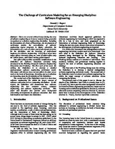
[18] or computer simulators [6], automated detection and segmentation of large lesions in CT colonography [19], and usable graphical user interface design for intuitive colonoscope control [20]. Since the beginning of the millennium an increasing research interest has been observed in the field of software engineering applications for gastrointestinal endoscopy. This interest is depicted in Figure 1, which shows the number of related scientific publications per quinquennium. The motivation for such applications is mainly the reduction of medical errors, and especially the false negative decisions in lesion detection. Pioneering works of this kind include systems for polyp detection in colonoscopy video [21, 22]. The advances over the first decade of evolution of these systems are thoroughly reviewed in [23].
Gastroenterology is one of the medical disciplines that have benefited more from the advances in computer science and medical software engineering. Recent applications in gastroenterology include medical decision support systems (MDSS) for prevention of adverse drug reactions [10]; diagnosis of celiac disease [11]; discrimination of the erosive reflux disease from the non-erosive for the provision of sodium phosphate [12]; diagnosis of complicated reflux and Barrett’s disease [13]; colonoscopy surveillance using natural language processing [14]; disease selfinspection and hospital registration recommendation [15]; colon and rectal surgery [16]; and, diagnosis of lymph node metastasis in gastric cancer [17]. Besides MDSS, other applications include computer-aided learning in capsule endoscopy using question games
*Address correspondence to this author at the Department of Computer Engineering, Technological Educational Institute of Central Greece, 3rd km Old National Road Lamias Athinas, GR 35100 Lamia, Greece; Tel: +30 22310 60159; Mob: +30 6932 252591; E-mail:
[email protected]
E-ISSN: 2308-6483/14
160
Number of publications
140 120 100 80 60 40 20 0 2000-2004
2005-2009
2010-2014
Years
Figure 1. Number of scientific publications on software engineering applications proposed in the broader field of computer science for endoscopy per quinquennium since 1 2000 . 1
Source: http://www.scopus.com, accessed 26-01-2014.
© 2014 Synergy Publishers
12
Global Journal of Gastroenterology & Hepatology, 2014 Vol. 2, No. 1
A contemporary approach to optical endoscopy is wireless capsule endoscopy (WCE). It is performed with a swallowable capsule that has an integrated miniature camera, wirelessly transmitting color video frames during its journey from the esophagus to the anus [3]. WCE is popular for the screening of the small intestine; however, capsules suitable for the examination of the esophagus and the colon are already available in the market. It enables reliable and efficient diagnosis of GI tract pathologies, including, Barrett’s esophagus, obscure bleeding, iron-deficiency anemia, and tumors of the small or the large bowel. Since their first appearance WCE systems have evolved and several variations of the original capsule have been researched [24]. The software engineering aspects of the research performed up to 2011 towards this direction, from a technical viewpoint, are reviewed in [25, 26], whereas reviews from a medical viewpoint include [27, 28]. In summary, all these studies converge to a summary of issues that are still to be resolved: a) low video quality; b) long reading times required by the endoscopists to review the WCE videos; c) low accuracy in the localization of the capsule with the conventional systems; d) inability to stabilize and navigate the capsule; and e) inability to perform biopsies. Issues (a-b) are considered as the main causes of the alarmingly low detection rate of lesions by physicians (less than 50%) [29], while issue (c) may also lead to inaccurate surgical interventions. Software engineering can effectively contribute in resolving these issues; however, issues (d-e) could be resolved only with the use of more efficient power sources, such as wireless power transmission [30, 31], and energyefficient ultra low-power electronics [32]. The rest of this paper is organized in four sections. The first three sections review the state of the art software engineering applications for endoscopic video quality enhancement, reading time reduction, and capsule endoscope localization, respectively. The last section provides a summary of conclusions. 2. ENDOSCOPIC VIDEO QUALITY ENHANCEMENT In order to keep the communication bandwidth and energy consumption low, wireless capsule endoscopes use CMOS (Complementary Metal Oxide Semiconductor) image sensors, which consume less energy, they are more easily integrable than CCD (Charge Coupled Device) sensors, but they produce images with more noise. In order to keep the energy
Dimitris K. Iakovidis
requirements low, both the spatial and the temporal resolution of the capsule endoscope are kept low, and (lossy or near-lossless) compression algorithms are used with a consequent further reduction of the video quality. Recently, a quality-preserving image compression algorithm has been based on a lowcomplexity color space [33], and significant noise reduction as compared with the state of the art has been achieved by an algorithm based on the dual tree complex wavelet transform [34]. Super-resolution algorithms can be used to increase the quality of the endoscopic video in terms of spatial (image) resolution, as indicated by preliminary results presented in [35]. Usually these algorithms combine the redundant information contained in consecutive video fames e.g. the same tissue is present in these frames at different scales, to produce a new image of higher resolution. Such algorithms are expected to perform better if the video frame rate is higher, since this would increase the redundancy of information between consecutive frames. Information redundancy between WCE video frames has been exploited for the automatic generation of panoramic images that provide a wider field of view to the endoscopists. it involves the extraction of visual features from each video frame, the detection of correspondences between the consecutive frames, and the geometric transformation of the frames so as to be ‘stitched’ together into a new more informative panoramic frame [36]. The complex folding structures, the diversity in the light reflectance properties of the objects in the gastrointestinal tract affecting illumination, the image contrast and the focus of the camera, result in difficulties in the interpretation of the image contents. Most recent of the few approaches proposed to cope with these issues is an algorithm that implements adaptive contrast diffusion [37], and methods that aim to contrast enhancement while preserving the color tones [38, 39]. Considering that color is an important feature for the discrimination of abnormalities (either lesions or blood) from normal tissue, a new real-time image processing technique for hemoglobin color enhancement has been proposed [40]. Image blurring artifacts, which can be caused by rapid changes of camera focus, can be alleviated by image de-blurring algorithms such as the one presented in [41]. A software approach to the reconstruction of threedimensional representations of the two-dimensional
Software Engineering Applications in Gastroenterology
Global Journal of Gastroenterology & Hepatology, 2014 Vol. 2, No. 1
endoscopic images [42] has been proposed as an image enhancement approach [43]. Experiments on phantom models showed that the three-dimensional reconstruction software was accurate and suitable for the enhancement of a significant proportion of vascular lesions but less so for inflammatory and protruding lesions.
sudden rough changes happen, the frame rate can decrease; thus enabling the assessment of possibly suspicious findings in detail.
3. ENDOSCOPIC VIDEO READING TIME REDUCTION The transit of a capsule endoscope lasts approx. 58 hours. The average number of frames transmitted by a conventional capsule during a typical 8-hour examination is of the order of 50,000, and the visual inspection of such a full-length video requires 45 to 120 minutes of undisrupted attention, depending on the experience of the examiner [44]. Such a manual examination process is undoubtedly time-consuming and does not guarantee that some abnormal regions are not missed, since abnormalities may be visible in only a few frames. In order to reduce the time required for visual inspection of the endoscopic videos a variety of software engineering applications have been proposed. Commercial software such as RAPID Reader [45] provides a straightforward but effective visualization of multiple consecutive frames simultaneously in a 2D arrangement. This enables the endoscopists to evaluate two, four or more frames at the same time, with a potential for a proportional reduction of the reading times. However, this potential is limited by human capabilities, as it is very difficult for humans to fully perceive the content of more than four different images projected simultaneously. Another commercially available approach to efficient visualization of WCE videos is QuickView. According to this approach only highlights from a whole video are presented. However, the validity of this approach has been strongly questioned and criticized [46, 47], since frames with important diagnostic information may be skipped. In another study [48] it has been noted that the QuickView mode is a safe diagnostic tool only when larger or diffuse lesions are suspected, such as those of Crohn’s or celiac disease. In order to avoid video frame skipping as with the QuickView visualization approach, a method for adaptive control of video display has been proposed in [49]. The principle behind this approach is that the WCE video can be played back at high frame rate in stable smooth frame sequences to save time and when
13
In [50] a method based on epitomes constructed from a set of consecutive frames has been proposed. it is based on prior knowledge regarding the normal and the possibly abnormal tissues, and the expected noninformative contents. The visual summarization obtained by the epitomes aims to be semantically organized, and its experimental evaluation showed that it can reduce the number of images down to less than 10% of the original videos. It should be noted that not all visualization methods aim to WCE reading time reduction; for example, the method proposed in [51] provides a visualization method for intestinal motility inspection. A significant reduction of 85% in the required WCE video reading time, with a zero diagnostic miss rate, has been reported in [52]. This was achieved with an unsupervised (i.e. without using prior knowledge) image mining methodology which is capable of detecting the most representative frames in the WCE video, automatically. It is worth noting that this approach does not remove frames from the WCE as its predecessor approach for video summarization [53]; instead, it introduces bookmarks indicating the frames that need to be examined more carefully by the endoscopist. This way, the endoscopist may inspect the rest of the video at faster display rates. In order to reduce the execution time of that methodology, a software implementation suitable for execution on parallel grid/cloud architectures, has been proposed in [54]. A strategy to abstract WCE video clips based on adjacent frame differences has been proposed in [55]. The frame differences were assessed in terms of lower-level image content i.e. color, texture and shape features, and linear discriminant analysis was used to select most representative frames based on an integrated distance metric used for the measurement of the adjacent frame differences. Another, also simple approach, has been proposed in [56], aiming at the selection of key-frames from the WCE video by application of video shot change detection algorithms. In [57] a camera motion estimation algorithm was used in order to detect WCE video frames that have obvious scene changes in temporal neighborhoods. By keeping only these frames an average compression ratio of 68% was possible. In a similar spirit optical flow analysis has been considered for the detection of
14
Global Journal of Gastroenterology & Hepatology, 2014 Vol. 2, No. 1
redundancy in WCE video frames [58], leading to an average reduction of 52.3%. Latest approaches that can be used for WCE video reading time reduction include a video summarization algorithm based on unsupervised frame clustering [59], and a summarization method based on panoramic visualization [36]. The former approach, features automatic determination of the number of clusters and discards frames considered as noise (non-typical); however, the results provided do not indicate whether frames containing abnormalities could be rejected as noise. The latter approach, involves the clustering of several consecutive video frames into single panoramic video frames. As discussed in the previous section, this provides a wider field of view for the endoscopists, but it also provides a way to reduce the number of frames to be examined. The experiments showed that it is possible to reduce the number of images down to less than 14.4% of the original videos. Other approaches such as the ones proposed in [60-62] aim to the identification of informative video frames and the rejection of non-informative ones e.g. frames containing turbid liquid and bubbles; thus, reduction of the WCE reading times can be achieved by the inspection of the informative frames only (or faster review of the non-informative ones). In the same spirit, one could argue that methods for automatic detection of abnormalities, such as bleeding [63-66], ulcers [67-69], and polyps [70-76] could also contribute to the reduction of the time required for the reading of the endoscopic videos; however, even if they would offer 100% accuracy, they wouldn’t absolve the physician from examining the whole video for other kinds of abnormalities. More general approaches, such as [77-79], aiming to the detection of any abnormality within the endoscopic video could potentially contribute to reading time reduction. 4. CAPSULE ENDOSCOPE LOCALIZATION The localization of a capsule endoscope within the body is typically performed by external wearable radiofrequency (RF) sensors. Commercially available systems provide only a rough estimation of capsule’s location through a graphic unitless representation via a graphical user interface of the video reading software [45]. Recent methods that promise more accurate localization of the capsule endoscopes, with low adverse health effects are based on magnetic sensor arrays. However, they are still in experimental phase and tested only in laboratory setups [80].
Dimitris K. Iakovidis
A limited number of software engineering applications has been proposed for capsule localization. These include applications that implement methods addressing only the estimation of the rotation angle of the capsule [81, 82], methods providing an estimation of whether the capsule faces the lumen tunnel or not [83], and temporal video segmentation methods. The latter are capable of inferring the part of the GI tract in which the capsule is located i.e. oesophagus, stomach, small intestine and colon, by applying supervised pattern recognition techniques for the identification either of the tissues of these parts [84] or of the transition points between these parts, such as the esogastric junction, the pylorus and the ileocecal valve [85-87]. The study of the color components of endoscopic videos indicate that color can be a significant feature for the discrimination of the different parts of the GI tract [88]. Relevant approaches have been proposed for topographic classification of the WCE video frames in compressed video [89], and unsupervised classification of the video frames [90]. Camera motion estimation approaches such as the model of deformable rings (MDR) presented in [91] could be used for the localization of the capsule endoscope within the GI tract. This approach enables a two-dimensional map representation of the internal surface of the GI tract for WCE content interpretation based on intestinal motility patterns. The feasibility of using capsule motion estimation for capsule endoscope localization has been verified in [92]. The method proposed in that study was inspired by visual odometry [93], which is a popular approach for the measurement of distances in robotics and in vehicular technologies research. Visual odometry is not affected by wheel slip in uneven terrain or other adverse conditions, and as compared to wheel odometry [94] it can provide more accurate trajectory estimates, with relative position error ranging from 0.1 to 2% [95]. Important issues in software capsule localization methods include the motility of the GI tract and the presence of intestinal content, both of which can interfere with the vision-based measurements. A starting point to cope with these issues is to consider recent methods for intestinal motility assessment [51, 96, 97], and methods for recognition of intestinal content [61, 62]. 5. CONCLUSIONS This paper provided an overview of the state of the art in software engineering applications in
Software Engineering Applications in Gastroenterology
Global Journal of Gastroenterology & Hepatology, 2014 Vol. 2, No. 1
gastroenterology, focusing on the advances of the last quinquennium in endoscopy. Although several review studies have been performed earlier in this domain, either from a technical or from a medical viewpoint, this study identified and highlighted contemporary research directions significant for efficient and effective clinical practice in gastroenterology.
[13]
Bertolini S, Maoli A, Rauch G, Giacomini M. Entropy-driven decision tree building for decision support in gastroenterology. Stud Health Technol Inform 2013; 186: 937.
[14]
Wagholikar K, Sohn S, Wu S, et al. Clinical decision support for colonoscopy surveillance using natural language processing. In: Healthcare Informatics, Imaging and Systems Biology (HISB), 2012 IEEE Second International Conference on. IEEE 2012 p. 12-21.
[15]
Mu B, Xiao F, Yuan S. A rule-based disease self-inspection and hospital registration recommendation system. In: Software Engineering and Service Science (ICSESS), 2012 IEEE 3rd International Conference on. IEEE 2012 p. 212-5.
[16]
McCoy AB, Melton GB, Wright A, Sittig DF. Clinical decision support for colon and rectal surgery: an overview. Clin Colon Rectal Surg. 2013; 26: 23-30. http://dx.doi.org/10.1055/s-0033-1333644
[17]
Zhou ZG, Liu F, Jiao LC, et al. A bi-level belief rule based decision support system for diagnosis of lymph node metastasis in gastric cancer. KBS 2013; 54: 128-36.
[18]
Postgate A, Haycock A, Thomas-Gibson S, et al. Computeraided learning in capsule endoscopy leads to improvement in lesion recognition ability. Gastrointest Endosc 2009; 70: 3106. http://dx.doi.org/10.1016/j.gie.2008.11.043
[19]
Grigorescu SE, Nevo ST, Liedenbaum MH, et al. Automated detection and segmentation of large lesions in CT colonography. IEEE Trans Biomed Eng 2010; 57: 675-84. http://dx.doi.org/10.1109/TBME.2009.2035632
[20]
Kuperij N, Reilink R, Schwartz MP, Stramigioli S, Misra S, Broeders IA. Design of a user interface for intuitive colonoscope control. In: Intelligent Robots and Systems (IROS), 2011 IEEE/RSJ International Conference on. IEEE 2011 p. 937-42.
[21]
Karkanis SA, Iakovidis DK, Maroulis DE, Magoulas GD, Theofanous N. Tumor recognition in endoscopic video images using artificial neural network architectures. In: Euromicro Conference, 2000. Proceedings of the 26th. vol. 2. IEEE 2000 p 423-9.
[22]
Karkanis SA, Iakovidis DK, Maroulis DE, Karras DA, Tzivras M. Computer-aided tumor detection in endoscopic video using color wavelet features. IEEE Trans Biomed Eng 2003; 7: 141-52. http://dx.doi.org/10.1109/TITB.2003.813794
Most of the applications reviewed are still in an early stage, and have been experimentally evaluated either on simulated or limited data. Their validation on real, large volume data, is a common challenge that still needs to be addressed. REFERENCES [1]
Haux R. Medical informatics: past, present, future. Int J Med Inform 2010; 79: 599-610. http://dx.doi.org/10.1016/j.ijmedinf.2010.06.003
[2]
Iakovidis DK, Papageorgiou E. Intuitionistic fuzzy cognitive maps for medical decision making. IEEE Trans Inf Technol Biomed 2011; 15: 100-7. http://dx.doi.org/10.1109/TITB.2010.2093603
[3]
Iddan G, Meron G, Glukhovsky A, Swain P. Wireless capsule endoscopy. Nature 2000; 405: 417. http://dx.doi.org/10.1038/35013140
[4]
Iakovidis DK, Tsevas S, Savelonas MA, Papamichalis G. Image analysis framework for infection monitoring. IEEE Trans Inf Technol Biomed 2012; 59: 1135-44. http://dx.doi.org/10.1109/TBME.2012.2185049
[5]
Tanaka M, Wada S, Nakamura M. Toward in silico medicine. In: Computational Biomechanics. Springer; 2012 p. 181-7. http://dx.doi.org/10.1007/978-4-431-54073-1_5
[6]
Cantù P, Penagini R. Computer simulators: The present and near future of training in digestive endoscopy. Dig Liver Dis 2012; 44: 106-10. http://dx.doi.org/10.1016/j.dld.2011.09.008
15
[7]
Wang W, Krishnan E. Big data and clinicians: a review on the state of the science. JMIR 2014; 2: e1.
[8]
Sultan N. Making use of cloud computing for healthcare provision: Opportunities and challenges. Int J Inform Manage 2014; 34: 177-84. http://dx.doi.org/10.1016/j.ijinfomgt.2013.12.011
[23]
Liedlgruber M, Uhl A. Computer-aided decision support systems for endoscopy in the gastrointestinal tract: a review. IEEE Rev Biomed Eng 2011; 4: 73-88. http://dx.doi.org/10.1109/RBME.2011.2175445
[9]
Sun J, Reddy CK. Big data analytics for healthcare. In: Proceedings of the 19th ACM SIGKDD international conference on Knowledge discovery and data mining. ACM 2013 p. 1525.
[24]
Ciuti G, Menciassi A, Dario P. Capsule endoscopy: from current achievements to open challenges. IEEE Rev Biomed Eng 2011; 4: 59-72. http://dx.doi.org/10.1109/RBME.2011.2171182
[10]
Bertsche T, Pfaff J, Schiller P, et al. Prevention of adverse drug reactions in intensive care patients by personal intervention based on an electronic clinical decision support system. Intensive Care Med 2010; 36: 665-72. http://dx.doi.org/10.1007/s00134-010-1778-8
[25]
Karargyris A, Bourbakis N. Wireless capsule endoscopy and endoscopic imaging: A survey on various methodologies presented. IEEE Eng Med Biol Mag 2010; 29: 72-83. http://dx.doi.org/10.1109/MEMB.2009.935466
[11]
Tenório JM, Hummel AD, Cohrs FM, Sdepanian VL, Pisa IT, de Fátima Marin H. Artificial intelligence techniques applied to the development of a decision–support system for diagnosing celiac disease. Int J Med Inform 2011; 80: 793802. http://dx.doi.org/10.1016/j.ijmedinf.2011.08.001
[26]
Mackiewicz M. Capsule endoscopy—state of the technology and computer vision tools after the first decade. New techniques in gastrointestinal endoscopy InTech, New York. 2011.
[27]
Fisher LR, Hasler WL. New vision in video capsule endoscopy: current status and future directions. Nat Rev Gastroenterol Hepatol 2012; 9: 392-405. http://dx.doi.org/10.1038/nrgastro.2012.88
[28]
Koulaouzidis A, Rondonotti E, Karargyris A. Small-bowel capsule endoscopy: a ten-point contemporary review. World J Gastroenterol 2013; 19: 3726-46. http://dx.doi.org/10.3748/wjg.v19.i24.3726
[12]
Imperiale TF, Sherer EA, Balph JAD, Cardwell JD, Qi R. Provider acceptance, safety, and effectiveness of a computer-based decision tool for colonoscopy preparation. Int J Med Inform 2011; 80: 726-33. http://dx.doi.org/10.1016/j.ijmedinf.2011.07.001
16
Global Journal of Gastroenterology & Hepatology, 2014 Vol. 2, No. 1
Dimitris K. Iakovidis
[29]
Zheng Y, Hawkins L, Wolff J, Goloubeva O, Goldberg E. Detection of lesions during capsule endoscopy: physician performance is disappointing. Am J Gastroenterol 2012; 107: 554-60. http://dx.doi.org/10.1038/ajg.2011.461
[46]
Günther U, Daum S, Zeitz M, Bojarski C. Capsule endoscopy: comparison of two different reading modes. Int J Colorectal Dis 2012; 27: 521-5. http://dx.doi.org/10.1007/s00384-011-1347-9
[47]
[30]
Pan G, Xin W, Yan G, Chen J. A video wireless capsule endoscopy system powered wirelessly: design, analysis and experiment. Meas Sci Technol 2011; 22: 1-9. http://dx.doi.org/10.1088/0957-0233/22/6/065802
Smirnidis A, Koulaouzidis A, Douglas S, Plevris J. PTU-143 Quickview in capsule endoscopy: is it enough? Gut 2012; 61(Suppl 2): A244. http://dx.doi.org/10.1136/gutjnl-2012-302514c.143
[48]
[31]
Chen W, Yan G, Wang Z, Jiang P, Liu H. A wireless capsule robot with spiral legs for human intestine. Int J Med Robot 2013; epub ahead of print.
Kyriakos N, Karagiannis S, Galanis P, et al. Evaluation of four time-saving methods of reading capsule endoscopy videos. Eur J Gastroenterol Hepatol 2012; 24: 1276-80.
[49]
[32]
Jiang H, Chen X, Wang Z. SoC for Capsule Endoscope. In: Ultra-Low Power Integrated Circuit Design. Springer; 2014 p. 219-32. http://dx.doi.org/10.1007/978-1-4419-9973-3_10
Vu H, Echigo T, Sagawa R, et al. Controlling the display of capsule endoscopy video for diagnostic assistance. IEICE Transact Inform Sys 2009; 92: 512-28. http://dx.doi.org/10.1587/transinf.E92.D.512
[50]
[33]
Khan TH, Wahid KA. White and narrow band image compressor based on a new color space for capsule endoscopy. Signal Process Image Commun 2014; In press.
[34]
Gopi VP, Palanisamy P. Capsule endoscopic image denoising based on double density dual tree complex wavelet transform. Int J Imag Robot 2013; 9: 48-60.
Chu X, Poh CK, Li L, et al. Epitomized summarization of wireless capsule endoscopic videos for efficient visualization. In: Medical Image Computing and Computer-Assisted Intervention–MICCAI 2010. Springer; 2010. p. 522-9. http://dx.doi.org/10.1007/978-3-642-15745-5_64
[51]
Drozdzal M, Segu S, Vitrià J, Malagelada C, Azpiroz F, Radeva P. Adaptable image cuts for motility inspection using WCE. Comput Med Imaging Graph 2013; 37: 72-80. http://dx.doi.org/10.1016/j.compmedimag.2012.09.002
[35]
Häfner M, Liedlgruber M, Uhl A. POCS-based superresolution for HD endoscopy video frames. In: Proc. Computer Based Medical Systems 2013 p. 185-90.
[52]
[36]
Iakovidis DK, Spyrou E, Diamantis D. Efficient homographybased video visualization for wireless capsule endoscopy. In: Bioinformatics and Bioengineering (BIBE), 2013 IEEE 13th International Conference on. IEEE 2013 p. 1-4.
Iakovidis DK, Tsevas S, Polydorou A. Reduction of capsule endoscopy reading times by unsupervised image mining. Comput Med Imaging Graph 2010; 34: 471-8. http://dx.doi.org/10.1016/j.compmedimag.2009.11.005
[53]
[37]
Li B, Meng MQH. Wireless capsule endoscopy images enhancement via adaptive contrast diffusion. J Vis Commun Image Represent 2012; 23: 222-8. http://dx.doi.org/10.1016/j.jvcir.2011.10.002
Tsevas S, Iakovidis DK, Maroulis D, Pavlakis E. Automatic frame reduction of wireless capsule endoscopy video. In: BioInformatics and BioEngineering, 2008. BIBE 2008. 8th IEEE International Conference on. IEEE 2008 p. 1-6.
[54]
[38]
Vu H, Echigo T, Yagi K, et al. Image-Enhanced capsule endoscopy preserving the original color tones. In: Abdominal Imaging. Computational and Clinical Applications. Springer; 2012 p. 35-43. http://dx.doi.org/10.1007/978-3-642-28557-8_5
Ioannis K, Tsevas S, Maglogiannis I, Iakovidis DK. Enabling distributed summarization of wireless capsule endoscopy video. In: Imaging Systems and Techniques (IST), 2010 IEEE International Conference on. IEEE 2010 p. 17-21.
[55]
Zhao Q, Meng MH. A strategy to abstract WCE video clips based on LDA. In: Robotics and Automation (ICRA), 2011 IEEE International Conference on. IEEE 2011 p. 4145-50.
[56]
Fu Y, Meng M, Liu H, Yu C, Yan T, Li T. Key-frame selection in WCE video based on shot detection. In: in 2012 10th World Congress on Intelligent Control and Automation (WCICA), IEEE 2012 p. 5030-4.
[39]
Okuhata H, Nakamura H, Hara S, Tsutsui H, Onoye T. Application of the real-time Retinex image enhancement for endoscopic images. In: Engineering in Medicine and Biology Society (EMBC), 2013 35th Annual International Conference of the IEEE. IEEE 2013 p. 3407-10.
[40]
Han Z, Ma S, Wang X, Li Z, Xie T. New real-time endoscopy image processing technology of hemoglobin color enhancement. In: Proc. of SPIE Vol. vol. 8192; 2011 p. 819257–1.
[57]
Liu H, Pan N, Lu H, Song E, Wang Q, Hung CC. Wireless capsule endoscopy video reduction based on camera motion estimation. J Digit Imaging 2013; 26: 287-301. http://dx.doi.org/10.1007/s10278-012-9519-x
[41]
Liu H, Lu WS, Meng MH. De-blurring wireless capsule endoscopy images by total variation minimization. In: Communications, Computers and Signal Processing (PacRim), 2011 IEEE Pacific Rim Conference on. IEEE 2011 p. 102-6.
[58]
Lee HG, Choi MK, Shin BS, Lee SC. Reducing redundancy in wireless capsule endoscopy videos. Comput Biol Med 2013; 43: 670-82. http://dx.doi.org/10.1016/j.compbiomed.2013.02.009
[59]
[42]
Koulaouzidis A, Karargyris A. Three-dimensional image reconstruction in capsule endoscopy. World J Gasteroenterol 2012; 18: 4086-90. http://dx.doi.org/10.3748/wjg.v18.i31.4086
Ismail M, Bchir O, Emam AZ. Endoscopy video summarization based on unsupervised learning and feature discrimination. In: Visual Communications and Image Processing (VCIP), 2013. IEEE 2013 p. 1-6.
[60]
[43]
Koulaouzidis A, Karargyris A, Rondonotti E, et al. Threedimensional representation software as image enhancement tool in small-bowel capsule endoscopy: A feasibility study. Dig Liver Dis 2013; 45: 909-14. http://dx.doi.org/10.1016/j.dld.2013.05.013
Bashar MK, Kitasaka T, Suenaga Y, Mekada Y, Mori K. Automatic detection of informative frames from wireless capsule endoscopy images. Med Image Anal 2010; 14: 44970. http://dx.doi.org/10.1016/j.media.2009.12.001
[61]
[44]
Delvaux M, Gay G. Capsule endoscopy: technique and indications. Best Pract Res Clin Gastroenterol 2008; 22: 81337. http://dx.doi.org/10.1016/j.bpg.2008.06.003
Sun Z, Li B, Zhou R, Zheng H, Meng MQH. Removal of noninformative frames for wireless capsule endoscopy video segmentation. In: Automation and Logistics (ICAL), 2012 IEEE International Conference on. IEEE 2012 p. 294-9.
[62]
[45]
Wireless Capsule Endoscopy Software. http://www.givenimaging.com/en-int/Innovative-Solutions/CapsuleEndoscopy/Software/Pages/default.aspx.
Segui S, Drozdzal M, Vilarino F, et al. Categorization and segmentation of intestinal content frames for wireless capsule endoscopy. IEEE Transact Inform Technol Biomed 2012; 16: 1341-52. http://dx.doi.org/10.1109/TITB.2012.2221472
Software Engineering Applications in Gastroenterology
Global Journal of Gastroenterology & Hepatology, 2014 Vol. 2, No. 1
[63]
Pan G, Yan G, Qiu X, Cui J. Bleeding detection in wireless capsule endoscopy based on probabilistic neural network. J Med Syst 2011; 35: 1477-84. http://dx.doi.org/10.1007/s10916-009-9424-0
[79]
Manivannan S, Wang R, Trucco E, Hood A. Automatic normal-abnormal video frame classification for colonoscopy. In: Biomedical Imaging (ISBI), 2013 IEEE 10th International Symposium on. IEEE 2013 p. 644-7.
[64]
Pan GB, Xu F, Chen JL. Bleeding detection in wireless capsule endoscopy using color similarity coefficient. Appl Mech Materials 2012; 195: 307-12. http://dx.doi.org/10.4028/www.scientific.net/AMM.195196.307
[80]
Than TD, Alici G, Zhou H, Li W. A review of localization systems for robotic endoscopic capsules. IEEE Trans Biomed Eng 2012; 59: 2387-99. http://dx.doi.org/10.1109/TBME.2012.2201715
[81]
[65]
Brzeski A, Blokus A, Cychnerski J. An overview of image analysis techniques in endoscopic bleeding detection. Int J Innov Res Comput Commun Eng 2013; 1: 1350-7.
[66]
Figueiredo IN, Kumar S, Leal C, Figueiredo PN. Computerassisted bleeding detection in wireless capsule endoscopy images. Comput Methods Biomech Biomed Eng Imaging Vis 2013; 1: 198-210.
Liu L, Hu C, Cai W, Meng MH. Capsule endoscope localization based on computer vision technique. In: Engineering in Medicine and Biology Society, 2009. EMBC 2009. Annual International Conference of the IEEE. IEEE 2009 p. 3711-4.
[82]
Spyrou E, Iakovidis DK. Homography-based orientation estimation for capsule endoscope tracking. In: Imaging Systems and Techniques (IST), 2012 IEEE International Conference on. IEEE 2012 p. 101-5.
[83]
Bao G, Pahlavai K. Motion estimation of the endoscopy capsule using region-based Kernel SVM classifier. Accepted by IEEE EIT, Rapid City, SD, May 9-11, 2013.
[84]
Mackiewicz M, Berens J, Fisher M. Wireless capsule endoscopy color video segmentation. IEEE Trans Med Imaging 2008; 27: 1769-81. http://dx.doi.org/10.1109/TMI.2008.926061
[67]
[68]
Yu L, Yuen PC, Lai J. Ulcer detection in wireless capsule endoscopy images. In: Pattern Recognition (ICPR), 2012 21st International Conference on. IEEE 2012 p. 45-8. Eid A, Charisis VS, Hadjileontiadis LJ, Sergiadis GD. A curvelet-based lacunarity approach for ulcer detection from Wireless Capsule Endoscopy images. In: Computer-Based Medical Systems (CBMS), 2013 IEEE 26th International Symposium on. IEEE 2013 p. 273-8.
17
[69]
Jebarani W, Daisy VJ. Assessment of Crohn’s disease lesions in Wireless Capsule Endoscopy images using SVM based classification. In: Signal Processing Image Processing & Pattern Recognition (ICSIPR), 2013 International Conference on. IEEE 2013 p. 303-7.
[85]
Cunha JS, Coimbra M, Campos P, Soares JM. Automated topographic segmentation and transit time estimation in endoscopic capsule exams. IEEE Trans Med Imaging 2008; 27: 19-27. http://dx.doi.org/10.1109/TMI.2007.901430
[70]
Szczypinski P, Klepaczko A, Strzelecki M. An intelligent automated recognition system of abnormal structures in WCE images. In: Hybrid Artificial Intelligent Systems. Springer; 2011 p. 140-7. http://dx.doi.org/10.1007/978-3-642-21219-2_19
[86]
Hwang S, Celebi M. Multilevel wireless capsule endoscopy video segmentation. In: Proc. SPIE; 2010 p. 76234D.
[87]
Li B, Meng MQH, Lau JY. Computer-aided small bowel tumor detection for capsule endoscopy. Artif Intell Med 2011; 52: 11-6. http://dx.doi.org/10.1016/j.artmed.2011.01.003
Zhou R, Li B, Sun Z, Hu C, Meng MQH. Wireless capsule endoscopy video automatic segmentation. In: Robotics and Biomimetics (ROBIO), 2012 IEEE International Conference on. IEEE; 2012. p. 825–830.
[88]
Karargyris A, Bourbakis N. Detection of small bowel polyps and ulcers in wireless capsule endoscopy videos. IEEE Trans Biomed Eng 2011; 58: 2777-86. http://dx.doi.org/10.1109/TBME.2011.2155064
Vu H, Yagi Y, Echigo T, et al. Color analysis for segmenting digestive organs in VCE. In: Pattern Recognition (ICPR), 2010 20th International Conference on. IEEE 2010 p. 246871.
[89]
Li B, Meng MH. Tumor recognition in wireless capsule endoscopy images using textural features and SVM-based feature selection. IEEE Trans Inf Technol Biomed 2012; 16: 323-9. http://dx.doi.org/10.1109/TITB.2012.2185807
Marques N, Dias E, Cunha J, Coimbra M. Compressed domain topographic classification for capsule endoscopy. In: Engineering in Medicine and Biology Society, EMBC, 2011 Annual International Conference of the IEEE. IEEE 2011 p. 6631-4.
[90]
Shen Y, Guturu P, Buckles BP. Wireless capsule endoscopy video segmentation using an unsupervised learning approach based on probabilistic latent semantic analysis with scale invariant features. IEEE Trans Inform Technol Biomed 2012; 1: 98-105.
[91]
Szczypinski PM, Sriram RD, Sriram PV, Reddy DN. Á model of deformable rings for interpretation of wireless capsule endoscopic videos. Med Image Anal 2009; 13: 312-24. http://dx.doi.org/10.1016/j.media.2008.12.002
[92]
Spyrou E, Iakovidis DK. Video-based measurements for wireless capsule endoscope tracking. Meas Sci Technol 2014; 25: 015002. http://dx.doi.org/10.1088/0957-0233/25/1/015002
[93]
Nistér D, Naroditsky O, Bergen J. Visual odometry. In: Computer Vision and Pattern Recognition, 2004. CVPR 2004. Proceedings of the 2004 IEEE Computer Society Conference on. vol. 1. IEEE; 2004. p. I–652.
[94]
Karargyris A, Koulaouzidis A. Capsule-odometer: A concept to improve accurate lesion localisation. World J Gastroenterol 2013; 19: 5943-6. http://dx.doi.org/10.3748/wjg.v19.i35.5943
[95]
Scaramuzza D, Fraundorfer F. Visual odometry [tutorial]. IEEE Robot Autom Mag 2011; 18: 80-92. http://dx.doi.org/10.1109/MRA.2011.943233
[71]
[72]
[73]
[74]
[75]
[76]
[77]
[78]
Li B, Meng MQH. Automatic polyp detection for wireless capsule endoscopy images. Expert Syst Appl 2012; 39: 10952-8. http://dx.doi.org/10.1016/j.eswa.2012.03.029 Li BP, Meng MQH. Comparison of several texture features for tumor detection in CE images. J Med Syst 2012; 36: 2463-9. http://dx.doi.org/10.1007/s10916-011-9713-2 Figueiredo IN, Kumar S, Figueiredo PN. An intelligent system for polyp detection in wireless capsule endoscopy images. Computational Vision and Medical Image Processing IV: VIPIMAGE 2013. 2013;p. 229. Hwang S. Bag-of-visual-words approach to abnormal image detection in wireless capsule endoscopy videos. In: Advances in Visual Computing. Springer 2011 p. 320-7. http://dx.doi.org/10.1007/978-3-642-24031-7_32 Hu E, Nosato H, Sakanashi H, Murakawa M. Anomaly detection for capsule endoscopy images using higher-order Local Auto Correlation features. In: Systems, Man, and Cybernetics (SMC), 2012 IEEE International Conference on. IEEE 2012 p. 2289-93.
18
[96]
Global Journal of Gastroenterology & Hepatology, 2014 Vol. 2, No. 1
Vilarino F, Spyridonos P, DeIorio F, Vitria J, Azpiroz F, Radeva P. Intestinal motility assessment with video capsule endoscopy: automatic annotation of phasic intestinal contractions. IEEE Trans Med Imaging 2010; 29: 246-59. http://dx.doi.org/10.1109/TMI.2009.2020753
Received on 27-01-2014
Dimitris K. Iakovidis
[97]
Gallo G, Granata E. LBP based detection of intestinal motility in WCE images. In: SPIE Medical Imaging. International Society for Optics and Photonics 2011 p. 79614T-79614T.
Accepted on 28-01-2014
Published on 21-02-2014
http://dx.doi.org/10.12970/2308-6483.2014.02.01.3
© 2014 Dimitris K. Iakovidis; Licensee Synergy Publishers. This is an open access article licensed under the terms of the Creative Commons Attribution Non-Commercial License (http://creativecommons.org/licenses/by-nc/3.0/) which permits unrestricted, non-commercial use, distribution and reproduction in any medium, provided the work is properly cited.