condition, object-based and space-based attention showed significant activations ...... In the monkey, neural activity has been demonstrated in this region when ...
Brain (1997), 120, 2013–2028
Space-based and object-based visual attention: shared and specific neural domains G. R. Fink1, R. J. Dolan1,2, P. W. Halligan3,4, J. C. Marshall4 and C. D. Frith1 1Wellcome
Department of Cognitive Neurology, Institute of Neurology, and 2Royal Free Hospital School of Medicine, London, and 3Rivermead Rehabilitation Centre, and 4Neuropsychology Unit, University Department of Clinical Neurology, The Radcliffe Infirmary, Oxford, UK
Correspondence to: Gereon R. Fink, MD, Wellcome Department of Cognitive Neurology, Institute of Neurology, 12 Queen Square, London WC1N 3BG, UK
Summary Visual attention can be primarily allocated to either where an object is in space (with little emphasis on the structure of the object itself) or to the structure of the object (with little emphasis on where in space the object is located). Using PET measures of regional cerebral blood flow (rCBF) to index neural activity, we investigated the shared and specific functional anatomy underlying both of these types of visual attention in a controlled non-cueing non-blocked paradigm that involved identical stimuli across the conditions of interest. The interaction of eye movements with these attentional systems was studied by introducing fixation or free vision as an additional factor. Relative to the control condition, object-based and space-based attention showed significant activations of the left and right medial superior parietal cortex and the left lateral inferior parietal cortex, the left prefrontal cortex and the cerebellar vermis. Significant
differential activations were observed during object-based attention in the left striate and prestriate cortex. Space-based attention activated the right prefrontal cortex and the right inferior temporal–occipital cortex. Differential neural activity due to free vision or fixation was observed in occipital areas only. Significant interactions of free vision/fixation on activations due to object-based and space-based attention were observed in the right medial superior parietal cortex and left lateral inferior parietal cortex, respectively. The study provides direct evidence for the importance of the parietal cortex in the control of object-based and spacebased visual attention. The results show that object-based and space-based attention share common neural mechanisms in the parietal lobes, in addition to task specific mechanisms in early visual processing areas of temporal and occipital cortices.
Keywords: visual attention; PET; parietal cortex; prestriate cortex; visual neglect Abbreviations: EOG 5 electrooculogram; rCBF 5 regional cerebral blood flow; SPM 5 statistical parametric mapping
Introduction The functional and neuroanatomical basis of dissociable prestriate pathways for visual object and spatial processing in humans and primates is well established (Ungerleider and Mishkin, 1982; Haxby et al., 1991). The posterior parietal cortex is primarily concerned with spatial processing (‘dorsal stream’), and the inferior temporal cortex with object processing (‘ventral stream’). It has also been argued that there may be a similar dissociation between object and spatial processing domains within the primate prefrontal cortex (Wilson et al., 1993). Patients with chronic visual neglect (usually following right hemispheric damage) can produce an adequate © Oxford University Press 1997
representation of the right half of a scene presented whilst leaving out figures on the left side. It has also been observed that some patients with neglect may omit the left half of several objects displayed across a scene (Driver and Halligan, 1991; Marshall and Halligan, 1993; Walker, 1995). Such observations suggest two modes for the allocation of visual attention: attention to location (space-based) and attention to the structure of an object (object-based). Space-based attention is concerned with the position of an object as a whole, relative to a predefined spatial reference (e.g. the point of fixation, another object in the visual field, or the subject’s midsagittal plane). Object-based attention involves
2014
G. R. Fink et al.
the analysis of the parts of an object as they contribute to the recognition of the whole (Posner, 1980; Duncan, 1984). The notion of an object-based attentional system is supported by human behaviourial studies; superior performance has been demonstrated in detecting attributes of the same object (compared with detection of attributes of different objects) even when their absolute spatial positions are controlled (Duncan, 1984). The suggestion of two separable attentional systems is also supported by experimental data from primate studies; selective activation of neurons in the supplementary frontal eye-fields has been demonstrated in a short-term memory task involving objectbased spatial awareness (Olson and Gettner, 1995). Psychological evidence for dissociations is not incompatible with the possibility that the neural domains of object-based and space-based attention might overlap to some extent. Cerebral lesion sites in patients suffering from objectbased or space-based attention deficits usually overlap (Marshall and Halligan, 1993). An important caveat of lesions studies is that damage leading to chronic visual neglect is typically large, and tends to involve extensive regions of the temporal–parietal cortex. The issue is further complicated by the fact that patients may exhibit both forms of visual neglect. In this study, we investigated the functional anatomy of object-based and space-based attention in the intact human brain. We used PET to index, in normal human subjects, neural activity associated with object-based and space-based attention by measuring associated changes in relative regional cerebral blood flow (rCBF) (Raichle, 1987). The specific aim of the present study was to investigate whether there are unique neural mechanisms involved in attending to the structure of an object or the position of the object in space, when identical stimuli are employed. The sole task requirement for subjects was to attend to either where the object appeared in space (space-based attention) or where, within the object, a specific feature occurred (object-based attention). We also used two levels of ocular control: fixation or free vision. Based on the results from human lesion studies we predicted that the source of both space-based and objectbased visual attention (as defined here) would be temporal– parietal (Vallar, 1993). Previous functional imaging studies on selective visual attention (Heinze et al., 1994; Fink et al., 1996, 1997b) also suggest that object-based and space-based attention have a modulatory effect on early visual processing.
Methods Subjects Twelve normal healthy male volunteers (aged 23–49 years) were recruited. All were right-handed with no history, past or current, of neurological or psychiatric illness. Informed written consent was obtained from all subjects. The study involved administration of 4.5 mSv effective dose equivalent of radioactivity per subject. Permission to administer
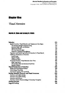
Fig. 1 Study design. Each box represents an experimental condition; it also represents the screen of the video display unit with the stimuli used across the experimental conditions (spacebased and object-based tasks, upper and middle row; control task, lower row). During the tasks, subjects were either instructed explicitly that they were allowed to move their eyes (overt attention) or to maintain fixation on the central dot throughout stimulus presentation (covert attention). In the object-based condition, subjects were required to attend to and report whether the square was on the left or the right of the object (i.e. left or right of the line irrespective of the line’s position relative to the central dot). In the space-based condition, subjects were required to attend to and report whether the object (i.e. the line) appeared left or right of the centre of the screen as marked by the central dot. In the control tasks, subjects were asked to attend to and report, whether a short or a long line was present. In the figure scan numbers refer to the counterbalanced order of scans for subject 1 (of the group of 12 subjects studied). Equivalent counterbalancing was performed for all other subjects, both within and between subjects.
radioactivity was obtained from the Administration of Radioactive Substances Advisory Committee of the Department of Health, UK. The study was approved by the local ethics committee of the National Hospital for Neurology and Neurosurgery, London, UK.
Paradigm design Examples of the figures used as stimuli during the objectbased attention tasks, space-based attention tasks and control tasks are shown in Fig. 1. In the object-based and spacebased attention tasks, identical stimuli were displayed across conditions. Subjects were instructed to attend to the requested level (object-based or space-based). In the space-based task, subjects were asked to attend to and report whether an object (i.e. an individual line, 55 mm long, with a square attached, appearing either at 0 or at 55 mm left or right of the dot) appeared left or right of a fixation dot located at the centre of the screen. In the object-based task, subjects were asked
Space- and object-based attention to attend and report whether the square was on the left or the right of the object (i.e. left or right of the line). The square was always centred 55 mm left or right of the central dot. In both conditions, the verbal response ‘left’ or ‘right’ was given by the subject. In the control task, individual long (220 mm) or short (110 mm) lines were presented in a pseudo-random sequence. Each line was centred in the middle of the screen, superimposed on the fixation dot. Subjects were asked to attend to and report, whether a short or a long line was presented. The verbal response by the subject was therefore ‘short’ or ‘long’. Stimuli were kept as simple as possible to reduce engagement of processes irrelevant to the current experiment. We investigated a further experimental factor by allowing or disallowing eye movements. In the conditions with eye movements, subjects initially fixated on the dot but were instructed explicitly that they were allowed to move their eyes on stimulus onset. In the other conditions, subjects were instructed to maintain fixation on the central dot throughout stimulus presentation. Prior to PET scanning, all subjects underwent a familiarization session. This ensured that they were fully familiar with the stimuli and tasks. Eye movements were monitored during the familiarization session and during all experimental conditions using routine electrooculogram (EOG) measurements with bitemporal electrode placement. These recordings showed that during the familiarization session, as well as during the PET rCBF measurements, subjects maintained fixation in the covert attention tasks and that they moved their eyes in the free vision conditions. The figures were presented during the PET rCBF measurements in black on a white background. A stimulus appeared every 1.5 s and remained for 150 ms. A 17-inch video display unit was used at a viewing distance of 40 cm. The figures appeared in a quasi-random sequence that did not permit the same stimulus to appear on more than five successive trials. Each sequence of stimulus presentations began 10 s prior to PET scanning and lasted for 120 s. The experiment involved 12 sequential relative rCBF measurements per subject; the six testing conditions were presented in a (fully factorial) 233 design with two repeats per condition (Fig. 1). To control for time effects the experimental conditions were fully counterbalanced both within and between subjects. The experiment employs a fully crossed factorial design in which one factor has three levels (object-based, spacebased, control) and the other factor has two levels (with eye movements, without eye movements). This gives a total of six different conditions, and therefore only two replications for each of these conditions across the twelve scans (per subject). However, this small number of replications of conditions is not associated with a loss of statistical power. By virtue of the factorial design, the various contrasts (i.e. the main effects and interaction terms) are all based on at least eight scans (per subject). Furthermore, 12 subjects were studied (i.e. each contrast is based on at least 96 scans).
2015
PET-scanning Relative rCBF was measured by recording the regional distribution of cerebral radioactivity following the i.v. injection of 15O-labelled water; 15O is a positron emitter with a half-life of 2.1 min (Mazziotta et al., 1985). The PET measurements were carried out using a Siemens/CPS ECAT EXACT HR 1 (model 962) PET scanner (CTI, Knoxville, Tenn., USA) with a total axial field of view of 155 mm covering the whole brain. Data were acquired in threedimensional mode (Townsend et al., 1991) with inter-detector collimating septa removed and a ‘Neuro-Insert’ installed to limit the acceptance of events originating from out-of-fieldof-view activity (from the whole body). For each measurement of relative rCBF, 9 mCi of H215O were given i.v. as a slow bolus over 20 s (Silbersweig et al., 1993). Twelve consecutive PET scans were collected, each beginning with a 30 s background scan before the delivery of the slow bolus. Emission data were thereafter collected sequentially over 90 s after tracer arrival in the brain, and corrected for background. This process was repeated for each emission scan, with 8 min between scans to allow for adequate decay of radioactivity. All emission scan data were corrected for the effects of radiation attenuation (e.g. by the skull) by means of a transmission scan taken prior to the first relative rCBF measurement. The corrected data were reconstructed into 63 transverse planes (separation 2.4 mm) and into 1283128 pixels (size 2.1 mm) by three-dimensional filtered back projection using a Hann filter of cutoff frequency 0.5 cycles per pixel and applying a scatter correction. The resolution of the resulting PET images was 6 mm (at full width half maximum).
MRI In separate sessions, an MRI of each subject’s brain was obtained (i) to exclude the possibility of morphological/ pathological abnormalities and (ii) for stereotactic normalization into the standard anatomical space (see below). This imaging was performed with a 2-Tesla system (VISION, Siemens, Germany) using a three-dimensional T1 weighted imaging technique producing 108 transaxial slices (13131.5 mm) which gave high grey–white matter contrast.
Image processing All calculations and image manipulations were performed on a SPARC workstation (SUN Computers). PROMATLAB software (Mathworks, USA) was used to calculate and display images. Statistical parametric mapping (SPM) software (SPM96; Wellcome Department of Cognitive Neurology, London, UK) was used for image realignment, image normalization, smoothing, and to create statistical maps of significant relative rCBF changes (Friston et al., 1995a, b).
2016
G. R. Fink et al. Table 1 Brain activity associated with object-based tasks and space-based tasks Region
(A) (Object-based 1 space-based) . control Superior medial parietal [1] (BA 7/19) Lateral inferior parietal (BA 40/7) Cerebellar vermis [3] Dorsolateral prefrontal cortex [2] (BA 9) (B) Control . (object-based 1 space-based) Posterior occipital cortex [4] (BA 17/18)
Side
Coordinates
Z-score
x
y
z
L R L
–12 8 –36
–66 –66 –44
52 52 48
6.5 3.7 4.3
M L
4 –50
–68 30
–16 30
5.2 4.0
L R
–22 34
–96 –94
–12 –2
4.0 5.6
Numbers in square brackets refer to labels in Fig. 2. BA 5 Brodmann area (based on the atlas of Talairach and Tournoux, 1988); L 5 left; R 5 right; M 5 midline. Coordinates (in standard stereotactic space, Talairach and Tournoux, 1988) refer to maximally activated foci as indicated by the highest Z-score within an area of activation associated with object-based attention relative to the control task (and vice versa) as follows: x 5 distance (mm) to right (1) or left (–) of the midsagittal (interhemispheric) line; y 5 distance anterior (1) or posterior (–) to the vertical plane (VAC) through the anterior commissure (AC); z 5 distance above (1) or below (–) the intercommissural (AC–PC) line.
Realignment, transformation and smoothing of PET images SPM96 software (Friston et al., 1995a) was used to realign all PET scans to the first emission scan to correct for head movement. A mean relative rCBF image was created for each subject. Each individual’s MRI and PET mean image (serving as a template for the individual PET images) were then transformed into a standard stereotactic anatomical space (Talairach and Tournoux, 1988; Friston et al., 1995a) using linear proportions and a non-linear sampling algorithm. The PET images were thereafter filtered using a low-pass Gaussian filter (resulting in an image resolution of 12 mm) to reduce the variance due to individual anatomical variability and to improve signal-to-noise ratio (Friston et al., 1995a). The resulting pixel size in stereotactic space was 232 mm with an interplane distance of 4 mm. Data were thereafter expressed in terms of standard stereotactic coordinates (x, y, z) as defined in Table 1).
Statistical analysis Following stereotactic normalization and image smoothing, statistical analysis was performed. The main effects of test conditions (object-based and space-based attention), their interactions with one another and with eye-movements (allowed or disallowed) were estimated on a pixel-by-pixel basis using SPM96 (Friston et al., 1995b). Task related differences in global CBF, within and between subjects, were removed by treating global activity as the covariate (Friston et al., 1995b). This removed systematic state-dependent differences in global blood flow associated with the different conditions which can obscure task related regional alterations in activity. For each pixel in stereotactic space the ANCOVA
(analysis of covariance) generated a condition specific adjusted mean rCBF value (arbitrarily normalized to 50 ml/ 100 ml/min) and an associated adjusted error variance (Friston et al., 1995b). This allowed the planned comparisons of the mean blood flow distributions across all sets of conditions. For each pixel, across all subjects and all scans, the mean relative rCBF values were calculated separately for each of the main effects. Comparisons of the means were made using the t statistic and thereafter transformed into normally distributed Z statistics. The resulting set of z-values constituted a statistical parametric map (SPM{Z} map) (Friston et al., 1995b). For the contrasts of interest, the significance of these statistical parametric maps was assessed by comparing the expected and observed distribution of the t statistic under the null hypothesis of no differential activation effect on rCBF. Only activations that were within the established visual processing stream (i.e. ventral and dorsal), the eye movement system or the attentional system, and which were significant in the above described sense at P , 0.001 or better, are reported. Other (non-predicted, i.e. outside the visual processing pathways, the eye movement system or the attentional system) activations were observed but are only reported if they were significant at P , 0.05, corrected for multiple comparisons. The data were analysed for the two main effects (object-based and space-based attention, with and without eye-movements) and their interaction; these comparisons were intended to identify those cortical areas concerned with the properties in question (i.e. object-based and space-based attention, overt and covert attention) and to assess whether eye movements or fixation interact with object-based and space based attention. To image the commonalities of object-based and space-based attention, the main effects of these two tasks combined were compared
Space- and object-based attention with the control task. To assess hemispheric asymmetries in rCBF responses, Hemisphere3Condition interactions were identified using SPM96.
Localization of activations The stereotactic coordinates of the pixels of local maximum significant changes in relative rCBF within areas of significant relative rCBF change associated with the different tasks were determined. The anatomical localization of these local maxima was assessed by reference to the standard stereotactic atlas of Talairach and Tournoux (1988) and the Montreal Neurological Institute template based on an average of 305 MRIs. Additional validation of this method of localization was obtained after superimposition of the SPM{Z} maps on the group mean MRI calculated after each individual’s MRI had been stereotactically transformed into the same standard stereotactic space (Friston et al., 1995a).
Results Areas common to object-based and space-based attention (relative to control) Table 1A summarizes the regions showing increases in relative rCBF common to these two directed attention tasks (each with and without eye movements) when compared with the respective control tasks. Figure 2 provides a pictorial representation in the form of SPM{Z} maps of these areas. Significant increases in relative rCBF were observed in the left and right medial and left lateral parietal cortex (P , 0.001, corrected for multiple comparisons), in the left dorsolateral prefrontal cortex (P , 0.001, uncorrected), and the cerebellar vermis (P , 0.01, corrected; Table 1A and Fig. 2A). Increases in relative rCBF that were common to the control tasks (with and without eye movements) relative to the two experimental conditions (object-based and space-based tasks, each with and without eye movements) were observed in the left (P , 0.001, uncorrected) and right inferior occipital cortex (P , 0.001, corrected).
Object-based attention (relative to space-based attention) Table 2A summarizes the principal areas with increases in relative rCBF associated with directing attention to the attribute within the object (with and without eye movements combined). Figure 3A provides a pictorial representation in the form of SPM{Z} maps of the areas with relative rCBF increases. A significant increase in relative rCBF was observed in the left posterior occipital cortex, including the striate and prestriate cortex (P , 0.001, uncorrected; Table 2A and Fig. 3A). As shown in the blood flow plot, adjusted rCBF is greater in the object-based task with eye movements (OO) than in the space-based task with eye movements (OS),
2017
and greater in the object-based task with fixation (CO) than in the space-based task with fixation (CS).
Space-based attention (relative to object-based attention) Relative increases in rCBF, associated with directing attention to where an object occurred relative to the central dot (with and without eye movements combined), were observed in the right inferior temporal and fusiform gyrus (P , 0.001, uncorrected; Table 2B and Fig. 3B) and the right dorsolateral prefrontal cortex (P , 0.001, uncorrected; Table 2B and Fig. 3B). No significant activations were observed in the parietal cortex.
Effects of allowing and disallowing eye movements Visual inspection of the EOG records showed that fixation was maintained during the fixation condition and that eye movements were made in the free vision condition. Table 3A summarizes the regions with increases in relative rCBF associated with free vision (object-based with eye movements 1 space-based with eye movements 1 control with eye movements . object-based without eye movements 1 spacebased without eye movements 1 control without eye movements). Figure 4A provides a pictorial representation in the form of a SPM{Z} map. Significant increases in relative rCBF were observed in the occipital cortex bilaterally, including striate and prestriate cortex (P , 0.001, corrected; Table 3A and Fig. 4A). Table 3B summarizes the regions showing increases in relative rCBF associated with disallowing eye movements (object-based without eye movements 1 space-based without eye movements 1 control without eye movements . objectbased with eye movements 1 space-based with eye movements 1 control with eye movements). Figure 4B provides the respective pictorial representation in the form of a SPM{Z} map. Significant increases in relative rCBF were observed in the inferior occipital cortex bilaterally extending into the fusiform gyri on the left (P , 0.001, uncorrected; Table 3B and Fig. 4B) and right (P , 0.001, corrected; Table 3B and Fig. 4B).
Interactions A significant interaction between eye movements and the activations engendered by object-based and space-based tasks (space-based with eye movements – object-based with eye movements versus space-based without eye movements – object-based without eye movements) was observed in the right superior parietal cortex (P , 0.001, uncorrected; Table 4 and Fig. 5) and the left inferior parietal cortex (P , 0.001, uncorrected; Table 4 and Fig. 5). As shown in the blood flow plots, there is a differential blood flow response dependent
2018
G. R. Fink et al.
Space- and object-based attention upon whether or not subjects moved their eyes. In the objectbased conditions, adjusted rCBF is greater during the covert attention task, while in the space-based conditions adjusted rCBF is greater during the overt attention task. Therefore, this interaction involves an augmentation of activation in the left inferior parietal cortex and the right superior parietal cortex when eye movements were disallowed during the object-based task, or when they were allowed during the space-based task (Fig. 5).
2019
left fusiform gyrus (P , 0.001, uncorrected; Table 5 and Fig. 6); no significant activations were observed in the superior or inferior parietal cortex.
Hemispheric asymmetries The observed hemispheric asymmetries were not significant when tested for Hemisphere3Condition effects, suggesting that differences between hemispheres were relative rather than absolute.
Object-based attention without eye movements Significant activations associated with the object-based task without eye movements relative to the object-based task with eye movements were observed in the right superior parietal cortex (P , 0.001, uncorrected; Table 5 and Fig. 6), in the left inferior parietal cortex (P , 0.001, uncorrected; Table 5 and Fig. 6) and the right inferior occipital cortex (P , 0.001, uncorrected; Table 5 and Fig. 6).
Space-based attention without eye movements Significant activations associated with the space-based tasks without eye movements relative to the space-based task with eye movements were observed in the left (P , 0.001, uncorrected; Table 5 and Fig. 6) and right (P , 0.05, corrected; Table 5 and Fig. 6) inferior occipital cortex, the
Discussion We manipulated attention towards either space-based or object-based attributes of identical stimuli, to localize differentially activated brain regions in a group of normal volunteers. In addition, the commonalities of object-based and space-based attention were imaged in relation to a control task. Our findings are consistent with the hypothesis that object-based and space-based attention share common neural mechanisms in the lateral inferior and medial superior parietal areas; the demonstration of additional differentially activated prestriate and prefrontal areas also suggests the presence of task-specific neural mechanisms. The effects of the eye movement system on the object-based and space-based attentional systems were also studied. A significant interaction of fixation versus eye movements on object-based and space-
Table 2 Brain activity during object-based (relative to space-based) and space-based (relative to object-based) tasks Region
(A) Object-based . space-based Striate and prestriate cortex [1] (BA 17/18) (B) Space-based . object-based Inferior temporal/fusiform gyri [3] (BA 20) Dorsolateral prefrontal cortex [2] (BA 9)
Side
Coordinates
Z-score
x
y
z
L
–18
–86
12
3.6
R
58
–36
–30
3.7
R
20
54
30
3.8
Details as in Table 1. Numbers in square brackets refer to labels in Fig. 3.
Fig. 2 Relative rCBF increases (for the 12 subjects) associated with (A) all experimental conditions (OO 1 CS 1 OS 1 CO . OC 1 CC) and (B) the control conditions (OC 1 CC . OO 1 CS 1 OS 1 CO), where: OO 5 object-based task with eye movements; CS 5 space-based task without eye movements; OC 5 control task with eye movements; CC 5 control task without eye movements; OS 5 space-based task with eye movements; and CO 5 object-based task without eye movements. Areas of significant relative rCBF increases (P , 0.001, uncorrected) are shown as through-projections onto representations of standard stereotactic space (Talairach and Tournoux, 1988; Friston et al., 1995a). Sagittal, side view; transverse, view from above, coronal, view from the back. To detail the functional anatomy of the activations and their relationship to underlying anatomy, the respective SPM{Z} maps were superimposed upon the group mean MRI, that has been spatially normalized into the same anatomical space (Talairach and Tournoux, 1988; Friston et al., 1995a). The exact coordinates of the local maxima (identified by numbers in boxes) within the areas of activation and their Z statistics are given in Table 1. In addition, adjusted mean rCBF (arbitrarily adjusted to a mean of 50 ml/dl/min) and the individual rCBF values per condition are displayed for the respective pixel of maximally significant relative rCBF increase within the area of interest (A, left superior medial parietal cortex; B, right inferior occipital cortex). R 5 right, A 5 anterior, P 5 posterior, VAC 5 vertical plane through the anterior commissure. The numbers at axes refer to coordinates of standard stereotactic space (Talairach and Tournoux, 1988).
2020
G. R. Fink et al.
Fig. 3 Relative rCBF increases (for the 12 subjects) associated with (A) object-based attention (OO 1 CO . OS 1 CS) and (B) spacebased attention (OS 1 CS . OO 1 CO). The exact coordinates of the local maxima (identified by numbers in boxes) within the areas of activation and their Z statistics are given in Table 2. For further details see legend to Fig. 2.
Space- and object-based attention
2021
Table 3 Brain activity during visual attention tasks with eye movements allowed (relative to disallowed eye movements) or disallowed (relative to allowed eye movements) Region
Side
Coordinates x
y
Z-score z
(A) Allowed eye movements . disallowed eye movements Striate and prestriate cortex L (BA 17/18) R
–6 18
–76 –74
6 8
7.0 6.9
(B) Disallowed eye movements . allowed eye movements Inferior occipital cortex/fusiform gyrus L (BA 37) R
–50 30
–78 –96
–24 –16
4.1 5.9
Details as in Table 1.
based attention was observed in the superior medial parietal and the inferior lateral parietal cortex, but not in the inferior temporal/prestriate areas.
Top-down versus bottom-up effects Previous functional imaging (Heinze et al., 1994; Fink et al., 1996, 1997b) and electrophysiological studies in man and macaques (Moran and Desimone, 1985; Spitzer et al., 1988; Motter, 1993) have demonstrated modulation of early visual processing activity as a function of task requirements. To assess ‘top-down’ effects we used visual stimuli that were identical across conditions. The sole change across conditions (object-based/space-based tasks) was the instruction to the subjects to direct attention to and report the different appropriate attributes. With identical stimuli, differences in ‘higher-order’ areas like the medial and lateral parietal areas or prefrontal areas cannot be explained as ‘bottom-up’ (stimulus-driven) effects, but rather reflect differential engagement of higher order attentional systems.
Regional activations due to object-based and space-based attention: shared and differential functional anatomy The network of areas activated by both the object-based and space-based tasks (relative to the control task) included the medial superior parietal cortex bilaterally, the lateral inferior parietal cortex on the left, the dorsolateral prefrontal cortex on the left and the cerebellar vermis. Except for the latter, these areas have previously been implicated in visual attention (Mesulam, 1990). The areas differentially activated during object-based attention (the left striate and prestriate cortex) are part of the ventral pathway for face and object matching tasks (Haxby et al., 1994). Furthermore, we recently reported (Fink et al., 1997b) that these areas are activated in a directed attention task which involved processing of global aspects of hierarchically structured objects (i.e. objects made of smaller objects) within the same category. The differential right hemispheric activations observed during space-based
attention included the inferior temporal/fusiform gyrus and the dorsolateral prefrontal cortex. Right hemispheric specialization for space-based tasks has been reported previously (Heilman and Van Den Abell, 1980; Mesulam, 1990). Activation in the inferior temporal–occipital stream in a task involving spatial attention has also been reported (Corbetta et al., 1993). The involvement of the right prefrontal cortex in attentional tasks has been established in previous functional imaging studies (Corbetta et al., 1991; Pardo et al., 1991) and direct connections between the prefrontal and the posterior parietal cortex have been demonstrated neuroanatomically (Petrides and Pandya, 1984; Andersen, 1995).
Effects of eye-movements on the attentional systems The attentional and eye movement systems are closely linked (Rizzolatti, 1983; Sheliga et al., 1994, 1995; Kustov and Robinson, 1996). While we usually move our head and eyes to fixate an attention-capturing stimulus, attention can be oriented covertly towards a location in the visual field without moving the head or eyes. It has been proposed that shifts of visual attention activate the same neural networks as those programming saccadic eye movements, the only difference being a suppression of the executive decision to perform eye movements; see the ‘premotor theory of attention’ of Rizzolatti et al. (1987). Covert shifts of attention enhance the detection of events in the pre-cued space (Posner, 1980) both in terms of speed of performance and reduction of threshold for target detection. Normally, however, attentional shifts lead to eye movements that bring the percept of interest into focus. While the behavioural effects of attention on eyemovements and vice versa have been studied in great detail (Posner, 1980; Sheliga et al., 1995), little is known about the neural basis of such interactions. According to the ‘premotor theory of attention’ (Rizzolatti et al., 1987) one would expect no differences between conditions with and without eye movements (except for differential effects in visual processing areas due to peripheral or central retinal stimulation). Comparison of parts A and B
2022
G. R. Fink et al.
Fig. 4 Relative rCBF increases (for the 12 subjects) associated with (A) allowed eye movements (OO 1 OS 1 OC . CO 1 CS 1 CC) and (B) with disallowed eye movements (CO 1 CS 1 CC . OO 1 OS 1 OC). The exact coordinates of the local maxima within the areas of activation and their Z statistics are given in Table 3. For further details see legend to Fig. 2.
Space- and object-based attention in Fig. 4 confirms what was observed on the EOG: namely, that eye movements indeed took place in the free vision condition. The observed lack of differential activations in eye movement areas in our study suggests a similar degree of neural engagement in these regions in both conditions, and thus adds further support to the ‘premotor theory of attention’. This finding is supported by independent observations that the frontal eye fields are activated during fixation (Petit et al., 1995). The differential activations observed in the conditions with and without eye movements
2023
in striate and prestriate areas most likely reflect differential engagement of the respective retinotopic fields within V1/V2/V3, consistent with the spatial organization of early visual areas. A significant interaction of eye movements on objectbased and space-based attention was observed in the right superior parietal cortex and the left inferior parietal cortex. The rCBF data indicated that the interaction reflected an increase of rCBF during the object-based task without eye movements and a decrease during the space-based task with
Fig. 5 Relative rCBF increases (for the 12 subjects) reflecting an interaction of allowing/disallowing eye movements on object-based and space-based tasks (CO – OO . CS – OS). The exact coordinates of the local maxima (identified by numbers in boxes) within the areas of activation and their Z statistics are given in Table 4. For further details see legend to Fig. 2.
Table 4 Brain activity changes due to an interaction of allowing/dis allowing eye movements and object-based/space-based tasks Region
Side
Coordinates x
Superior parietal cortex [1] (BA 7) Inferior parietal cortex [2] (BA 40)
Z-score
y
z
R
18
–54
66
3.9
L
–48
–42
36
3.1
Details as in Table 1. Numbers in square brackets refer to labels in Fig. 5.
2024
G. R. Fink et al.
Fig. 6 Relative rCBF increases (for the 12 subjects) associated with (A) object-based attention without eye movements (CO . OO) and (B) space-based attention without eye movements (CS . OS). The exact coordinates of the local maxima (identified by numbers in boxes) within the areas of activation and their Z statistics are given in Table 5. For further details see legend to Fig. 2.
Space- and object-based attention eye movements. Further significant activations during the object-based task without eye movements relative to the object-based task with eye movements were observed in the right inferior occipital cortex. We have argued that the parietal areas activated are part of an attentional system common to both the space-based and object-based tasks. Thus, additional areas activated in the object-based task without eye movements have further functional implications. It is likely that subjects first oriented their attention covertly to the stimulus as a whole and then analysed the object for where the target (left or right of the object) occurred. The superior parietal activation may therefore reflect this covert shift of attention. The right inferior occipital cortex activation may reflect additional local processing of object-based attributes (Fink et al., 1997b). On the other hand, the space-based task without eye movements did not show any significant activations (relative to the space-based task with eye movements) in inferior or superior parietal regions; this suggests that no significant differential demand was imposed on this system during overt and covert attention tasks.
The parietal cortex Most functional imaging studies in humans implicate a superior region in the posterior parietal lobe for visual attention (Corbetta et al., 1991, 1993; Anderson et al., 1994). By contrast, non-human primate brain studies (Mountcastle et al., 1981; Lynch and McLaren, 1989; Andersen, 1995; Steinmetz and Constantinidis, 1995) and neuropsychological studies in patients (Milner and Goodale, 1995) strongly suggest that the inferior parts of the posterior parietal lobe are crucially important in visuospatial attention. There are several possible reasons for this discrepancy. Some of the activations observed in covert attention tasks to visual stimuli seem to lie within and around the intraparietal sulcus. In the monkey, neural activity has been demonstrated in this region when eye movements are made (Thier and
Andersen, 1996). Suppression of eye movements in covert attention tasks may involve this region in a similar way in humans. The region may also be activated irrespective of eye movements, as has been demonstrated for the superior colliculus in monkeys (Kustov and Robinson, 1996). It has also been proposed that the posterior parietal cortex may be involved in disengaging attention (Posner et al., 1984). In a study in which sensorially cued peripheral targets appeared across changing locations, superior parietal activations have been attributed to covert shifts of visuospatial attention (Corbetta et al., 1993). However, activation of this same area has also been found in a feature conjunction search task (Corbetta et al., 1995) and in a divided attention task involving global and local processing (Fink et al., 1997a). This suggests that the region is involved in more than shifts of spatial attention. We observed no additional superior parietal activation during the space-based attention task without eye movements over and above the activation seen in the space-based task with eye movements; there is, however, activation of the superior parietal cortex bilaterally in both object-based and space-based tasks relative to the control tasks. This suggests that this area is activated by attention to both stimulus location and stimulus features. In the current study, we also found activation in the left inferior parietal lobe, in particular in area 40. Most previous functional imaging studies of visuospatial attention have not reported activation in this area; we suggest that this area is activated when subjects are required to locate an object in peripheral space and/or when they must make a judgement about object-based properties. Failure of previous studies to detect activation in this area with visuospatial tasks (Corbetta et al., 1993) or object pattern orientation (Vandenberghe et al., 1996) may also be explained by the blocked design used in those studies (i.e. attention was directed to one side of the visual field in a given block of trials). In the right superior medial parietal cortex we observed an interaction of
Table 5 Brain activity during the object-based task with eye movements disallowed and the space-based task with eye movements disallowed Region
Side
2025
Coordinates x
y
Z-score z
(A) Object-based without eye movements . object-based with eye movements Superior parietal cortex [1] R 20 –52 66 (BA 7) Inferior parietal cortex [2] L –50 –44 34 (BA 40) Inferior occipital cortex [3] R 30 –100 –18 (BA 18/19)
3.2
(B) Space-based without eye movements . space-based with eye movements Inferior occipital cortex [4] L –36 –90 (BA 18/19) R 32 –96 Fusiform gyrus [5] L –50 –74 (BA 17/18)
3.7 4.7 4.0
Details as in Table 1. Numbers in square brackets refer to labels in Fig. 6.
–22 –16 –22
4.2
3.5
2026
G. R. Fink et al.
overt and covert attention on object-and space-based tasks similar to that seen in the left inferior parietal lobe. These findings suggest task-related attentional modulation of neural activity in posterior parietal cortex as has been demonstrated in animal studies (Mountcastle et al., 1981; Goldberg and Segraves, 1987; Assad and Maunsell, 1995).
Possible confounds It is possible that the control task (line length discrimination) preferentially activated the right hemisphere (Szatkowska et al., 1993) due to the subjects focusing their attention primarily to the left hemispace. We suggest that this interpretation is unlikely, since if it were true one would expect the activations in Fig. 2B to be more asymmetrical than they are; no significant differences were observed between hemispheres. In terms of task-demands, what we have called the spacebased task requires simply that the observer detects and reports the position in space of the stimulus-object as a whole. Specifically, the subject must state whether the stimulus occurs to the left or right of the fixation dot (which is itself aligned with the subject’s midsagittal plane). By contrast, the object-based condition could be described in three ways. The task could be performed by mechanisms of (i) object recognition, (ii) orientation detection, and (iii) the spatial positioning of parts of an object relative to the whole. To a considerable extent, proposals (i) and (ii) can be reduced to (iii) in the context of our experiment. Thus the stimulusobjects that we deployed did not differ in shape (i) (e.g. a square versus a triangle), but rather in orientation (ii). The assignment of an orientation to the stimuli would, however, depend upon parsing each stimulus into a line and a square and determining at which end of the line the square appeared on each trial (iii). It is in this sense that our experiment differs from previous studies of object-based attention where the task has been to discriminate, recognize, or name shapes. Nonetheless, it remains the case that the object-based task could have been performed in several partially distinct ways. Spatial coding of local parts relative to the whole object does not necessarily implicate the same processes as coding orientation of a global shape. If the experiment had involved stimuli in different orientations and the verbal response was, e.g. ‘horizontal’ or ‘vertical’, global processing would be a strong candidate to describe the task-demands. However, the design of our experiment should, by contrast, predispose the subject to engage in relational rather than global processing in the object-based task. Our instructions stress that the task is to respond ‘left’ or ‘right’ according to where the square is relative to the line.
terms of space- and object-based attention, but the tasks could equally well be described as involving different forms of spatial representation. The general issue is whether ‘the functional architecture of spatial attention’ is intrinsically modular (Umilta, 1995) or whether the same attentional mechanisms can be applied across different stimulusdomains. Bisiach (1993) has attempted to resolve this controversy by pointing out that ‘attention has to be viewed as a concept abstracted from, and supraordinate to, representation’. On this view, ‘there is no room for a principled conflict between representational and attentional interpretations of neglect’ (Bisiach, 1993). Our results show increased neural activity in medial and lateral parietal cortex during covert object-based attention (over and above activation of these areas due to the main effects of space- and object-based attention). The covert object-based condition involves forming a representation that includes both the fixation dot and the stimulus-object (parsed into a line and a square). This representation is more complex than that of the line and square without the fixation dot that suffices in the overt object-based condition. Our results accordingly suggest that more complex representations demand greater processing resources in areas implicated in spatial attention (Posner, 1980; Posner et al., 1984) and feature conjunction (Friedman-Hill et al., 1995).
Clinical implications Deficits in visual attention can result from a variety of lesions to both cortical and subcortical structures. Spatial manifestations of neglect are observed in the everyday behaviour of patients. Right hemispheric dominance for visuospatial attention is suggested by the greater incidence of chronic neglect after right hemispheric lesions, typically including the parietal lobe (Heilman and Van Den Abell, 1980; Mesulam, 1981). Nevertheless, patients with left hemisphere damage can show right-sided neglect in the acute stage, and a few manifest chronic right neglect (Halligan et al., 1991). The current study complements lesion studies in demonstrating the importance of the parietal cortex in the attentional control of space-based visual attention. It furthermore demonstrates that a similar network of medial and lateral parietal areas is involved in object-based visual attention. We propose that the parietal areas implicated in object-based and space-based attention tasks reflect activation of a common attentional network (Tipper and Behrmann, 1996). These findings are consistent with the clinical observation that patients with object-based neglect have lesions involving the dorsal visual stream (Gainotti et al., 1972; Marshall and Halligan, 1993).
Attentional versus representational demands
Acknowledgements
The current results also speak to a long-standing argument about the nature of visual neglect. Is the basic deficit one of representation or attention? We have couched our findings in
We wish to thank S. Young, Oxford, for the stimulus software and K. Friston, London, for advice on experimental design and statistical analysis. We are particularly grateful to R.
Space- and object-based attention
2027
Passingham, Oxford, for instructive discussions on the parietal lobe. We also wish to thank S. Grootonk, A. Brennan, A. Carrol, J. Galliers and G. Lewington for help with PET scanning. P.W.H. and J.C.M. are supported by the Medical Research Council. C.D.F., R.J.D. and G.R.F. are supported by the Wellcome Trust.
Gainotti G, Messerli P, Tissot R. Qualitative analysis of unilateral spatial neglect in relation to laterality of cerebral lesions. J Neurol Neurosurg Psychiatry 1972; 35: 545–50.
References Andersen RA. Encoding of intention and spatial location in the posterior parietal cortex. [Review]. Cereb Cortex 1995; 5: 457–69.
Haxby JV, Grady CL, Horwitz B, Ungerleider LG, Mishkin M, Carson RE, et al. Dissociation of object and spatial visual processing pathways in human extrastriate cortex. Proc Natl Acad Sci USA 1991; 88: 1621–5.
Anderson TJ, Jenkins IH, Brooks DJ, Hawken MB, Frackowiak RSJ, Kennard C. Cortical control of saccades and fixation in man: a PET study. Brain 1994; 117: 1073–84.
Goldberg ME, Segraves MA. Visuospatial and motor attention in the monkey. Neuropsychologia 1987; 25: 107–18. Halligan PW, Cockburn J, Wilson BA. The behavioural assessment of visual neglect. Neuropsychol Rehab 1991; 1: 5–32.
Assad JA, Maunsell JH. Neuronal correlates of inferred motion in primate posterior parietal cortex. Nature 1995; 373: 518–21.
Haxby JV, Horwitz B, Ungerleider LG, Maisog JM, Pietrini P, Grady CL. The functional organization of human extrastriate cortex: a PET-rCBF study of selective attention to faces and locations. J Neurosci 1994; 14: 6336–53.
Bisiach E. Mental representation in unilateral neglect and related disorders: the twentieth Bartlett Memorial Lecture. Q J Exp Psychol 1993; 46A: 435–61.
Heilman KM, Van Den Abell T. Right hemisphere dominance for attention: the mechanism underlying hemispheric asymmetries of inattention (neglect). Neurology 1980; 30: 327–30.
Corbetta M, Miezin FM, Dobmeyer S, Shulman GL, Petersen SE. Selective and divided attention during visual discriminations of shape, color, and speed: functional anatomy by positron emission tomography. J Neurosci 1991; 11: 2383–402.
Heinze HJ, Mangun GR, Burchert W, Hinrichs H, Scholz M, Mu¨nte TF, et al. Combined spatial and temporal imaging of brain activity during visual selective attention in humans. Nature 1994; 372: 543–6.
Corbetta M, Miezin FM, Shulman GL, Petersen SE. A PET study of visuospatial attention. J Neurosci 1993; 13: 1202–26. Corbetta M, Shulman GL, Miezin FM, Petersen SE. Superior parietal cortex activation during spatial attention shifts and visual feature conjunction. Science 1995; 270: 802–5. Driver J, Halligan PW. Can visual neglect operate in object-centred co-ordinates? An affirmative single-case study. Cogn Neuropsychol 1991; 8: 475–96. Duncan J. Selective attention and the organization of visual information. J Exp Psychol Gen 1984; 113: 501–17. Fink GR, Halligan PW, Marshall JC, Frith CD, Frackowiak RSJ, Dolan RJ. Where in the brain does visual attention select the forest and the trees? Nature 1996; 382: 626–8. Fink GR, Halligan PW, Marshall JC, Frith CD, Frackowiak RSJ, Dolan RJ. Neural mechanisms involved in the processing of global and local aspects of hierarchically organized visual stimuli. Brain 1997a; 120: 1779–92. Fink GR, Marshall JC, Halligan PW, Frith CD, Frackowiak RSJ, Dolan RJ. Hemispheric specialization for global and local processing: the effect of stimulus category. Proc R Soc Lond B Biol Sci 1997b; 264: 487–94. Friedman-Hill SR, Robertson LC, Treisman A. Parietal contributions to visual feature binding: evidence from a patient with bilateral lesions. Science 1995; 269: 853–5. Friston KJ, Ashburner J, Frith CD, Poline J-B, Heather JD, Frackowiak RSJ. Spatial registration and normalization of images. Hum Brain Mapp 1995a; 3: 165–89. Friston KJ, Holmes AP, Worsley KJ, Poline J-B, Frith CD, Frackowiak RSJ. Statistical parametric maps in functional imaging: a general linear approach. Hum Brain Mapp 1995b; 2: 189–210.
Kustov AA, Robinson DL. Shared neural control of attentional shifts and eye movements. Nature 1996; 384: 74–7. Lynch J, McLaren JW. Deficits of visual attention and saccadic eye movements after lesions of parietooccipital cortex in monkeys. J Neurophysiol 1989; 61: 74–90. Marshall JC, Halligan PW. Visuo-spatial neglect: a new copying test to assess perceptual parsing. J Neurol 1993; 240: 37–40. Mazziotta JC, Huang SC, Phelps ME, Carson RE, MacDonald NS, Mahoney K. A non-invasive positron computed tomography technique using oxygen-15 labeled water for the evaluation of neurobehavioral task batteries. J Cereb Blood Flow Metab 1985; 5: 70–8. Mesulam MM. A cortical network for directed attention and unilateral neglect. [Review]. Ann Neurol 1981; 10: 309–25. Mesulam MM. Large-scale neurocognitive networks and distributed processing for attention, language, and memory. [Review]. Ann Neurol 1990; 28: 597–613. Milner AD, Goodale MA. The visual brain in action. Oxford: Oxford University Press, 1995. Moran J, Desimone R. Selective attention gates visual processing in the extrastriate cortex. Science 1985; 229: 782–4. Motter BC. Focal attention produces spatially selective processing in visual cortical areas V1, V2, and V4 in the presence of competing stimuli. J Neurophysiol 1993; 70: 909–19. Mountcastle VB, Andersen RA, Motter BC. The influence of attentive fixation upon the excitability of the light-sensitive neurons of the posterior parietal cortex. J Neurosci 1981; 1: 1218–25. Olson CR, Gettner SN. Object-centered direction selectivity in the macaque supplementary eye field. Science 1995; 269: 985–8.
2028
G. R. Fink et al.
Pardo JV, Fox PT, Raichle ME. Localization of a human system for sustained attention by positron emission tomography. Nature 1991; 349: 61–4.
Szatkowska I, Grabowska A, Nowicka A. Hemispheric asymmetry in stimulus size evaluation. Acta Neurobiol Exp (Warsz) 1993; 53: 257–62.
Petit L, Tzourio N, Orssaud C, Pietrzyk U, Berthoz A, Mazoyer B. Functional neuroanatomy of the human visual fixation system. Eur J Neurosci 1995; 7: 169–74.
Talairach J, Tournoux P. Co-planar stereotaxic atlas of the human brain. Stuttgart: Thieme, 1988.
Petrides M, Pandya DN. Projections to the frontal cortex from the posterior parietal region in the rhesus monkey. J Comp Neurol 1984; 228: 105–16. Posner MI. Orienting of attention. Q J Exp Psychol 1980; 32: 3–25. Posner MI, Walker JA, Friedrich FJ, Rafal RD. Effects of parietal injury on covert orienting of attention. J Neurosci 1984; 4: 1863–74. Raichle ME. Circulatory and metabolic correlates of brain function in normal humans. In: Mountcastle VB, Plum F, Geiger SR editors. Handbook of Physiology, Sect. 1, Vol. 5, Pt 2. Bethesda (MD): American Physiological Society, 1987: 643–74. Rizzolatti G. Mechanisms of selective attention in mammals. In: Ewert JP, Capranica RR, Ingle DJ, editors. Advances in vertebrate neuroethology. New York: Plenum Press, 1983: 261–97. Rizzolatti G, Riggio L, Dascola I, Umilta C. Reorienting attention across the horizontal and vertical meridians: evidence in favor of a premotor theory of attention. Neuropsychologia 1987; 25: 31–40. Sheliga BM, Riggio L, Rizzolatti G. Orienting of attention and eye movements. Exp Brain Res 1994; 98: 507–22. Sheliga BM, Riggio L, Rizzolatti G. Spatial attention and eye movements. Exp Brain Res 1995; 105: 261–75. Silbersweig DA, Stern E, Frith CD, Cahill C, Schnorr L, Grootoonk S, et al. Detection of thirty-second cognitive activations in single subjects with positron emission tomography: a new low-dose H215O regional cerebral blood flow three-dimensional imaging technique. J Cereb Blood Flow Metab 1993; 13: 617–29.
Thier P, Andersen RA. Electrical microstimulation suggests two different forms of representation of head-centered space in the intraparietal sulcus of rhesus monkey. Proc Natl Acad Sci USA 1996; 93: 4962–7. Tipper SP, Behrmann M. Object-centered not scene-based visual neglect. J Exp Psychol Hum Percept Perform 1996; 22: 1261–78. Townsend DW, Geissbuller A, Defrise M, Hoffman EJ, Spinks TJ, Bailey DL, et al. Fully three-dimensional reconstruction for a PET camera with retractable septa. IEEE Trans Med Imag 1991; 10: 505–12. Umilta C. Domain-specific forms of neglect. J Clin Exp Neuropsychol 1995; 17: 209–19. Ungerleider LG, Mishkin M. Two cortical visual systems. In: Ingle DJ, Goodale MA, Mansfield RJW, editors. Analysis of visual behaviour. Cambridge (MA): MIT Press, 1982: 549–86. Vallar G. The anatomical basis of spatial hemineglect in humans. In: Robertson IH, Marshall JC, editors. Unilateral neglect: clinical and experimental studies. Hove (UK): Lawrence Erlbaum, 1993: 27–59. Vandenberghe R, Dupont P, De Bruyn B, Bormans G, Michiels J, Mortelmans L, et al. The influence of stimulus location on the brain activation pattern in detection and orientation discrimination. Brain 1996; 119: 1263–76. Walker R. Spatial and object-based neglect. Neurocase 1995; 1: 371–83.
Spitzer H, Desimone R, Moran J. Increased attention enhances both behavioral and neuronal performance. Science 1988; 240: 338–40.
Wilson FAW, Scalaidhe SP, Goldman-Rakic PS. Dissociation of object and spatial processing domains in primate prefrontal cortex [see comments]. Science 1993; 260: 1955–8. Comment in: Science 1993; 260: 1876.
Steinmetz MA, Constantinidis C. Neurophysiological evidence for a role of posterior parietal cortex in redirecting visual attention. Cereb Cortex 1995; 5: 448–56.
Received April 22, 1997. Revised April 15, 1997. Accepted June 20, 1997