The Journal of Neuroscience, July 11, 2007 • 27(28):7363–7364 • 7363
Journal Club Editor’s Note: These short reviews of a recent paper in the Journal, written exclusively by graduate students or postdoctoral fellows, are intended to mimic the journal clubs that exist in your own departments or institutions. For more information on the format and purpose of the Journal Club, please see http://www.jneurosci.org/misc/ifa_features.shtml.
Spatial Updating in a Three-Dimensional World Suzanne Ryan,1 Åsa Pellijeff,1 Catherine Preston,1 and Kirsten McKenzie2 1
School of Psychology and 2Institute of Neuroscience, University of Nottingham, Nottingham NG7 2RD, United Kingdom
Review of Genovesio et al. (http://www.jneurosci.org/cgi/content/full/27/12/3268)
Our visual experience of the world around us remains stable despite constant changes in the relationship between direction of gaze and the location of visual objects in our environment brought about by eye movements. Presaccadic activity, first observed in the monkey lateral intraparietal (LIP) area, is thought to underlie this phenomenon through a process in which the responsiveness of posterior parietal neurons shifts from the location of the current receptive field to its future, postsaccadic, location (Duhamel et al., 1992). This predictive spatial remapping may lead to perisaccadic changes in visual perception that are responsible for spatial constancy. In a delayed-saccade task, Barash et al. (1991) observed activity in area LIP not only presaccadically, but also coincident with the eye movement and postsaccadically. Although the anticipatory activity was thought to be associated with saccade planning, the postsaccadic activity was not greatly discussed. Heiser and Colby (2006) have suggested that spatial updating activity in individual neurons in area LIP occurs for all twodimensional (2D) saccade directions, presumably aiding our perception of spatial constancy throughout the entire visual field. Given this, and the threedimensional nature of our visual environReceived May 14, 2007; revised June 7, 2007; accepted June 7, 2007. Correspondence should be addressed to Suzanne Ryan, School of Psychology, University of Nottingham, Nottingham NG7 2RD, UK. E-mail:
[email protected]. DOI:10.1523/JNEUROSCI.2201-07.2007 Copyright©2007SocietyforNeuroscience 0270-6474/07/277363-02$15.00/0
ment, with visual objects located at numerous locations in depth, it seems reasonable to assume that spatial remapping should also take place in this plane. In a recent Journal of Neuroscience study, Genovesio et al. (2007) attempted to explore the role of postsaccadic activity in area LIP. Their goal was to differentiate between two potential explanations for its functional significance in terms of coding current eye position or alternatively the vector of the previous eye movement. This task is unusual in that it considers eye movements made in depth and the spatial updating associated with this, whereas previous studies have tended to consider only movements made in two dimensions (Wexler, 2005). Genovesio and colleagues trained rhesus monkeys to perform saccadic eye movements to targets located at different depths. Target depth was measured by the degree of vergence of the eyes, with a total of eight different locations separated in 1° steps, from 13° (closest position) to 6° (farthest position) of vergence angle [Genovesio et al. (2007), their Fig. 1a (http://www.jneurosci.org/cgi/content/full/ 27/12/3268/F1)]. The fixation point was presented centrally, and the target appeared 500 – 800 ms later in a peripheral location at the predefined point of maximal neural activity. Initiation of the saccade was prompted by the disappearance of the fixation point (go-signal), which occurred after a variable delay of 800 –1200 ms. All fixation points and peripheral targets appeared at identical degrees of eccentricity
from the eyes but varied between the eight different depth positions, allowing a consideration of the effects of variations in the size and direction of retinal disparity between the initial and final vergence angles for each saccade. The authors focused on data for saccades made from the closest and farthest fixation points. A diagram illustrating the relative locations of the target locations and the near and far initial starting points is shown in Figure 1. Two main groups of cells were identified. In the first, postsaccadic activity was modulated by the initial eye position (35% of neurons), whereas in the second group (18% of neurons), it was modulated by final eye position. Around onethird of the neurons influenced by initial eye position were additionally modulated by final eye position. A preference for the nearest target location in depth in the activity of two different classes of neurons was additionally noted by the authors. The effects of both initial and final eye position are demonstrated by the activity of an example neuron [Genovesio et al. (2007), their Fig. 2, left (http://www.jneurosci.org/cgi/ content/full/27/12/3268/F2)]. Although the modulation for final eye position can easily be discriminated in terms of a variation in the discharge rate of this neuron for target locations at differing depths, i.e., a gradual increase from target 8 –1, the exact nature of the modulation by initial eye position is less clear. For this particular neuron, there appears to be a preference for “far” initial fixation points; however, no indication is given in the results as to
Ryan et al. • Journal Club
7364 • J. Neurosci., July 11, 2007 • 27(28):7363–7364
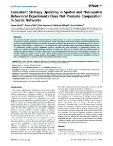
how many of the neurons were similarly modulated or instead showed a preference for “near” initial eye positions. Furthermore, this modulation by initial eye position could alternatively be explained by the type of eye movement required in each case, i.e., all of the eye movements made from near fixation points are diverging, whereas all of those made from far initial fixation points require a converging movement (Fig. 1). Such a possibility was not discussed by the authors. To investigate potential associations between postsaccadic activity and eye movement preparation, three epochs were considered: delay (300 ms before the go-signal), early (100 –300 ms) postsaccadic, and late (500 –700 ms) postsaccadic. The effect of initial eye position and target location was considered for each of these epochs to assess whether their respective influence on discharge rate varied throughout the trial. Whereas a modulation by target position was apparent in both the early and late postsaccadic periods, for the example neuron shown, the effect of initial eye position was restricted to only the early postsaccadic epoch [Genovesio et al. (2007), their Fig. 3 (http://www.jneurosci.org/cgi/ content/full/27/12/3268/F3)]. In the majority of neurons, a higher correlation was seen between activity in the delay and early (compared with late) postsaccadic periods [Genovesio et al. (2007), their Fig. 4 (http://www.jneurosci.org/cgi/content/ full/27/12/3268/F4)]. This study is important because, as mentioned above, previous investigations of saccade-related spatial updating focused on movements within a 2D plane, whereas in reality saccades are made in a three-dimensional (3D) visual environment. In addition to this, there are no differences in terms of perceptual experience for eye movements made to any location regardless of the hemifield, direction, or depth, i.e., spatial constancy is maintained throughout, suggesting that spatial updating must occur for all possible saccade vectors. Spatial updating in 3D (cf. Wexler,
Figure 1. Schematic figure of the task showing diverging eye movements made from a near initial eye position (a) and converging eye movements made from a far initial eye position (b), to eight possible targets located at the same retinal eccentricity but differing depths, as measured by vergence angle (v). FP, Fixation point; T, target.
2005) may be particularly important for reaching movements, for which it is essential that the location of an object in depth is accurately represented. If the preference for nearer target locations noted in this study holds true for the majority of neurons, this could perhaps be interpreted as an ecological adaptation whereby stimuli locations in peripersonal space, i.e., within reaching distance, invoke greater neural activity than those located out of reach. Such an idea could perhaps be tested by comparing neural activity for target locations located within either peripersonal or extrapersonal space. This study also differs from much of the previous work in considering how the predicted sensory consequences of a saccade are integrated with sensory reafference after the movement, rather than investigating anticipatory activity. Although presaccadic remapping is thought to be based on an efference copy of the motor command, the exact extraretinal signals used for postsaccadic integration processes are still a matter of uncertainty. The authors concluded that both vector subtraction and eye position signals may be used, which makes sense theoretically because this would presumably be more accurate than relying exclusively on only one of these sources of information. It will be interesting to investigate
postsaccadic activity in other areas that have been linked to saccade-related spatial updating processes, e.g., the frontal eye fields, superior colliculus, and visual cortex (Merriam et al., 2007). The current task could also be applied in humans using functional imaging and could be extended to include double or multistep saccade sequences, which have been used to investigate spatial updating in two dimensions.
References Barash S, Bracewell RM, Fogassi L, Gnadt JW, Andersen RA (1991) Saccade-related activity in the lateral intraparietal area. I. Temporal properties; comparison with area 7a. J Neurophysiol 66:1095–1108. Duhamel JR, Colby CL, Goldberg ME (1992) The updating of the representation of visual space in parietal cortex by intended eye movements. Science 255:90 –92. Genovesio A, Brunamonti E, Giusti MA, Ferraina S (2007) Postsaccadic activities in the posterior parietal cortex primates are influenced by both eye movement vectors and eye position. J Neurosci 27:3268 –3273. Heiser LM, Colby CL (2006) Spatial updating in area LIP is independent of saccade direction. J Neurophysiol 95:2751–2767. Merriam EP, Genovese CR, Colby CL (2007) Remapping in human visual cortex. J Neurophysiol 97:1738 –1755. Wexler M (2005) Anticipating the threedimensional consequences of eye movements. Proc Natl Acad Sci USA 102:1246 –1251.