Brain and Cognition 41, 9–26 (1999) Article ID brcg.1999.1093, available online at http://www.idealibrary.com on
Spatial Working Memory in Asperger’s Syndrome and in Patients with Focal Frontal and Temporal Lobe Lesions Robin G. Morris,* A. Rowe,† N. Fox,‡ J. D. Feigenbaum,‡ E. C. Miotto,† and P. Howlin§ *Neuropsychology Unit, †Department of Psychology, and ‡Department of Clinical Psychology, Institute of Psychiatry, London, United Kingdom; and §Department of Psychology, St. Georges Hospital, London, United Kingdom Spatial working memory (SWM) was investigated in 15 patients with Asperger’s syndrome (AS) comparing their performance to 18 age- and IQ-matched control subjects. An additional comparison was made with 20 unilateral frontal excision patients [9 right (RFL); 11 left (LFL)] and with 38 unilateral temporal lobectomy patients [18 right (RTL); 18 left (LTL)], the frontal and temporal lobe patients having separate matched control groups. SWM was tested using the Executive Golf Task, a test that also measures spatial strategy formation. The AS group showed a substantial deficit on SWM, but no impairment in strategy formation. The LFL showed the same pattern of impairment, but with a less substantial deficit. The RFL group showed a large deficit, but some of this was accounted for by a strategy formation impairment. Of the temporal lobe lesions groups, only the RTL group was impaired on SWM, but this group showed normal strategy formation. It was concluded that the SWM deficit in AS may reflect a more general difficulty in accessing different types of representations in order to guide voluntary behavior, providing at least a partial explanation for the executive deficits found in AS. 1999 Academic Press
Key Words: Asperger’s syndrome; spatial working memory.
INTRODUCTION
The term Asperger’s syndrome (AS) is commonly used for patients who have many of the clinical features of autism, but demonstrate normal intelligence (Wing, 1981). Although there is a debate whether AS constitutes a separate syndrome or is on a continuum with autism, certain characteristic features are apparent, including more abstract stereotypies and preserved language function, with the exception of abnormal intonation patterns. The neuropsychological profile of AS has been found to differ significantly from that Address correspondence to Robin G. Morris, Institute of Psychiatry, De Crespigny Park, London SE5 8AF, UK. E-mail:
[email protected]. 9 0278-2626/99/$30.00 Copyright 1999 by Academic Press All rights of reproduction in any form reserved.
10
MORRIS ET AL.
of high functioning autism (HFA) (see, for example, Ozonoff, Pennington, & Rogers, 1991a, b), although a lack of uniformity in the usage of the term may have contributed to difficulties in interpreting different studies (Klin et al., 1995). In common, both AS and HFA appear to share a deficit in executive function, the ability to sequence mental and behavioral functions in a coordinated fashion. The earliest study in this area was conducted by Rumsey (1985), who found that HFA patients were significantly more perseverative than normal on the Wisconsin Card Sorting Test (WCST). A similar result was found in a further study by Rumsey and Hamburger (1988), with impairment in the number of categories achieved on the WCST and impaired planning on the Tower of Hanoi test observed in the context of relatively preserved language and memory function and normal visuospatial abilities. Executive function has also been found to be predictive of outcome in men with HFA (Szatmari et al., 1990). A study by Ozonoff et al. (1991a, b) showed comparable WSCT and Tower of Hanoi deficits in both AS and HFA patients. The neurocognitive underpinnings of this deficits have been debated, but the absence of unimodal impairment in both cases has led to the notion of more central cognitive deficits affecting executive function. This would be the result of abnormal development of either the prefrontal cortex (Ozonoff et al., 1991a, b) or a more widespread cortical neural network that subserves the analysis of complex information patterns (Minshew & Goldstein, 1993). While the neuroanatomical aspects of both conditions are not fully understood, parallels have been long been drawn in terms of the behavioral characteristics of patients with frontal lobe damage. For example, Damasio and Maurer (1978) suggest that high functioning autistic individuals show a similar ‘‘lack of initiative, concreteness of thought, inability to focus attention and lack of apathy.’’ They proposed that the site of abnormality was the ring of mesolimbic cortex in the frontal lobes. While neuroanatomical studies have failed to show consistent abnormalities in autism (see Piven, Arndt, Bailey, Havercamp, Andreasen, & Palmer, 1995; Saitoh, Courchesne, Egaas, Lincoln, & Schreibman, 1995) or AS (Berthier, Bayes, & Tolosa, 1993), neuropsychological studies that compare these groups with focal frontal lesions may help to determine the extent to which the pattern of executive deficit is similar across conditions. The prefrontal cortex has been implicated in storing information that is used directly to govern behavior (Goldman-Rakic, 1987). This stemmed initially from studies of spatial working memory (SWM) in nonhuman primates. Such studies have shown severe impairments in SWM performance associated with bilateral lesions to the region of the sulcus principalis (cytoarchitectonic areas 46 and 9) or the mid-dorsolateral cortex; additional electrophysiological recordings have shown neuronal activity associated specifically with the retention period of a delayed response task (Funahashi, Bruce, & Goldman-Rakic, 1989; Wilson, Scalaidehe, & Goldman-Rakic,
SPATIAL WORKING MEMORY IN AS
11
1993). More recently, such regions have been shown to have a retinotopic representation of spatial location and a specific spatial/executive module has been suggested (Goldman-Rakic, 1996). Such a system (differing in characteristics from the simple rehearsal and storage mechanism proposed in the Baddeley and Hitch working memory model; cf. Baddeley, 1986) may play a key role in the holding of internal representations or hypothetical configurations to guide planning and problem solving. A possible impairment in this system has been proposed by Ozonoff et al. (1991a) to account for the central deficit that underlies the measured impairments in executive function. The aim of the current study was to explore (1) whether AS patients are impaired in SWM; (2) whether a putative impairment was the similar in kind to that shown by patients with prefrontal cortical lesions; a further comparison was made with patients who have undergone unilateral temporal lobectomies, to elucidate further the neuranatomical basis of SWM in humans; and (3) to determine whether the impairment was primary or secondary to other types of executive impairment, for example, impairment in the use of strategy during working memory performance. The performance of a set of AS patients was compared directly with previously collected data for patients with unilateral focal frontal and temporal lesions (Feigenbaum, Polkey, & Morris, 1996; Miotto, Polkey, Bullock, & Morris, 1996). METHOD
Subjects Asperger patients. The group included 15 adults, all of whom had a diagnosis of Asperger’s syndrome according to the ICD10 research criteria, but without the requirement for delay or deviance of language development; the diagnosis was made by an experienced clinician (see Table 1 for details). They all showed grossly intact semantic or structural characteristics in relation to current language function. They were selected if their Wechsler Intelligence Quotient (IQ) was above 70 and if free from known neurological conditions or other major physical or medical problems. Nine were right and six left-handed (Annett, 1970). A brief background neuropsychological assessment was administered, including a short form of the Wechsler Adult Intelligence Scale-Revised (WAIS-R) (Canavan, Dunn, & McMillan, 1986). Asperger controls. The subjects were compared to 18 control subjects matched approximately for sex, handedness, Verbal IQ, and Performance IQ. The inclusion criteria were a WAIS-R IQ over 70 and no psychiatric disorder, neurological impairment, or current other physical illness.1
1 The Asperger’s syndrome patients showed a higher proportion of left handers than normal. The controls for this group were selected to match in terms of proportion of left handers. This led to the decision to maintain separate control subjects for the Asperger’s syndrome and frontal and temporal lesion patient groups. The Asperger control patients, however, appear to be relatively worse on strategy formation than the FL and TL controls, as confirmed by a t test comparing the Asperger controls to a combined FL and TL control group on collapsed level 6 and 8 data (t(1, 18) ⫽ 24.2, p ⬍ .001).
12
MORRIS ET AL.
TABLE 1 Background Characteristics of Patient and Control Groups
AS (n ⫽ 15) ASCONT (n ⫽ 18) RFL (n ⫽ 9) LFL (n ⫽ 11) FCONT (n ⫽ 22) RTL (n ⫽ 20) LTL (n ⫽ 20) TLCONT (n ⫽ 20)
Sex M/F
Handedness Left/Right
Age (years)
14/1
9/6
16/1
11/7
3/6
7/2
4/7
8/3
9/13
17/5
7/13
0/20
8/12
0/20
9/11
0/20
29.5 (19–49) 29.4 (19–45) 35.3 (19–69) 35.3 (19–69) 36.0 (24–62) 35.0 (18–49) 34.0 (20–50) 36.0 (18–59)
NART-R
VIQ
PIQ
—
99.0 (81–129) 106.3 (96–137) —
100.1 (84–137) 105.8 (78–136) —
—
—
— 102.2 (80–121) 102.2 (80–121) 103.5 (82–112) 104.5 (80–124) 105.0 (91–120) 110.8 (89–129)
— —
—
—
—
—
—
Note. (Groups: Asperger’s syndrome (AS); Asperger’s control (ASCONT); right frontal (RFL); left frontal (LFL); frontal control (FCONT); right temporal lobe (RTL); left temporal lobe (LTL); temporal lobectomy control (TLCONT).
Frontal lobe excision patients. There were 20 patients who had undergone unilateral frontal lobe neurosurgery (11 left; 9 right) (see also Miotto et al., 1996). The verification of the locus and the extent of the lesion were established both by neurosurgeons’ drawings at the time of the operation and by use of CT and MRI scanning. Of the 11 left frontal (LF) patients, 6 had frontal lobectomies for the relief of intractable epilepsy, 2 for the removal of an astrocytoma, 1 for a traumatic lesion associated with insertion of a shunt, and 1 for removal of an arteriovenous malformation. Of the right frontal (RF) group, 4 had undergone treatment for the relief of epilepsy, there were 2 cases where a meningioma was removed, 1 case of an anterior communicating aneurysm that had been clipped, and 1 case where a glioma had been removed (for diagrams showing the extent of cortical excision, see Miotto et al., 1996). Temporal lobe excision patients. These included 40 patients who had undergone unilateral temporal lobectomies (20 left and 20 right hemisphere operations) (see also Feigenbaum et al., 1996). They had undergone neurosurgery for the treatment of epilepsy, involving a standard en bloc resection (Polkey, 1989), which involves removal of 5.5 to 6.5 cm of medial temporal tissue from the anterior pole, including the amygdala and approximately the anterior two thirds of the hippocampus. Effectively, the anterior temporal lobe is removed completely, with perirhinal and entorhinal cortices nonexistent on the operated side (see Morris et al. (1999) and Nunn et al. (1999) for further details of the operation and magnetic resonance imaging analysis). These patients were studied after a minimum of 6 months postoperatively. Focal lesion control groups. Separate control groups were used for the frontal and temporal surgical lesions groups, in each case matched approximately for age and intelligence, as estimated using the National Adult Reading Test—Revised (NART-R) (see Table 1). The same inclusion criteria were used for these control groups.
SPATIAL WORKING MEMORY IN AS
13
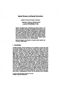
FIG. 1. Illustration of the layout for the Executive-Golf Task at the eight-hole level. Note the golfer in the distance.
Procedure The spatial working memory paradigm was based upon the task devised originally by Morris et al. (1988; Owen et al., 1996). This task, termed the Executive-Golf Task was administered on a DELL 386P/50 desktop computer with a TAXAN 775 14-in. monitor fitted with a touchsensitive screen and written in Qbasic-45, using three-dimensional graphics. On the screen is a representation of a golf course and a golfer (see Fig. 1). On the golf course are a specified number of ‘‘golf holes’’ into which the golfer must ‘‘putt’’ an equal number of golf balls. Initially, the subject is instructed to guess which hole the golfer is going to putt the ball into, the guess signaled by touching the chosen hole. If this is correct, the computer emits a ‘‘positive’’ tone and the golfer is seen to putt the ball into the hole. If incorrect, the golf hole flag turns red and a ‘‘negative’’ tone is given. The subject continues trying each hole in turn until the correct one is found. The subject is instructed to then search for another hole into which the golfer is predicted to putt a further ball. They are also warned not to return to holes that have already had balls putted into them and that this counts as an error. The subject proceeds with a series of searches until all the balls have been placed in the separate holes. At the end of this ‘‘game’’ a further trial is administered according to the overall design. The task was divided into four increasing levels of difficulty, starting with instruction trials with just two holes, then test trials with three, four, six, and eight holes (in practice, only the level 4–8 data were analyzed, since level 3 trials produced minimal errors). At level 2, there were two trials; with levels 3, 4, 6, and 8, four trials. The sequence of correct holes was determined by a pseudorandom function based on the pattern of searching by the subject.
RESULTS
For each analysis the patient groups were compared to their relevant control groups. Thus the data were divided into either two (AS versus controls)
14
MORRIS ET AL.
or three groups (LFL versus RFL versus controls; LTL versus RTL versus controls). Analyses of variance were conducted (ANOVA and MANOVA) on the data, in each case using a square root transformation to produce approximate homogeneity of variance across the cells of the group by level of difficulty variable. Spatial Working Memory Errors Two types of memory errors were recorded by the computer for each trial: (1) Within-Search Errors, where the subject ‘‘tried’’ the same hole more than once during a search for the correct hole, and (2) Between-Search Errors, in which the subject ‘‘tried’’ a hole that has already been successful on a previous search. For each measure, the mean number made in each trial at each difficulty level was computed for subsequent analysis. First, the AS patients and their respective controls were compared on their number of Within-Search Errors using a MANOVA, with Group as the between-subjects factor and difficulty level (four, six, and eight holes) as the within-subject factor (see Fig. 2a). This revealed a main effect of Group (F ⫽ 8.2, df ⫽ 1, 31, p ⬍ .001) and of Difficulty (F ⫽ 27.2, df ⫽ 2, 62; p ⬍ .0001), with a significant Group X Difficulty interaction (F ⫽ 3.4, df ⫽ 2, 62, p ⬍ .05). Further analyses revealed the AS group to be significantly impaired on levels 6 and 8 only (respectively: (F(1, 31) ⫽ 5.32, p ⬍ .05); (F(1, 31) ⫽ 6.57, p ⬍ .05)). For the Between-Search Errors (see Fig. 3a) the MANOVA again showed a main effect of Group (F ⫽ 21.8, df ⫽ 1, 31, p ⬍ .0001) and of Difficulty (F ⫽ 132.3, df ⫽ 2, 62; p ⬍ .0001), with a significant Group X Difficulty interaction (F ⫽ 5.5, df ⫽ 2, 62, p ⬍ .01). Here, the AS group were impaired at levels 4, 6, and 8 (respectively: (F(1, 31) ⫽ 12.02, p ⬍ .01); (F(1, 31) ⫽ 19.38, p ⬍ .001); (F(1, 31) ⫽ 15.97, p ⬍ .001)). Thus, on both measures, the AS group were impaired, the extent of impairment increasing with the difficulty level of the task. For the unilateral frontal lobe lesion patients a MANOVA on the WithinSearch Errors showed no significant main effects or interactions. As shown in Fig. 2b, there were very few errors made by any subjects at all the levels of difficulty. For the Between-Search Errors (see Fig. 3b) there was a significant main effect of Group (F ⫽ 13.8; df ⫽ 2, 39; p ⬍ .01) and a Group X Difficulty interaction (F ⫽ 9.5; df ⫽ 4, 78, p ⬍ .001). This reflected the impairments of the two patient groups, emerging with the higher difficulty levels. One-way ANOVA’s at the two highest Difficulty levels revealed sig-
FIG. 2. Graphs showing mean numbers of Within-Search Errors. On the top is the comparison of the Asperger’s syndrome (AS) and control group (ASCONT). In the middle is the comparison between the right and left prefrontal lesions patients (RFL; LFL) and relevant controls (FCONT); At the bottom are the right and left unilateral temporal lobectomy (RTL; LTL) and control data (TLCONT). Standard errors are given.
SPATIAL WORKING MEMORY IN AS
15
16
MORRIS ET AL.
nificant differences (level 6: F ⫽ 9.0; df ⫽ 2, 39; p ⬍ .001; level 8: F ⫽ 14.6; df ⫽ 2, 39; p ⬍ .001). Posthoc LSD tests revealed that the RFL and LFL patients were significantly impaired relative to their controls at levels 6 and 8, but at level 8 the RFL were also significantly worse than the LFL group. These data were also analyzed with respect to the location of the lesion, since there was significant heterogeneity of lesion size and position within the frontal lobe group (see Miotto et al., 1996, for diagrams showing the extent of the cortical lesions). To investigate these factors, the patients were classified according to whether the overall area of the lesion exceeded 4.5 cm 2 or not, based on the surgeons’ drawings. Eight patients had large and 12 small lesions. A series of ANOVA’s were used to compare the resultant two groups on the between-subject errors for levels 4, 6, and 8, but this did not reveal any significant differences. The patients were also split according to the location of the lesion, generating three groups (orbitofrontal ⫽ 6; dorsolateral ⫽ 7; lesions involving both regions ⫽ 7) (a further two patients could not be categorized in this fashion). One-way ANOVA’s were performed on the between-subject data, with posthoc comparisons using LSD. This revealed no significant differences. For the Unilateral Temporal Lobectomy Patients the MANOVA on the Within-Search Errors (see Fig. 2c) showed no main effect of group, but a significant effect of Difficulty (F ⫽ 30.7, df ⫽ 2, 114, p ⬍ .0001) and no interaction between the factors. In common with the frontal lobe patients and controls, there were very few errors, with some emerging at the highest level. For the Between-Search Errors (see Fig. 3c) the main effect of Group was not significant, but there was a main effect of Difficulty (F ⫽ 230.7, df ⫽ 4, 114, p ⬍ .001) and a Group X Difficulty interaction (F ⫽ 2.5, df ⫽ 4, 114; p ⬍ .05). Further one-way ANOVA’s were used to explore these interactions, comparing the three groups at the different difficulty levels. This showed trends toward significant effects of Group at level 6 (F ⫽ 3.1, df ⫽ 2, 57, p ⫽ .053) and level 8 (F ⫽ 2.4, df ⫽ 2, 57, p ⫽ .10). Posthoc Least Significant Difference analyses at each level revealed the RTL group to be significantly impaired in comparison to the controls at levels 6 and 8, but no other differences. Strategy Formation As indicated above, the subjects were able to aid their performance on the task by adopting a particular strategy. The most efficient one was to
FIG. 3. Graphs showing the mean number of Between-Search Errors, the error bars showing standard errors. (Groups: Asperger’s syndrome (AS); Asperger’s control (ASCONT); right frontal (RFL); left frontal (LFL); frontal control (FCONT); right temporal lobe (RTL); left temporal lobe (LTL); temporal lobectomy control (TLCONT).
SPATIAL WORKING MEMORY IN AS
17
18
MORRIS ET AL.
FIG. 4. An example of an efficient search strategy used by a left temporal lobectomy subject (after Feigenbaum et al., 1996).
follow a predetermined search sequence in which the subject begins each search always at the same location and then maintains the same search path, but skipping holes that have been successful on previous searches. A measure of this strategy is the number of different starting locations used in each trial, where a low number indicates efficient use of the strategy. An example of this is given in Fig. 4, where the locations are coded numerically. In this example, the subject starts with location 8 until this is successful (second search). They then switch to the next location used in the first search (7), skipping 8. This starting position is used consistently for four searches until the subject serendipitously drops the strategy, switches to 6, and is successful. Figure 5 shows the strategy data for the different groups at difficulty levels 6 and 8. For the AS versus controls comparison, a MANOVA with Group as the between-subjects factor and Difficulty as the within-subject factor showed only a main effect of difficulty (F ⫽ 103.4, df ⫽ 1, 31, p ⬍ .001). The same analysis for the unilateral frontal lobe lesions showed a significant main effect of Group (F ⫽ 6.7; df ⫽ 1, 39; p ⬍ .01) and a significant interac-
SPATIAL WORKING MEMORY IN AS
19
tion between Group and Difficulty (F ⫽ 5.2; df ⫽ 3, 79; p ⬍ .01). Subsequent ANOVA’s at levels 6 and 8 revealed significant effects of group (level 6: F ⫽ 5.0; df ⫽ 2, 39; p ⬍ .05; level 8: F ⫽ 6.9; df ⫽ 2, 39; p ⬍ .01). Posthoc LSD tests showed that the RFL group showed significantly worse strategy formation than the controls and the LFL group at both levels 6 and 8; the LFL showed normal strategy formation. As a further exploration, the strategy formation measure was included as covariate when exploring the spatial working memory deficit in the RFL group, to investigate whether there might be a causal relationship between the two. When comparing the RFL and LFL groups, the significant difference in spatial working memory disappeared, but the difference remained significant, when comparing the RFL groups and their controls (F ⫽ 9.2; df ⫽ 1, 30, p ⬍ .01). The patients were also split into those with small and large lesions, as described above. No differences in strategy formation were observed. A further split into the three lesion location groups again did not reveal any group differences. Finally, when analyzing strategy formation for the unilateral temporal lobectomy patients, the MANOVA revealed no significant main effect of Group, a significant effect of Difficulty (F ⫽ 179.4, df ⫽ 1, 57, p ⬍ .0001), but no interaction. DISCUSSION
In summary, the AS patients showed a clear deficit in spatial working memory, as indicated by both the within- and between-search errors. This can be compared with the unilateral frontal lesion patients, where both RFL and LFL groups were impaired, but with only the RFL group showing a similar level of deficit. Of the unilateral temporal lobectomy patients only the RTL group were impaired, the level of performance being similar to that of the LFL group. The AS patients were not impaired in strategy formation, with only the RFL group showing a deficit on this measure. In the case of the RFL group, strategy formation impairment accounted for a significant portion of the spatial working memory deficit, but this clearly was not the case in the AS group. Thus it appears that the pattern of spatial working memory impairment does not follow exactly that observed in the focal brain lesion patients. The spatial working memory deficit was numerically the largest of the five patient groups, yet there was no strategy formation impairment. Impairments in executive function in both AS and HFA have been reported previously and this study suggests impairment in one facet of this function, namely, the ability to hold information temporally in mind, necessary to guide and coordinate cognition and behavior (Goldman-Rakic, 1996). Previous studies have shown, for example, difficulties on the Tower of Hanoi task, in which the patient has to rearrange a number of discs, threaded into three rods, to achieve a goal position. In the study by Ozonoff et al. (1991b)
20
MORRIS ET AL.
the measure from this task was combined with the Wisconsin Card Sorting Task to provide a composite score that successfully discriminated the AS group from the control subjects. These tasks invoke a range of different cognitive components, including, for example, working memory and response inhibition. A further decomposition of these components would help establish the nature of the executive impairment in AS. Perhaps, as suggested by the large impairment measured in the current study, a core aspect is impairment in working memory, as had indeed been suggested originally by Ozonoff et al. (1991b). Further experiments in which, for example, the working memory demand of the Tower of Hanoi plan are measured directly would be of interest in exploring the link between these factors. Rogers and Pennington (1991) have speculated on what causes the social deficits found in AS, given that this group do not appear to have theory of mind difficulties (Bowler, 1992; Ozonoff et al., 1991b). One possibility is that a combination of emotion perception and executive function deficits leads to symptoms. The former may relate to a wider prosodic/pragmatic deficit, while the latter may limit the flexibility of thought needed in social interaction. Failure to hold in mind pertinent information during social interaction could be explained by a deficit in working memory, another factor that could be explored directly in a more naturalistic setting, for example, by testing in a group setting the ability to keep track of what which participants have contributed to a conversation (Alberoni, Baddeley, Della Sala, Logie, & Spinnler, 1992). In the current study, the task was specifically spatial in nature, and further investigation is needed to explore whether the working memory deficit applies to other types of material or modalities. The link between executive dysfunction and social impairment has also been made in relation to children with prefrontal cortical lesions, for example, the case JC described by Eslinger, Biddle, and Grattan (1997), whose working memory impairments appeared to contribute to ‘‘emerging difficulties in his understanding of group interactions, social discourse and the social behaviour of others’’ as an early teenager. On the Executive-Golf Task, two types of errors were recorded; the Within-Search Error, in which the subject selects a location that has been tried previously within any particular search, and the Between-Search Error, which involves returning to a location that was successful on a previous search. A notable finding was that the AS patients were the only group to show significant within-search errors. This suggests that these patients are not able to remember simple sequences of spatial locations. Previously, MiFIG. 5. Graphs showing the means for the Strategy Formation Score. A high score means poorer strategy use. The error bars showing standard errors. (Groups: Asperger’s syndrome (AS); Asperger’s control (ASCONT); right frontal (RFL); left frontal (LFL); frontal control (FCONT); right temporal lobe (RTL); left temporal lobe (LTL); temporal lobectomy control (TLCONT).
SPATIAL WORKING MEMORY IN AS
21
22
MORRIS ET AL.
otto et al. (1996) suggested that this type of memory is likely to be the equivalent of the Visuo-Spatial Scratchpad, involved in the retention of small amounts of visuospatial information in immediate memory (Logie & Marchetti, 1991). To avoid between-search errors, the locations have to be held in memory for a much longer time period, with the possibility of interference emerging between searches because of the overlapping requirements to monitor the immediate sequence within a search and avoid an accumulating set to locations. It is possible that the prefrontal cortex is responsible for the latter function, hence the large and consistent deficit in the prefrontal surgical lesion patients. Avoidance of between-search errors is likely to involve a long-term memory component, particularly with the level 8 condition, and this may account for the small, but significant deficit in the RTL group, who tend to show deficits in spatial long-term memory tasks (Abrahams et al., 1997, 1999; Morris et al., 1996, 1999). Since this deficit is relatively slight, this might imply that most of the mnemonic demand of the task relates to working memory or executive function. The current study compares patients with AS with those with focal frontal and temporal lobe lesions. This comparison is guided partly by the known executive deficits associated with AS and also past theorizing about the involvement of the temporal lobes in the etiology of autism (cf. Bachevalier, 1994). Little is known about the neurobiology of AS, but the executive deficits may call into question the integrity of prefrontal cortical functioning. The link between spatial working memory and the prefrontal cortex comes not only from studying patients with surgical lesions (Miotto et al., 1996; Owen et al., 1996), but through functional neuroimaging. Activation Positron Emission Tomography studies have indicated prefrontal activation during memory location ( Jonides, Smith, Koeppe, Awh, Minoshima, & Mintun, 1993) and this appears to be dissociated, in terms of the precise neuronal structures involved, from memory for other types of material (McCarthy, Puce, Constable, Krystal, Gore, & Goldman-Rakic, 1996). In AS, structural neuroimaging studies have not been conducted to date, in order to reveal any structural abnormalities in the prefrontal cortex, nor has functional neuroimaging been used to determine whether regions of prefrontal hypoperfusion exist, associated with executive function. It remains a possibility that a parallel can be drawn with schizophrenia, where prefrontal hypoperfusion has been demonstrated in the absence of convincing structural abnormalities (Weinberger, Berman, & Daniel, 1991), and also there exist deficits in spatial working memory (Keefe, Roitman, Harvey, Blum, DuPre, Prieto, Davison, & Davis, 1995). In this case, the abnormalities in prefrontal cortical functioning have been more recently related to faulty frontal–temporal lobe connectivity (Bullmore, Horwitz, Morris, Curtis, Maguire, Sharma, Williams, Murray, & Brammer, 1997; Frith, Friston, Herold, Silberswieg, Fletcher, Cahill, Dolan, Frackowiak, & Liddle, 1995; Weinberger, 1991), a possibility that exists for AS.
SPATIAL WORKING MEMORY IN AS
23
Alternative neuroanatomical hypotheses have been put forward in relation to autism, where damage to the hippocampus has been proposed to explain at a neurodevelopmental level abnormalities in language, pragmatics, and the construction of flexible structures for meaning (DeLong, 1991). This type of theorizing also makes the link between autism and amnesia, partly because of the known involvement of the hippocampus in memory function. For example, Boucher and Warrington (1976) have drawn attention to the apparent similarities between the amnesic syndrome and autism in children, with the latter showing impairments in learning unrelated word pairs. More recent studies have tended not to support this link, partly because autistic children are not deficient in cued recall (Boucher & Lewis, 1989; Boucher & Warrington, 1976), in contrast to amnesic patients. This applies also to patients with AS, who show normal cued recall, even though, in common with autistic individuals, they fail to use semantic information to aid recall (Bowler, Matthews, & Gardiner, 1997). The current results relate to spatial working memory, but they show at least that the performance of the AS patients is very different from that of the temporal lobectomy patients, albeit with unilateral hippocampal lesions. One potential explanation for the spatial working memory deficit is that it is part of a more general impairment in nonverbal abilities in AS. For example, Klin, Volkmar, Sparrow, Cicchetti, and Rourke (1995) compared patients with AS and HFA on Verbal and Performance Intelligence. They found that while the Verbal Intelligence of the AS was significantly higher than that for the HFA, the reverse was true for Performance Intelligence. They draw attention to the similarities of the neuropsychological profile in AS with the Non-Verbal Disabilities Syndrome (Rourke, 1989). Selective case studies have been reported in which hypoperfusion of the right hemisphere in AS has been detected using single photon emission computed tomography (McKelvey, Lambert, Mottron, & Shevell, 1995). The current sample, however, did not show a difference between Verbal and Performance Intelligence and, as such, is in keeping with other groups of AS patients (e.g., Ozonoff et al., 1991b; Szatmari et al., 1990). The discrepancy with the Klin et al. (1995) sample may relate to their more stringent definition of the syndrome, including additional criteria relating to motor function and allabsorbing special skills or activities. The current patients showed the spatial working memory deficit even in the absence of a low Performance Intelligence, indicating that these aspects are not related, at least in this group of AS patients. In this sense, the spatial working memory deficit cannot simplistically be related to abnormalities in the right hemisphere, the region implicated in the nonverbal disability syndrome (Brumback, Harper, & Weinberg, 1996). Thus it appears that the AS patients have a deficit on spatial working memory performance not accounted for by problems with strategy formation. This deficit extents to simple within-search errors and more complex
24
MORRIS ET AL.
between-search errors and was found in the context of intellectual functioning within the average range. Perhaps difficulties in holding in mind information to guide and direct behavior is a major aspect of executive function in this group, an area which needs further exploration in the future. REFERENCES Abrahams, S., Pickering, A., Polkey, C. E., & Morris, R. G. 1997. Spatial memory deficits in patients with unilateral damage to the right hippocampal formation. Neuropsychologia, 35, 11–24. Abrahams, S., Morris, R. G., Polkey, C. E., Jarosz, J. M., Cox, T. C. S., Graves, M., & Pickering, A. 1999. Hippocampal involvement in spatial and working memory: A structural MRI analysis of patients with unilateral mesial temporal lobe sclerosis. Brain and Cognition, 41, 39–65. Alberoni, M., Baddeley, A. D., Della Sala, S., Logie, R. H., & Spinnler, H. 1992. Keeping track of a conversation: impairments in Alzheimer’s disease. International Journal of Geriatric Psychiatry, 7, 639–646. Annett, M. 1970. A classification of hand preference by association analysis. British Journal of Psychology, 61, 427–430. Bachevalier, J. 1994. Medial temporal lobe structures and autism: a review of clinical and experimental findings. Neuropsychologia, 32, 627–648. Baddeley, A. D. 1986. Working memory: Theory and practice. Oxford: Clarendon Press. Berthier, M. L., Bayes, A., & Tolosa, E. S. 1993. Magnetic resonance imaging in patients with concurrent Tourette’s disorder and Asperger’s syndrome. Journal of the American Academy of Child and Adolescent Psychiatry, 32(3), 633–639. Brumback, R. A., Harper, C. R., & Weinberg, W. A. 1996. Non-verbal learning disabilities, pervasive developmental disorder—Should we care? Journal of Child Neurology, 11(6), 427–429. Boucher, J., & Warrington, E. K. 1976. Memory deficits in early infantile autism: Some similarities to the amnesic syndrome. British Journal of Psychology, 67, 73–87. Boucher, J., & Lewis, V. 1989. Memory impairment and communication in relatively able autistic children. Journal of Child Psychology and Psychiatry, 30, 99–122. Bowler, D. 1992. ‘‘Theory of Mind’’ in Aspergeis syndrome. Journal of Child Psychology and Psychiatry, 33, 877–893. Bowler, D. M., Matthews, N. J., & Gardiner, J. M. 1997. Asperger’s syndrome and memory: Similarity to autism but not amnesia. Neuropsychologia, 35(1), 65–70. Bullmore, E. T., Horwitz, B., Morris, R. G., Cutis, V. A., McGuire, P. K., Sharma, T., Williams, S. C. R., Murray, R. M., & Brammer, M. J. 1997. Causally connected cortical networks for language in functional (MR) brain images of normal and schizophrenic subjects. [submitted manuscript] Canavan, A. G., Dunn, G., & McMillan, T. M. 1986. Principal components of the WAIS— R. British Journal of Clinical Psychology, 25(2), 81–85. Damasio, A. R., & Maurer, R. G. 1978. A neurological model for childhood autism. Archives of Neurology, 35, 777–786. Eslinger, P. J., Biddle, K. R., & Grattan, L. M. 1997. Cognitive and social development in children with prefrontal cortical lesions. In N. A. Krasnegor, S. R. Lyon, & P. S. Goldman-Rakic (Eds.), Development of the prefrontal cortex: Evolution, neurobiology and behaviour (pp. 295–335).
SPATIAL WORKING MEMORY IN AS
25
Feigenbaum, J. D., Polkey, C. E., & Morris, R. G. 1996. Deficits in spatial working memory after unilateral temporal lobectomy in man. Neuropsychologia, 34(3), 163–176. Frith, C. D., Friston, K. J., Herold, S., Silbersweig, D., Fletcher, P., Cahill, C., Dolan, R. J., Frackowiak, R. S. J., & Liddle, P. F. 1995. Regional brain activity in chronic schizophrenic patients during the performance of a verbal fluency task. British Journal of Psychiatry, 167, 243–349. Funahashi, S., Bruce, C., & Goldman-Rakic, P. S. 1989. Mmemonic coding of visual space by neurons in the monkey’s dorsolateral prefrontal cortex revealed by an oculomotor delayed-response task. Journal of Neurophysiology, 8, 4693–4706. Goldman-Rakic, P. S. 1987. Circuitry of primate prefrontal cortex and regulation of behavior by representational memory. In V. B. Mountcastle, F. Plum, & S. R. Geiger (Eds.), Handbook of physiology: The nervous system (pp. 373–417). Bethesda, MD: Am. Physiol. Soc. Goldman-Rakic, P. S. 1996. The prefrontal landscape: Implications of functional architecture for understanding human mentation and the central executive. Philosophical Transactions of the Royal Society London B: Biological Science, 351(1346), 1445–1453. Jonides, J., Smith, E. E., Koeppe, R. A., Awh, E., Minoshima, S., & Mintun, M. A. 1993. Spatial working memory in humans as revealed by PET. Nature, 353, 623–625. Keefe, R. S. E., Lees Roitman, S. E., Harvey, P. D., Blum, C. S., Du Pre, R. L., Prieto, D. M., Davidson, M., & Davis, K. L. 1975. A pen-and-paper human analogue of monkey prefrontal cortex activation task: Spatial working memory in patients with schizophrenia. Schizophrenia Research, 16, 87–110. Klin, A., Volkmar, F. R., Sparrow, S. S., Cicchetti, D. V., & Rourke, B. P. 1995. Validity and neuropsychological characterisation of Asperger’s syndrome: Convergence with nonverbal learning disabilities syndrome. Journal of Child Psychology and Psychiatry, 36(7), 1127–1140. McCarthy, G., Puce, A., Constable, R. T., Krystal, J. H., Gore, J. C., & Goldman-Rakic, P. 1996. Activation of human prefrontal cortex during spatial and nonspatial working memory tasks measured by functional MRI. Cerebral Cortex, 6(4), 600–611. McKelvey, J. R., Lambert, R., Mottron, L., & Shovell, M. I. 1995. Right hemisphere dysfunction in Asperger’s syndrome. Journal of Child Neurology, 10(4), 310–314. Minshew, N. J., & Goldstein, G. 1993. Is autism an amnestic disorder? Evidence from the California Verbal Learning Test. Neuropsychology, 7, 209–216. Miotto, E. C., Bullock, P., Polkey, C. E., & Morris, R. G. 1996. Spatial working memory and strategy formation in patients with frontal lobe excisions. Cortex, 32, 613–630. Morris, R. G., Downes, J. J., Sahakian, B. J., Evender, J. L., Heald, A., & Robbins, T. W. 1988. Planning and spatial working memory in Parkinson’s disease. Journal of Neurology, Neurosurgery, and Psychiatry, 51, 757–766. Morris, R. G., Pickering, A., Abrahams, S., & Feigenbaum, J. D. 1996. Space and the hippocampal formation in humans. Brain Research Bulletin, 40, 487–490. Morris, R. G., Nunn, J. A., Abrahams, S., Feigenbaum, J. D., & Recce, M. 1999. The Hippocampus and spatial memory in humans. In N. Burgess, K. J. Jeffrey, & J. O’Keefe (Eds.), The hippocampal and parietal foundations of spatial cognition. Oxford: Oxford Univ. Press. Nelson, H. E., & Willison, J. R. 1991. National Adult Reading Test (NART): Test manual (2nd ed.). Windsor: NFER-Nelson. Nunn, J. A., Polkey, C. E., & Morris, R. G. 1999. Differential spatial memory impairment after right temporal lobectomy demonstrated using temporal titration. Brain. [in press] Owen, A. M., Morris, R. G., Sahakian, B. J., Polkey, C. E., & Robbins, T. W. 1996. Double
26
MORRIS ET AL.
dissociations of memory and executive functions in working memory tasks following frontal lobe excisions, temporal lobe excisions or amygdalo-hippocampectomy in man. Brain, 119(5), 1597–1615. Ozonoff, S., Pennington, B. F., & Rogers, S. J. 1991a. Executive function deficits in highfunctioning autistic individuals: Relationship to theory of mind. Journal of Child Psychology and Psychiatry, 32(7), 1081–1105. Ozonoff, S., Pennington, B. F., & Rogers, S. J. 1991b. Asperger’s Syndrome: Evidence of an empirical distinction from high-functioning autism. Journal of Child Psychology and Psychiatry, 32, 1107–1122. Polkey, C. E. 1989. Surgery for epilepsy. Archives of Diseases of Childhood, 64, 185–187. Piven, J., Arndt, S., Bailey, J., Havercamp, S., Andreasen, N. C., & Palmer, P. 1995. An MRI study of brain size in autism. American Journal of Psychiatry, 152(8), 1145–1149. Rogers, S. J., & Pennington, B. F. 1991. A theoretical approach to the deficits in infantile autism. Development and Psychopathology, 3(2), 137–162. Rourke, B. 1989. Nonverbal learning disabilities: The syndrome and the model. New York: Guilford Press. Rumsey, J. M. 1985. Conceptual problem-solving in highly verbal, nonretarded autistic men. Journal of Autism and Developmental Disorders, 15, 23–36. Rumsey, J. M., & Hamburger, S. D. 1988. Neurosychological findings in high-function men with infantile autism, residual state. Journal of Clinical Experimental Neuropsychology, 10, 201–221. Saitoh, O., Courchesne, E., Egaas, B., Lincoln, A. J., & Schreibman, L. 1995. Cross-sectional area of the posterior hippocampus in autistic patients with cerebellar and corpus callosum abnormalities. Neurology, 45(2), 317–324. Szatmari, P., Tuff, L., Finlayson, M. A. J., & Bartolucci, G. 1990. Asperger’s syndrome and autism: Neurocognitive aspects. Journal of the American Academy of Child and Adolescent Psychiatry, 29, 130–136. Weinberger, D. R. 1991. Anteromedial temporal-prefrontal connectivity: A functional neuroanatomical system implicated in schizophrenia. In Carroll, B. J., & Barrett, J. (Eds.), Psychopathology and the brain. New York: Raven Press, pp. 25–43. Weinberger, D. R., Berman, K. F., & Daniel, D. G. 1991. Prefrontal cortex dysfunction. In H. S. Levin, H. M. Eisenberg, & A. L. Benton (Eds.), Frontal lobe function and dysfunction. Oxford: Oxford Univ. Press. Wilson, F. A. W., Scalaide, S. P. O., & Goldman-Rakic, P. S. 1993. Dissociation of object and spatial processing domains in primate prefrontal cortex. Science, 296, 1955–1958. Wing, L. 1981. Apserger’s syndrome: A clinical account. Psychological Medicine, 11, 115– 130.


![[PDF] Download Aspergers and Adulthood: A Guide to Working ...](https://m.moam.info/img/260x300/pdf-download-aspergers-and-adulthood-a-guide-to-wo_64785f27097c474e708cbd6c.jpg)