APPLIED AND ENVIRONMENTAL MICROBIOLOGY, Oct. 2005, p. 5908–5919 0099-2240/05/$08.00⫹0 doi:10.1128/AEM.71.10.5908–5919.2005 Copyright © 2005, American Society for Microbiology. All Rights Reserved.
Vol. 71, No. 10
Specific Detection, Isolation, and Characterization of Selected, Previously Uncultured Members of the Freshwater Bacterioplankton Community† Frederic Gich, Karin Schubert, Alke Bruns,‡ Herbert Hoffelner,§ and Jo ¨rg Overmann* Bereich Mikrobiologie, Ludwig-Maximilians-Universita ¨t Mu ¨nchen, Maria-Ward-Str. 1a, D-80638 Mu ¨nchen, Germany Received 9 February 2005/Accepted 6 May 2005
High-throughput cultivation was combined with rapid and group-specific phylogenetic fingerprinting in order to recover representatives of three freshwater bacterioplankton communities. A total of 570 bacterial cultures were obtained by employing the most probable number and MicroDrop techniques. The majority of the cultured bacteria were closely related to previously uncultured bacteria and grouped with the ␣-Proteobacteria, -Proteobacteria, Actinobacteria, Firmicutes, or Flavobacteria-Cytophaga lineage. Correspondingly, the natural bacterioplankton community was analyzed by high-resolution phylogenetic fingerprinting of these five bacterial lineages. 16S rRNA gene fragments were generated for each lineage and subsequently separated by denaturing gradient gel electrophoresis. By the combination of five group-specific PCR protocols, the total number of 16S rRNA gene fingerprints generated from the natural communities was increased sixfold compared to conventional (eubacterial) fingerprinting. Four of the environmental ␣-Proteobacteria 16S rRNA gene sequences obtained from the natural community were found to be identical to those of bacterial isolates. One of these phylotypes was detected in 14 different cultures and hence represented the most frequently cultured bacterium. Three of these 14 strains were characterized in detail. Their complete 16S rRNA gene sequences showed only 93% similarity to that of Sandaracinobacter sibiricus, the closest relative described so far. The novel phylotype of bacterium is a strict aerobe capable of using numerous organic carbon substrates and contains bacteriochlorophyll a bound to two different photosynthetic light-harvesting complexes. Dot blot hybridization revealed that the strains occur in lakes of different trophic status and constitute up to 2% of the microbial community.
High-throughput approaches for extinction culturing in small volumes of low-nutrient media have recently been established and applied to bacterioplankton samples (9, 10, 14). As an alternative separation method, bacterial cells have been encapsulated in gel microdroplets and cultivated in flowthrough systems monitoring bacterial growth by flow cytometry (62). Using the MicroDrop microdispenser system (9), large arrays of small cultures in microtiter plates can be inoculated with 170-pl droplets containing single bacterial cells, thereby generating 96 cultures in less than 1 minute. Due to the high number of replicates at a high dilution, highly enriched or even pure cultures are obtained at a much higher frequency than with conventional most probable number (MPN) dilutions (9). In order to analyze the large number of isolates generated, efficient and at the same time sufficiently sensitive screening techniques are required. Fingerprinting of 16S rRNA gene fragments by denaturing gradient gel electrophoresis (DGGE) provides a means for the rapid comparison of isolates with environmental sequences (8, 9, 10, 26, 40). When conventional eubacterial PCR primers are employed, however, bacteria that constitute ⱕ9% of complex microbial communities may not be detected by DGGE (51). Based on available diversity estimates (between 300 and 600 species in as little as 2.2 liters of water) (1, 26, 56) and based on the theoretical model of log-normal distribution of species abundance, most species are expected to occur at a very low abundance in marine bacterioplankton communities (16) and thus will escape detection by conventional DGGE analyses. Indeed, previous analyses of such
Planktonic bacteria mediate a significant proportion of the carbon turnover in freshwater lakes (17). So far, however, a more comprehensive understanding of the functions of freshwater bacteria has been hindered by the limited information on their physiology. Very few (in some cases as little as 0.001%) of the cells multiply in laboratory media (3). Even in improved, dilute culture media, the cultivation success of freshwater bacteria may not exceed 1.4% (9, 45). Culture-independent 16S rRNA-based studies indicate that the previously uncultured fraction comprises numerous unknown bacteria and entirely novel phylogenetic groups (20, 23, 54, 58, 64). In order to be able to link the structure and function of freshwater bacterioplankton, cultivation approaches thus need to be directed toward the isolation of its characteristic members. Recently, a few typical planktonic -Proteobacteria and Actinobacteria could be isolated by employing novel cultivation approaches (10, 24, 25).
* Corresponding author. Mailing address: Bereich Mikrobiologie, Ludwig-Maximilians-Universita¨t Mu ¨nchen, Maria-Ward-Str. 1a, D-80638 Mu ¨nchen, Germany. Phone: 49-89-2180-6123. Fax: 49-89-2180-6125. E-mail:
[email protected]. † Supplemental material for this article may be found at http://aem .asm.org/. ‡ Present address: Becton Dickinson GmbH, Tullastr. 8-12, D-69126 Heidelberg, Germany. § Present address: Max von Pettenkofer Institut fu ¨r Hygiene und Medizinische Mikrobiologie, Pettenkoferstr. 9a, D-80336 Mu ¨nchen, Germany. 5908
VOL. 71, 2005
ISOLATION OF NOVEL FRESHWATER BACTERIA
Group
5⬘-CGCCCGCCGCGCCCCGCGCCCGGCCCGCCGCCCCCGCCCCGTGCCAGCAGCCGCGG-3⬘ 5⬘-GGTAAGGTTCTGCGCGTT-3⬘ 5⬘-CRCGTGTAGCAGTGA-3⬘ 5⬘-CGCCCGCCGCGCCCCGCGCCCGGCCCGCCGCCCCCGCCCCAGCTGACGACAGCCAT-3⬘ 5⬘-CGCCCGCCGCGCCCCGCGCCCGGCCCGCCGCCCCCGCCCCGTGCCAGCAGCCGCGG-3⬘ 5⬘-ACCTTCCTCCGAGTTRAC-3⬘ 5⬘-AGAGTTTGATCCTGGCTCAG-3⬘ 5⬘-GCCTTGCGACCATACTCCC-3⬘ 5⬘-CGCCCGCCGCGCCCCGCGCCCGGCCCGCCGCCCCCGCCCCGCAGTAGGGAATCTTCSR-3⬘ 5⬘-CCGTCAATTCCTTTGAGTTT-3⬘ 5⬘-CGCCCGCCGCCGCCCCGCGCCCGGCCCGCCGCCCCCGCCCCGTACTGAGACACGGACCA-3⬘ 5⬘-CCGTCAATTCCTTTGAGTTT-3⬘ 5⬘-CGCCCGCCGCGCCCCGCGCCCGGCCCGCCGCCCCCGCCCCCTCCTACGGGAGGCAGCAG-3⬘ 5⬘-CCGTCAATTCCTTTGAGTTT-3⬘
Primer sequenceb
31 41 44 3 31 33 31 42 36 40 35, 26 40 40 40
Reference(s)
TABLE 1. Set of PCR primers used for the development of specific amplification protocols and DGGE fingerprinting of seven different phylogenetic groups of bacteria
Designationa
␣-Proteobacteria
-Proteobacteria
Actinobacteria
Planctomyces
Firmicutes
Flavobacteria-Cytophaga
All groups
f, forward; r, reverse; GC, GC clamp. Degenerated oligonucleotides are indicated according to IUPAC-IUB notation: R ⫽ A/G; S ⫽ C/G.
GC-517f Alf968r Beta680f GC-1055r GC-517f AB1165r 8f PLA886r GC-LGC354f 907r GC-CFB319f 907r GC-341f 907r
a
MATERIALS AND METHODS Study sites and sample collection. Three freshwater lakes of different trophic status were sampled. The oligotrophic alpine Walchensee (47°35⬘N, 11°19⬘E; near Garmisch-Partenkirchen) is located 802 m above sea level, has a surface area of 1.640 ha, and is characterized by an extraordinary relative depth (maximum depth, 190 m; mean depth, 79 m). Samples were obtained at a site located 30 m from the west shore. The mesotrophic prealpine lake Starnberger See (25 km southwest of Munich) is located 584 m above sea level and has a surface area of 5.636 ha, a maximum depth of 128 m, and a mean depth of 53 m. Samples were obtained from the head of a pier located on the east shore near the town of Ammerland (47°55⬘N, 11°02⬘E). The eutrophic Zwischenahner Meer (53°12⬘N, 8°0⬘E) is a small (surface area, 520 ha) and shallow (maximum depth, 5 m; mean depth, 3.3 m) lake located in northern Germany close to Oldenburg at an altitude of 6 m above sea level. Zwischenahner Meer contains high concentrations of humic substances (5) (see Table 3). This lake was sampled from the head of a pier on its east shore. Water samples were collected on 17 July and 25 October 2001 from Walchensee and Starnberger See at a depth of 3 m using a bilge pump connected to isoversinic tubing. The inlet of the tubing consisted of two polyvinyl chloride cones spaced 1 cm apart as described previously (43). Samples from Zwischenahner Meer were collected on 15 July and 14 October 2001 0.2 m beneath the lake surface. Since the entire water column of this shallow lake mixes frequently even during summer (26), surface samples are representative of the water column. One-liter samples were prefiltered through a 20-m-mesh plankton net, kept at 4°C in the dark, and processed within 10 h after being sampled. Water temperature and conductivity were determined with a WTW LF95 conductivity meter equipped with a TetraCon 96 electrode (WTW, Weilheim, Germany). The pH was measured with a Knick pH meter 763. Total cell counts. Subsamples were fixed in 2% (vol/vol) glutaraldehyde, and total cell numbers were determined by epifluorescence counting after staining with 4⬘,6-diamidino-2-phenylindole (DAPI) as described previously (8). Cultivation of planktonic bacteria. Synthetic freshwater buffered with 10 mM HEPES (5) was supplemented with formate, acetate, propionate, succinate, citrate, ␣-ketoglutarate, and pyruvate (200 M each; all Na⫹ salts); an amino acid mixture containing all 20 amino acids (200 M each); Tween 80 (0.001% [vol/vol]); and glucose (40 M) (10). Aliquots of 200 l of growth medium were dispensed into sterile 96-well polystyrene microtiter plates (Corning Inc., Corning, N.Y.). The wells were inoculated by employing the MicroDrop AutoDrop microdispenser system version 5.50 (MicroDrop GmbH, Norderstedt, Germany), which generates very small droplets of 170-pl volume in a reproducible manner. The number of bacterial cells per droplet was calculated from the total cell count and the known volume of individual droplets. Based on previous determination of the average culturability of the bacterioplankton in the different lakes (as done by MPN analyses), each well was inoculated with a number of droplets so that it statistically received 0.5 viable cells. This procedure resulted in a large number of positive wells containing highly enriched or pure cultures (9). On each microtiter plate, 12 wells were left without inoculation and served as a control for contamination during manipulation of the cultures. However, contamination of these control wells was never observed. A total of 420 individual growth tests (wells) for each sample were inoculated. In parallel, MPN series were performed in microtiter plates as described by Bruns et al. (9). After 6 weeks of incubation at 15°C, growth was monitored by turbidimetry. Growth rates of selected isolates were determined in synthetic freshwater media supplemented with peptone or yeast extract at 5, 1, 0.1, 0.01, and 0.001% (wt/vol) at a temperature of 15°C. Growth was followed by measurement of the optical density at 580 nm. Nucleic acid extraction. Bacterial cultures in microtiter plates were harvested by centrifugation at 15,000 ⫻ g for 20 min at a temperature of 4°C. Cell pellets were lysed by six freeze-thaw cycles (each cycle consisting of an incubation for 3 min at ⫺80°C and an incubation for 3 min at 100°C). Aliquots of 0.2 l of the resulting crude extracts were directly used for PCR amplifications of 16S rRNA gene fragments. Natural lake water samples were extracted using a modification of the protocol of Fuhrman et al. (19). One liter of sample was filtered through 47-mm, 0.1-m-pore-diameter isopore polycarbonate membrane filters (Millipore,
b
bacterioplankton assemblages by PCR-DGGE have yielded a maximum of only 20 to 30 different sequence types (4, 39). Obviously, the resolution of the PCR-DGGE approach needs to be increased in order to take advantage of its speed and high throughput capacity.
5909
Based on E. coli numbering. Determined by a combination of (i) database analysis using the RDP Probe_Match routine (http://rdp.cme.msu.edu/probematch/search.jsp) and (ii) the empirical detection of 16S rRNA partial gene sequences of nontarget phylogenetic groups as described in Results. c As reported for target group using the specific primer as a FISH probe in the original publications (cited in Table 1) or in reference (32), or determined by the RDP Probe_Match routine. b
90 95.1 96 56.4 84.7 Coveragec (%)
Total no. of cycles Specificity b
a
588 GC-CFB319f (1.0) 907r (1.0) 10 cycles at 65°C for 45 s; 25 cycles at 60°C for 45 s 25 Few -Proteobacteria
612 GC-LGC354f (1.0) 907r (1.0) 10 cycles at 58°C for 45 s; 20 cycles at 53°C for 45 s 30 At decreased stringency: one subgroup of Planctomyces 52.1 896 8f (1.0) PLA886r (0.2) 10 cycles at 68°C for 30 s; 20 cycles at 63°C for 30 s 30 Exclusively Planctomyces 704 GC-517f (1.0) AB1165r (1.0) 10 cycles at 62°C for 45 s; 20 cycles at 57°C for 45 s 30 Exclusively Actinobacteria 430 Beta680f (1.0) GC-1055r (1.0) 10 cycles at 56°C for 1 min; 20 cycles at 51°C for 1 min 30 Exclusively -Proteobacteria 507 GC-517f (0.2) Alf968r (0.2) 10 cycles at 68°C for 20 s; 30 cycles at 62°C for 20 s 40 Few Actinobacteria, ␥- or ␦-Proteobacteria Fragment length (bp) Forward primer (M)b Reverse primer (M) Annealing
Firmicutes Planctomyces Actinobacteria -Proteobacteria ␣-Proteobacteria
a
Condition
Eschborn, Germany). The filters were cut in 5-mm strips with a sterile scalpel on a sterile petri dish, and the strips were transferred to sterile 12-ml polypropylene tubes. After the addition of 4 ml of STE buffer (10 mM Tris-HCl [pH 8.0], 1 mM EDTA, 100 mM NaCl) and 0.1 volume of 10% (wt/vol) sodium dodecyl sulfate (SDS), the filters were boiled for 10 min in a water bath. The liquid phase was transferred to a fresh polypropylene tube, and the filters were washed with 2 ml of TE buffer (10 mM Tris-HCl, 1 mM EDTA [pH 8.0]). Both liquid phases were combined and extracted with 4 ml of phenol-chloroform–isoamyl alcohol (25:24:1), followed by a chloroform extraction. After centrifugation at 5,000 ⫻ g for 10 min for phase separation, the water phase was transferred to a Centricon-50 dialysis filtration unit (Millipore), and the DNA was purified further by washing it with 2 ml TE buffer according to the instructions of the manufacturer. DNA was recovered by standard ethanol precipitation and resuspended in 50 l of 10 mM Tris-HCl, pH 8.0. DNA concentrations were determined with the fluorescent dye PicoGreen (Molecular Probes, Eugene, Oreg.). High-resolution phylogenetic fingerprinting of natural bacterioplankton. After determining the phylogenetic affiliation of the isolated bacteria, a set of PCR primer pairs were selected targeting each of the major phylogenetic groups present in the culture collection (Table 1). The PCR products generated were sufficiently short (430 to 700 base pairs) (Table 2) to be separated in the subsequent DGGE (40). The specific primers for ␣-Proteobacteria and Planctomyces and the specific primer targeting gram-positive bacteria with low GC content (the Firmicutes group) were designed using specific probe sequences from probeBase (32). For the Firmicutes, a degenerated primer was designed from the three probes available in the database, and hence, covering the entire known diversity of this group. Appropriate PCR conditions were established by testing genomic DNAs of representative strains from nine different bacterial divisions or subdivisions: Rhodospirillum rubrum DSMZ 467T (␣-Proteobacteria), Rhodocyclus tenuis DSM109T (-Proteobacteria), Escherichia coli DSMZ 499 (␥-Proteobacteria), Mycobacterium phlei DSM 43239T (Actinobacteria), Rhodopirellula baltica (Planctomyces), Bacillus subtilis DSMZ 10 (Firmicutes), Acidobacterium capsulatum DSMZ 11244 (Acidobacteria), Flavobacterium aquatile DSMZ 1132 (Flavobacteria-Cytophaga), and Desulfovibrio desulfuricans CSN (␦-Proteobacteria). In order to establish specific amplification conditions for each target group, DNAs of members of the other groups served as negative controls. 16S rRNA gene sequences were amplified in a GeneAmp PCR System 9700 (Applied Biosystems, Foster City, CA). Each PCR mixture contained 50 mM KCl, 10 mM Tris-HCl (pH 8.3), 5 mM MgCl2, 0.001% (wt/vol) gelatin, 50 M of each deoxyribonucleoside triphosphate, 1 M of each primer, 0.8 g/l bovine serum albumin (Sigma-Aldrich, Steinheim, Germany), and 1.25 U of AmpliTaq Gold DNA polymerase (Applied Biosystems, Foster City, CA). Reaction mixtures were adjusted to a final volume of 50 l with sterile distilled water. For each primer set, one of the primers contained a 40-bp-long GC clamp at its 5⬘ end (Table 1) in order to obtain stable melting behavior of the generated DNA fragments during the subsequent DGGE. All amplifications were performed by a step-down PCR (44). A start at 95°C for 5 min routinely preceded the cycling reactions. Melting and primer extension were always carried out at 94°C for 0.5 min and 72°C for 1 min, respectively. Annealing temperatures and numbers of cycles varied depending on the primers used (Table 2). For comparison, 16S rRNA gene fragments were amplified using the conventional eubacterial primer pair GC341f and 907r (40) and the reaction conditions described previously (44). Denaturing gradient gel electrophoresis. PCR products were separated by DGGE in 6% (wt/vol) polyacrylamide gels containing a linear gradient of 30 to 70% denaturant as described earlier (44) using a Ingeny phorU system (Ingeny International BV, Goes, The Netherlands). Polyacrylamide gels were stained with SYBRGold (MoBiTec, Go ¨ttingen, Germany) for 45 min. DNA bands of interest were excised from the gel with a sterile scalpel, and the DNA was eluted for 45 min at 65°C in 20 l of 10 mM Tris-HCl buffer at a pH of 8.0. Gel slices containing very small amounts of DNA were electroeluted as described previously (44). Sequencing and phylogenetic analyses. One to 5 l of the eluate was used for reamplification using corresponding primers without a GC clamp. The PCR products were separated from free PCR primers using the QIAquick Spin Kit (QIAGEN GmbH, Hilden, Germany) and sequenced directly using an ABI Prism Big Dye Terminator Cycle Sequencing Ready Reaction Kit (Applied Biosystems GmbH, Weiterstadt, Germany) and an ABI Prism 310 Genetic Analyzer (Applied Biosystems, Foster City, CA). Bands which resulted in ambiguous sequences were analyzed after being cloned. The TOPO TA Cloning Kit (Invitrogen, Carlsbad, CA) was used; recombinants were picked at random and grown overnight in liquid LB media according to the manufacturer’s instructions, and the plasmids were extracted using the QIAprep Miniprep Kit (QIAGEN GmbH, Hilden, Germany). The presence of the insert was verified by restriction
APPL. ENVIRON. MICROBIOL.
Flavobacteria-Cytophaga
GICH ET AL.
TABLE 2. PCR conditions for specific amplification of 16S rRNA gene fragments of six phylogenetic groups of bacteria
5910
VOL. 71, 2005
ISOLATION OF NOVEL FRESHWATER BACTERIA
5911
TABLE 3. Total cell numbers and physicochemical characteristics of the lakes sampled (n.a., not available). Characteristic
Trophic state Mixing type NO3-N [mg liter⫺1] P-total [g liter⫺1] Chlorophyll a [g liter⫺1] DOC [mg liter⫺1] Dated ⫺1
Conductivity (25°C) [S cm ] Salinity (25°C) [mg liter⫺1] Temp [°C] pH Total cell count [106 ml⫺1] (Mean ⫾ SD)
Walchenseea
Starnberger Seeb
Zwischenahner Meer c
Oligotrophic Dimictic 0.58 5.0 1.5 1.2
Mesotrophic Monomictic 0.32 9.0 2.0 NAe
Eutrophic Polymictic NA NA 13.7–63.4 20.9
17.07.01
25.10.01
17.07.01
25.10.01
17.07.01
14.10.01
263 128 16.5 8.5 1.8 ⫾ 0.6
267 130 11.8 8.4 9.9 ⫾ 1.8
299 146 19.8 8.9 5.8 ⫾ 2.3
287 140 13.4 8.7 23.7 ⫾ 4.2
331 161 17.9 8.3 5.3 ⫾ 2.7
266 298 14.6 8.0 33.0 ⫾ 6.1
a Means for 2001. Sources: B. Lenhart, Wasserwirtschaftsamt Weilheim, personal communication, and http://www.wwa-wm.bayern.de/ebene2/daten/seenportraets /walchensee/wm_seenportraets_walchensee.htm. b Means for 2001. Source: http://www.wasserwirtschaftsamt-muenchen.de/app/starnberger_see?slidesstarnberger_see_langzeit⫽1. c Reference 5. d Day.month.year. e NA, not analyzed.
enzymatic digestion with EcoRI (Fermentas GmbH, St. Leon-Rot, Germany), and the corresponding clones were sequenced. 16S rRNA sequences were analyzed using the ARB software package (34). Sequences of the closest relatives were retrieved from the GenBank database by employing BLAST 2.0.4 (2) and imported into the ARB database. The Fast Aligner v. 1.03 tool was used for automatic sequence alignment. The latter was checked and manually corrected based on secondary-structure information. Small (ⱕ3 nucleotide positions) differences between the 16S rRNA gene sequences could be caused by sequencing errors (21). Consequently, such nucleotide positions were considered real only if they could be confirmed either by compensatory changes of the paired nucleotide in double-stranded regions of the 16S rRNA molecule or by different melting behavior of the fragments in DGGE gels. A phylogenetic tree was constructed by maximum likelihood analyses using only sequences longer than 1,300 bp. Subsequently, the shorter sequences obtained from DGGE bands, as well as closely related shorter (500- to 1,300-bp) sequences from the database, were added by applying the parsimony tool, thereby maintaining the overall tree topology. Sequence similarities were calculated using the ARB distance matrix tool. Dot blot hybridization. The natural abundance of the most frequently cultured phylotype was assessed by a dot blot procedure. Ninety-liter water samples were taken on 29 July 2003 from Starnberger See and Walchensee and concentrated by tangential flow in a Pellicon system (Millipore, Eschborn, Germany) using a cassette with 0.1-m pore size. DNA was extracted from the resulting cell concentrate using the DNeasy Tissue kit (QIAGEN GmbH, Hilden, Germany). Genomic DNA from Sphingomonas cloacae DSM 14926 was prepared in the same way and used as a negative control. Dilution series of genomic DNA from the natural bacterioplankton community were prepared in TE buffer, and the DNA was denatured for 10 min at 95°C and vacuum blotted onto positively charged nylon membranes (Hybond N⫹; Amersham, Freiburg, Germany). The membrane was baked for 30 min at 120°C and prehybridized in 10 ml of DIG Easy Hyb buffer (Roche, Mannheim, Germany) at 53°C for 1 h. Probes were prepared from genomic DNAs of three phylogenetically identical strains (so36, so42, and wo26) by random-primed labeling with digoxigenin (DIG)-11-d-UTP using the DIG High Prime DNA labeling and detection starter kit II (Roche). Hybridization was carried out for 16 h in 3.5 ml prewarmed DIG Easy Hyb buffer containing 180 to 250 ng of denatured probe. After hybridization, the blots were washed twice for 15 min at room temperature in 2⫻ SSC (1 ⫻ SSC is 150 mM NaCl plus 15 mM sodium citrate [pH 7.0]) containing 0.1% SDS, followed by three stringent washing steps (twice in 1⫻ SSC plus 0.1% SDS and once in 0.5⫻ SSC plus 0.1% SDS; 15 min each at 56°C). The hybridization signal was detected with an X-ray film kit (WICO Rex B⫹; Linhardt Ro ¨ntgenbedarf, Munich, Germany), exposed for 50 min, and developed according to the instructions of the manufacturer. The image was digitized with a flatbed scanner. For quantification of the individual dots, the ZERO-Dscan software (Scanalytics, Billerica, Mass.) was employed.
Nucleotide sequence accession numbers. 16S rRNA gene sequences obtained in this study are available at the EMBL database under accession numbers AY902680 through AY902745.
RESULTS Physical and chemical characteristics of the lakes. The lakes chosen for the present study cover the whole range of trophic states. On the sampling dates, total bacterial numbers were lowest in Walchensee and reached the highest values in Zwischenahner Meer in October 2001 (Table 3). Conductivity and pH values were similar in all three lakes. Cultivation of bacterioplankton. A total of 570 bacterial cultures were obtained by the microdrop and MPN techniques. The cultures were subsequently screened by PCR-DGGE fingerprinting with eubacterial primers (9), and 112 cultures representing the most frequent melting types were selected for further analysis. The majority (i.e., 108 of 112) of the cultures sequenced were identified as members of five different phylogenetic groups, ␣-Proteobacteria, -Proteobacteria, high-GC gram-positive bacteria (class Actinobacteria), Firmicutes, and the Flavobacteria-Cytophaga (see Fig. 2 and Fig. S1 to S3 in the supplemental material). With the exception of the Firmicutes, these bacterial groups comprise typical freshwater species (10, 22, 23, 25, 58, 64). The 108 bacterial sequences represented a total of 73 different phylotypes (see Fig. 2 and Fig. S1 to S3 in the supplemental material). Culture-independent analysis of bacterioplankton by groupspecific phylogenetic fingerprinting. Specific primers corresponding to the five major phylogenetic groups cultivated were established and used to screen for the presence of the cultured 16S rRNA sequence types in the natural bacterioplankton community. Since only three of the cultured strains were affiliated with the ␥-subgroup of the Proteobacteria (see Fig. S1 in the supplemental material), no effort was made to establish a specific primer set for that group.
5912
GICH ET AL.
APPL. ENVIRON. MICROBIOL.
FIG. 1. 16S rRNA gene fragments of five different phylogenetic groups of planktonic bacteria separated by DGGE. A negative image of a SybrGold-stained gel is shown. The letters s, w, and z denote samples from Starnberger See, Walchensee, and Zwischenahner Meer, respectively. The letters j and o denote samples obtained in July and October 2001, respectively. ST, standard. Numbers indicate DNA bands that were excised and sequenced. The same numbers mark positions of identical sequences. The percentages on the left margin give the concentrations of denaturant.
When using the group-specific primer sets, a total of 80 different melting types could be resolved by DGGE (Fig. 1). The Bacteroidetes proved to be most diverse and comprised 25 phylotypes, followed by the Firmicutes and ␣-Proteobacteria with 20 and 17 phylotypes, respectively. Twelve fingerprints were detected among the -Proteobacteria and six among the Actinobacteria. For these two groups, almost identical fingerprint patterns were detected in the three different lakes and on different sampling dates. More pronounced differences between lakes and seasons existed for the ␣-Proteobacteria, Bacteroidetes, and Firmicutes fingerprints (Fig. 1). In order to compare the resolution attained by group-specific fingerprinting with that of the conventional DGGE method, fingerprint patterns were generated from the same DNA samples employing eubacterial primers. A total of 14 different eubacterial fingerprints were observed, which were rather similar for all six samples (Fig. 1). As expected, the combination of the group-specific PCR-DGGE fingerprinting methods thus provides a more comprehensive analysis of the natural community than eubacterial primers alone. Representative and discrete DGGE bands of different melting types were chosen and excised, eluted, and sequenced. Overall, 52 bands representing 47 different melting types were analyzed. Four of the group-specific melting types (bands 4, 12, 13, and 37) (Fig. 1) were detected in more than one lake. The 16S rRNA gene sequences were analyzed phylogenetically in order to (i) investigate the specificity of the amplification protocols and (ii) generate a database for the identification of representative members of the bacterioplankton communities among the cultured isolates. Direct sequencing of the eubacterial fingerprints yielded only three unambiguous sequences. Subsequent cloning and sequencing revealed that the remaining DNA bands contained a mixture of several 16S rRNA gene sequences. Most of the sequences obtained from eubacterial fingerprints were affiliated with freshwater Actinobacteria (see Fig. S3 in the supplemental material).
In contrast to the eubacterial fingerprints, group-specific fingerprints of the same melting type always contained single and identical 16S rRNA gene sequences. The phylogenetic analysis of fingerprints of -Proteobacteria and Actinobacteria confirmed that the respective PCR protocols were group specific, since none of the 12 sequences recovered clustered outside of the respective target groups (compare Fig. 1 and 2, and Fig. S1 to S3 in the supplemental material). As the target sequence of primer ALF968r is present not only in the ␣-Proteobacteria but also in a few ␥-Proteobacteria, ␦-Proteobacteria, and Actinobacteria (Table 2), it was expected that the PCRDGGE method used for ␣-Proteobacteria would recover sequences of these other phylogenetic groups. Accordingly, our phylogenetic analysis of the actual amplification products demonstrated that 3 out of 12 sequences analyzed (ENVSEQ_SJ4, ENVSEQ_ZO4 and ENVSEQ_WO9; see Fig. S3 in the supplemental material) clustered with the Actinobacteria. The fact that both sequence types were present in only faint bands (bands 4 and 9 in Fig. 1) indicates that either the amplification efficiencies or the natural abundances of both sequences were lower than those of ␣-Proteobacteria. Primers targeting the Bacteroidetes also retrieved five sequences of -Proteobacteria. Based on database analyses, the primers employed for the amplification of 16S rRNA genes of Firmicutes were expected to be highly specific. However, under PCR conditions which yielded amplification products from our natural bacterioplankton samples, only sequences of Planctomyces were recovered with the Firmicutes primer pair (see ENVSEQ sequences 33 through 40 in Fig. S4 in the supplemental material). The amplification of environmental sequences of Planctomyces could be due to low specificity of the PCR conditions or to the presence of the primer target sequence in novel types of planctomycete 16S rRNA gene sequences. Novel types of 16S rRNA gene sequences were recovered when an additional primer pair for Planctomyces was used (8F/PLA886r) (Table 2). Amplification products were separated by cloning. However, none of the 23 clones randomly selected for sequencing contained the
VOL. 71, 2005
ISOLATION OF NOVEL FRESHWATER BACTERIA
5913
FIG. 2. Maximum likelihood phylogenetic trees calculated for 16S rRNA gene sequences of ␣-Proteobacteria obtained in this study (given in boldface) and those of their closest relatives. Sequences were obtained from Starnberger See (s), Walchensee (w), and Zwischenahner Meer (z) and in July (j) or October (o) 2001. Sequences of cultured bacteria are marked “CULTURE”; sequences of the three strains investigated in detail are marked “STRAIN.” Sequences from bacteria cultured in MPN series are marked M. “ENVSEQ” denotes 16S rRNA gene sequences recovered by culture-independent DGGE fingerprinting; the numbers of these environmental sequences refer to those in Fig. 1. Environmental sequences and sequences of cultured bacteria that are identical are shaded in gray.
5914
GICH ET AL.
target sequence of the Firmicutes primer, indicating that the amplification of planctomycete sequences with the Firmicutes primer pair was caused by nonspecific primer binding. The failure to detect Firmicutes bacteria despite this decreased stringency of the PCR protocol thus indicates a very low abundance of members of this group in the three bacterioplankton assemblages. Interestingly, the primer combination 8F/PLA886r retrieved a subgroup of the Planctomyces (see ENVCLONE in Fig. S4 in the supplemental material) that (with one exception) was phylogenetically distinct from the sequences recovered by the Firmicutes primer set. Apparently, the two primer sets select for different subgroups of planctomycete sequences. Identification of typical representatives of the bacterioplankton among cultured bacteria. Out of the total of 75 individual 16S rRNA gene sequences obtained by the cultureindependent approach in the present study, 61 were most closely related to previously uncultured aquatic bacteria (Fig. 2; see Fig. S1 to S4 in the supplemental material). Of the latter, 23 sequences exhibited a sequence similarity of ⬎97% to sequences of previously uncultured bacteria and hence may represent the same species (49). In eight instances (see ENVSEQ_SO13, ENVSEQ_WO13, and ENVSEQ_ZO13 in Fig. S1 and ENVSEQ_SJ4, ENVSEQ_SJ29, ENVSEQ_SJ30, ENVSEQ_SJ41, and ENVSEQ_ZO4 in Fig. S3 in the supplemental material), the sequences were even identical to those of uncultured bacteria. In contrast to the large number of sequences related to previously uncultured phylotypes, only 14 sequences were related to cultured bacteria, and of these, 9 sequences could be affiliated with known species based on sequence similarity (Fig. 2; see Fig. S1 to S4 in the supplemental material). Again, most of the cultured relatives of our sequences are typical bacterioplankton species (e.g., Brevundimonas alba, Caulobacter sp., and Blastopirellula marina). Most of the planktonic bacteria cultivated in the present study represent novel phylotypes (Fig. 2; see Fig. S1 to S4 in the supplemental material). Eight of the isolates (Mzj2, zj97, wo51, so30, wo07, zo36, wo38, and wo56) represent the first cultivated representatives of a cluster of -Proteobacteria that had been known only from environmental clones (see Fig. S1 in the supplemental material). The closest cultured representatives of these previously uncultured bacteria were the aquatic bacterium R1-B14 (accession no. AB195746; 96% similarity to so30, wo38, and wo51), Methylophilus methylotrophus (AB193724; 95% similarity to Mzj2), and Methylophilus sp. (AY360349; 92% similarity to wo56). Another clade that is represented for the first time by a pure culture (zj04) was identified within the freshwater actinobacterial cluster acI (see Fig. S2 in the supplemental material) and was related to the gram-positive isolate Z34 (97% similarity) (10). Most remarkably, six of the environmental sequence types exactly matched those of bacteria isolated from the same bacterioplankton communities (Fig. 2; see Fig. S1 in the supplemental material). All sequences represented novel phylotypes. Four of the sequences clustered with the ␣-Proteobacteria (ENVSEQ_SJ2, ENVSEQ_SJ3, ENVSEQ_SO5, and ENVSEQ_ZJ11) (Fig. 2), whereas another (ENVSEQ_ZO15) belonged to a -proteobacterium (see Fig. S1 in the supplemental material). In addition, a dominant -Proteobacteria phylotype which had previously been detected by culture-independent methods in Zwischenahner Meer (see AF311985 in
APPL. ENVIRON. MICROBIOL.
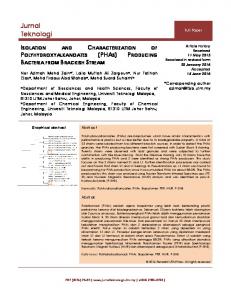
FIG. 3. Quantification of genomic DNAs of strains so36, so42, and wo26 in the natural bacterioplankton communities by dot blot hybridization. For nomenclature of strains, see the legend to Fig. 1. Different concentrations of genomic DNA of strain wo26 served as standards. DNA of Sphingomonas cloacae was used as a control for specificity. The values below the dots indicate the concentrations of genomic DNA employed (in ng l⫺1).
Fig. S2 in the supplemental material) (26) could be isolated from all three of the lakes (isolates so30, wo7, and zo36). One sequence (ENVSEQ_SJ2) was detected in 14 bacterial isolates and in fact represented the dominant cultured phylotype (Fig. 2). Three of these isolates (so36, so42, and wo26) had identical 16S rRNA gene sequences when analyzed over 1,350 base pairs. Abundances of the dominant cultured bacteria in the bacterioplankton. As determined by dot blot hybridization, the three strains so36, so42, and wo26 exhibited considerable genomic differences. Genome-genome similarity values ranged from 60.1% between strains so36 and so42 to 66.0% between so36 and wo26 (not depicted in Fig. 3). Since different strains of ␣-Proteobacteria with identical 16S rRNA gene sequences can exhibit considerable ecophysiological differences and coexist in the same bacterioplankton community (27), the abundances of the three strains in natural bacterioplankton were determined separately using genome-genome hybridization instead of fluorescent in situ hybridization (FISH) of the 16S rRNA. The abundances of the three strains in the natural community were 1.4% to 2.3% for strain so36, 0.8% to 0.9% for strain so42, and 0.5% to 0.7% for strain wo26 in Starnberger See and Walchensee, respectively (Fig. 3). Theoretically, cross-hybridization with genomic DNA of related but phylogenetically distinct bacteria could interfere with dot blot quantification and result in an overestimation of the natural abundances of our three strains. It appears unlikely,
VOL. 71, 2005
ISOLATION OF NOVEL FRESHWATER BACTERIA
FIG. 4. (A) Phase-contrast photomicrograph of cells of isolate so42. Bar, 5 m. (B) Absorption spectra of whole cells (solid line) and acetone extracts (dashed line) of isolate so42. The inset depicts longwavelength absorption maxima of photosynthetic complexes at 800 and 865 nm and shoulder at 837 nm.
5915
however, that the signals obtained by our dot blot approach were caused by genomic DNA of such other bacteria because of the following reasoning. As a control for hybridization stringency, the related Sphingomonas cloacae DSMZ 14926 was selected. The phylogenetic similarity between S. cloacae and strains so36, so42, and wo26 was 93.4% (Fig. 2), while the respective genome-genome cross-hybridization values were 11.4%, 13.6%, and 9.6%. If the signals obtained for the bacterioplankton communities were due to bacteria like S. cloacae, a significantly higher abundance of about 20% would be necessary to explain the observed dot blot hybridization signals. This high abundance, however, is very unlikely, given the fact that ␣-Proteobacteria constitute only a few percent of the total bacterioplankton community in freshwater lakes, including those investigated in the present study (10, 22). Our data therefore indicate that the most frequently cultured phylotype is indeed a detectable constituent of the natural bacterioplankton community in Starnberger See and Walchensee. Characterization of previously uncultured planktonic ␣Proteobacteria. The cultures of strains so36, so42, and wo26 consisted of thin gram-negative nonmotile rods (Fig. 4A). Cell width ranged from 0.24 to 0.32 m, cell length from 1.55 to 3.24 m, and calculated cell volumes from 0.14 to 0.15 m3 (Table 4). No change in cell volume occurred upon transfer of cultures from low-nutrient artificial freshwater medium to media containing high concentrations of complex substrates. The three isolates tested negative for cytochrome oxidase and
TABLE 4. Main morphological and physiological characteristics of isolates compared to S. sibiricus RB16-17 Property
Environment Cell shape Cell width (m) Cell length (m) Motility Color Gram staining Cytocrome oxidase Catalase In vivo absorption maxima carotenoids (nm) BChla Photosynthetic reaction center LHI (nm) LHII (nm) Metabolism Utilizationb Glucose (5 mM/5 mM) Maltose (5 mM/2.5 mM) Acetate (5 mM/12 mM) Pyruvate (5 mM/9 mM) Butyrate (2.5 mM/9 mM) Malate (5 mM/6 mM) Citrate (2 mM/5 mM) Succinate (10 mM/4 mM) Lactate (10 mM/9 mM) Ethanol (5 mM/22 mM) Methanol (5 mM/31 mM) Yeast extract (1%/0.1%) a b
so36
Rods 0.32 ⫾ 0.09 1.55 ⫾ 0.44
so42
Freshwater (planktonic) Rods 0.24 ⫾ 0.03 3.24 ⫾ 0.81 ⫺a Orange-red ⫺ ⫺ ⫹ 420, 460, 480
Rods 0.26 ⫾ 0.05 2.33 ⫾ 0.83
⫹ ⫹ 865 800, 837 (shoulder) Strictly aerobic ⫹ ⫺ ⫺ ⫺ ⫺ ⫺ ⫺ ⫺ ⫺ ⫺ ⫺ ⫹
⫹ ⫺ ⫹ ⫺ ⫹ ⫺ ⫺ ⫹ ⫹ ⫺ ⫺ ⫹
S. sibiricus RB16-17
wo26
Freshwater (benthic) Rods 0.3–0.5 1.5–2.5 ⫹ (3 subpolar flagella) Yellow-orange ⫺ ⫹ ⫺ 424, 450, 474 ⫹ ⫹ 867 ⫺ Strictly aerobic (facultative photoheterotroph)
⫹ ⫺ ⫹ ⫺ ⫹ ⫺ ⫺ ⫺ ⫺ ⫺ ⫹ ⫹
⫺, absent; ⫹, present. Concentrations used for the substrate test (isolates/S. sibiricus). Information on S. sibiricus taken from reference (61).
⫹ ⫹ ⫹ ⫹ ⫹ ⫺ ⫺ ⫺ ⫺ ⫺ ⫺ ⫹
5916
GICH ET AL.
APPL. ENVIRON. MICROBIOL.
FIG. 5. Effects of the peptone and the yeast extract on growth rates and lag phases of three ␣-Proteobacteria bacterioplankton isolates, so36, so42, and wo26, grown in synthetic freshwater mineral media. The error bars represent 1 standard deviation.
were catalase positive. They grew as single cells and formed red colonies on agar plates containing synthetic freshwater media. Our phylogenetic analysis grouped the strains with the family Sphingomonadaceae and identified Sandaracinobacter sibiricus RB16-17 as one of the closest cultured relatives (92.8% similarity) (Fig. 2). S. sibiricus is an aerobic phototrophic bacterium which forms an isolated phylogenetic branch among nonphotosynthetic members of the ␣-4 subgroup (23) of the Proteobacteria. By spectroscopy and high-performance liquid chromatography analysis, cell extracts of our three strains were found to contain bacteriochlorophyll a (Bchla). Absorption spectra of whole cells (Fig. 4B) revealed maxima that are consistent with the presence of two different photosynthetic light-harvesting complexes, LHI and LHII, in the cells (Table 4). Strains so36, so42, and wo26 were obligate aerobes and grew chemo-organoheterotrophically in the dark in media supplemented with Casamino Acids, yeast extract, and peptone, whereas no growth was observed with fermented rumen extract as a substrate. Contrary to most other members of the Sphingomonadaceae, the three isolates grew with different organic acids as the sole organic substrate but did not utilize most of the alcohols (9 of 10) or sugars (29 of 31) tested. The three
strains clearly differed with respect to carbon substrate utilization (Table 4). The growth responses of the three isolates were tested at different concentrations of yeast extract and peptone (Fig. 5). Complete inhibition of growth was observed at substrate concentrations of 5% (wt/vol). With yeast extract as a substrate, the maximum growth rate was observed at a concentration of 0.1% (wt/vol). In peptone-containing media, strains so42 and wo26 grew fastest at 1% (wt/vol). The minimum doubling times recorded for the strains were between 12.1 and 16.8 h. When precultures grown at low substrate concentrations were inoculated into the complex media, lag phases of up to 80 h occurred at higher substrate concentrations (Fig. 5).
DISCUSSION Cultivation of representative planktonic bacteria. Marine bacterioplankton communities have been estimated to contain between 340 and 570 species (1, 26, 56). In only a few instances have planktonic species been found to attain a pronounced dominance of 16 to 41% of the total cell count, like the marine ␣-Proteobacteria SAR11 clade (37) or the marine oligotrophic bacterium Sphingopyxis alaskensis (18, 55). Although the species richness of freshwater bacterioplankton is not yet known,
VOL. 71, 2005
the vast majority of bacterioplankton species are expected to constitute ⱕ1% of the bacterial numbers (16). This has been substantiated by FISH and quantitative PCR of natural plankton assemblages, which revealed a correspondingly low abundance of particular representatives of the -Proteobacteria or ␥-Proteobacteria (11, 53). Less abundant bacteria often exhibit the highest cell-specific activities (7, 63), which is most likely caused by selective grazing of bacteriovores on metabolically active and dividing bacteria (28) and in turn may lead to a dominance of grazingresistant groups, like the freshwater Actinobacteria (25, 46). For a better understanding of the functions of planktonic bacteria, the physiology of less abundant but potentially highly active and novel types of bacteria therefore needs to be elucidated. Accordingly, the aim of the present work was to identify representative members of the bacterioplankton community among a large number of isolates obtained in synthetic freshwater media containing low concentrations of 29 different carbon substrates after inoculation by the high-throughput MicroDrop technique. Many of the isolates recovered were found to be most closely related to freshwater bacteria, indicating that our cultivation approach is appropriate for the recovery of typical bacterioplankton community members. The majority of the isolates were members of the ␣- and -Proteobacteria, the Bacteroidetes, and the Actinobacteria. These four bacterial lineages typically occur in freshwater bacterioplankton (22, 23). In contrast, no representatives of the typically aquatic planctomycetes (54, 57) were detected in our culture collection, although our culture-independent analyses recovered many different 16S rRNA gene sequences of this lineage in the three lakes and planctomycetes constituted 9.1% of the bacterioplankton in Walchensee as determined by fluorescent in situ hybridization (J. Overmann, unpublished results). The lack of planctomycetes in our culture collection may be attributed, at least in part, to the fact that most of the known species are usually obtained in media containing complex carbon sources, like yeast extract or peptone, or in media with N-acetylglucosamine as the sole carbon and nitrogen source, as for the genus Pirellula (50). In the present study, 27 novel 16S rRNA gene sequences of this phylum were recovered, indicating a considerable diversity of the group, which is still to be explored. Our culture collection was dominated by previously unrecognized bacterial phylotypes exhibiting low sequence similarity to described species. Of the 105 isolates sequenced, 44 strains representing 20 novel phylotypes were affiliated with the Sphingomonadaceae (␣-4 subgroup of the Proteobacteria). So far, Sphingomonadaceae have been isolated from soil, the rhizosphere, plant surfaces, and human wounds, but rarely from freshwater sources, like rivers and pristine groundwater (59). By 16S rRNA-based culture-independent methods, members of this group have been detected in different freshwater lakes, however (23, 64). To date, most ␣-Proteobacteria ribotypes that are typical of freshwater bacterioplankton communities are still missing in culture collections (64). Our findings support the view that numerous novel types of freshwater planktonic bacteria can still be isolated by high-throughput methods in diluted growth media. In order to identify individual representative members of the bacterioplankton community among the novel isolates,
ISOLATION OF NOVEL FRESHWATER BACTERIA
5917
cultured phylotypes were systematically compared to the 16S rRNA gene sequences obtained by PCR-DGGE from the original bacterioplankton assemblages. In previous comparisons of this kind, cultured phylotypes could not be detected in 16S rRNA gene clone libraries of bacterioplankton (52). This has been attributed to (i) the failure of the most abundant bacteria to grow in standard media and (ii) incomplete coverage of the clone libraries (52). Therefore, we combined high-throughput cultivation in specially adapted media with high-resolution phylogenetic fingerprinting of the original bacterioplankton communities. Rapid, high-resolution phylogenetic fingerprinting of bacterioplankton. Group-specific fingerprinting significantly enhances the sensitivity of the PCR-DGGE method compared to eubacterial fingerprinting (15, 44). The combination of just five different group-specific fingerprinting methods used in the present study increased the number of different DGGE melting types sixfold compared to conventional eubacterial fingerprinting. Due to the increased resolution, phylotypes representing 0.5 to 2.3% of the bacterioplankton assemblage were repeatedly detected by our PCR-DGGE approach. This detection limit is 1 order of magnitude lower than conventional eubacterial fingerprinting, which in complex microbial communities may recover only phylotypes with an abundance of ⱖ9% (51). Although very rare phylotypes will still be missed by our group-specific fingerprinting, this approach yields an inventory of the phylotypes in natural bacterial communities that is sufficiently large for a cross comparison with large culture collections. Indeed, several detectable members of the original bacterioplankton assemblage were also present in our culture collection. Recovery of a novel phylotype of freshwater planktonic bacterium. The dominant cultured phylotype ENVSEQ_SJ2 grouped with the Sphingomonadaceae and has to be considered a novel genus and species based on the large phylogenetic distance to the closest validly described relative. Since the novel type of bacterium also constituted a detectable fraction of the bacterioplankton assemblage in the oligotrophic Walchensee, it may be adapted to low concentrations of dissolved organic carbon (DOC) (1.2 mg DOC liter⫺1) (Table 3). Typical oligotrophic bacteria are capable of growing at substrate concentrations of ⬃1 mg DOC liter⫺1 but cannot grow at ⬃1 g DOC liter⫺1 (12, 55). The growth of obligate oligotrophs, like Pelagibacter ubique strain HTCC1062, is inhibited by peptone at concentrations as low as 10 mg liter⫺1 (47) (corresponding to 4.3 mg DOC liter⫺1) (13), whereas oligotrophic marine ␥-Proteobacteria (the OMG group) do not grow at ⱖ360 mg DOC liter⫺1 (12). Doubling times of Pelagibacter ubique and the OMG bacteria range between 1.2 and 1.7 days (at 40 mg DOC liter⫺1) (47) and between 0.26 and 0.59 days (at 1 to 36 mg DOC liter⫺1) (12), respectively. In contrast, facultative oligotrophic bacteria like Sphingopyxis alaskensis RB 2256T exhibit doubling times of 0.18 to 0.22 days over a wide range of organic substrate concentrations (0.8 to 80 mg DOC liter⫺1) (18) and are also capable of growing at 10 g DOC liter⫺1 in complex media (55). By comparison, the isolates obtained in the present study grew chemo-organotrophically with doubling times of 1.8 to 8.5 days at low substrate concentrations (0.001% peptone or yeast extract, corresponding to 4.4 mg DOC liter⫺1) that mimic
5918
GICH ET AL.
substrate concentrations in lakes (1 to 3 mg DOC liter⫺1) (12, 38). Given the average doubling times determined for freshwater bacterioplankton (0.25 to 8.7 days depending on nutrient supply) (28, 48), our isolates thus represent rather slow-growing members of the bacterioplankton assemblage under purely chemo-organotrophic growth conditions and appear to be less well adapted to the low organic carbon concentrations in oligotrophic habitats. The cell volume of the novel isolates (0.14 to 0.15 m3) is slightly higher than that of typical freshwater (Polynucleobacter necessarius; 0.044 to 0.135 m3) (24) and marine (Sphingopyxis alaskensis; 0.05 to 0.09 m3) (55) ultramicrobacteria and falls in the size range which is readily removed by heterotrophic nanoflagellates and bacteriovorus crustacean zooplankters (29). Taken together, this susceptibility to protozoan grazing and the comparatively low rates of chemo-organotrophic growth suggest a low competitive advantage of the isolates under oligotrophic conditions. Aerobic phototrophic Bchla-containing bacteria have been detected in marine surface waters, where they can constitute up to 11% of the total bacterial community and account for up to 5% of the photosynthetic electron transport (6, 30). Until very recently, the freshwater members of this group had been isolated almost exclusively from nutrient-rich sediment environments, such as cyanobacterial mats of hot springs or subtropical ponds (60). This is also true for the closest cultured relative of our isolates, Sandaracinobacter sibiricus. Very recently, however, two planktonic Bchla-containing species were isolated from ultraoligotrophic Crater Lake, Oregon (45). All marine and freshwater isolates known to date are affiliated with the Rhodobacteraceae (␣-3 subgroup of the Proteobacteria) and the -1 subgroup of the Proteobacteria. In contrast, the isolates obtained in the present study are the first planktonic representatives of the ␣-4 subgroup of the Proteobacteria detected in situ, as well as isolated in pure culture. Apparently, planktonic aerobic Bchla-containing bacteria are more diverse than realized so far and are more widespread in freshwater environments. Photosynthetic generation of ATP permits aerobic phototrophic bacteria to increase the fraction of organic carbon substrates assimilated into cell material and hence to increase the growth yield in comparison to purely chemoheterotrophic bacteria (60). The presence of the novel lineage of aerobic Bchla-containing bacteria in lakes of all trophic states (oligotrophic through eutrophic) suggests that these bacteria may have a selective advantage not exclusively under oligotrophic conditions. Future research will reveal whether photosynthetic electron transfer occurs under natural conditions and represents a significant energy source for freshwater bacterioplankton. The LHII complexes detected in our isolates are present only in the aerobic phototrophic bacteria that are not closely related, namely, Erythromicrobium ramosum, Erythromicrobium ezovicum, Erythromicrobium hydrolyticum, and Citromicrobium bathyomarinum, but are missing in Sandaracinobacter sibiricus (60, 61). In addition, the isolates did not utilize some of the carbon substrates typical of Sandaracinobacter sibiricus. Therefore, they represent a phylogenetically, physiologically, and biochemically novel type of bacterium. As shown here, previously unknown types of bacteria can be recovered by the combination of high-throughput cultivation
APPL. ENVIRON. MICROBIOL.
techniques with efficient high-resolution phylogenetic fingerprinting. Our results substantiate the concept that novel isolates can exhibit distinct, and previously unrecognized, physiological traits and consequently need to be characterized for a more comprehensive understanding of bacterioplankton function. ACKNOWLEDGMENTS We thank Martina Sterz and Kajetan Vogl for help with sampling and are indebted to Ann-Katrin Manske for assistance with the phylogenetic analyses. Matthias Horn from the Institute of Ecology and Conservation Biology (University of Vienna, Vienna, Austria) is acknowledged for kindly providing the sequence of probe ALF968 when it was not available in the database. We thank Frank Oliver Glo ¨ckner for providing genomic DNA of Rhodopirellula baltica. This work was funded by BMBF (Bundesministerium fu ¨r Bildung, Wissenschaft, Forschung und Technologie) grant no. BIOLOG/ 01LC0021 to J. Overmann. REFERENCES 1. Acinas, S. G., V. Klepac-Ceraj, D. E. Hunt, C. Pharino, I. Ceraj, D. L. Distel, and M. F. Polz. 2004. Fine-scale phylogenetic architecture of a complex bacterial community. Nature 430:551–554. 2. Altschul, S. F., T. L. Madden, A. A. Scha ¨ffer, J. Zhang, W. Miller, and D. J. Lipman. 1997. Gapped BLAST and PSI-BLAST: a new generation of protein database search programs. Nucleic Acids Res. 25:3389–3402. 3. Amann, R., W. Ludwig, and K.-H. Schleifer. 1995. Phylogenetic identification and in situ detection of individual microbial cells without cultivation. Microbiol. Rev. 59:143–169. 4. Bano, N., and J. T. Hollibaugh. 2002. Phylogenetic composition of bacterioplankton assemblages from the Arctic Ocean. Appl. Environ. Microbiol. 68:505–518. 5. Bartscht, K., H. Cypionka, and J. Overmann. 1999. Evaluation of cell activity and of methods for the cultivation of bacteria from a natural lake community. FEMS Microbiol. Ecol. 28:249–259. 6. Be´ja `, O., M. T. Suzuki, J. F. Heidelberg, W. G. Nelson, C. M. Preston, T. Hamada, J. A. Eisen, C. M. Fraser, and E. F. DeLong. 2002. Unsuspected diversity among marine aerobic anoxygenic phototrophs. Nature 415: 630–633. 7. Bernard, L., C. Courties, P. Servais, M. Troussellier, M. Petit, and P. Lebaron. 2000. Relationships among bacterial cell size, productivity, and genetic diversity in aquatic environments using cell sorting and flow cytometry. Microb. Ecol. 40:148–158. 8. Bruns, A., H. Cypionka, and J. Overmann. 2002. Cyclic AMP and acyl homoserine lactones increase the cultivation efficiency of heterotrophic bacteria from the central Baltic Sea. Appl. Environ. Microbiol. 68:3978–3987. 9. Bruns, A., H. Hoffelner, and J. Overmann. 2003. A novel approach for high throughput cultivation assays and the isolation of planktonic bacteria. FEMS Microbiol. Ecol. 45:161–171. 10. Bruns, A., U. Nu ¨bel, H. Cypionka, and J. Overmann. 2003. Effect of signal compounds and incubation conditions on the culturability of freshwater bacterioplankton. Appl. Environ. Microbiol. 69:1980–1989. 11. Burkert, U., F. Warnecke, D. Babenzien, E. Zwirnmann, and J. Pernthaler. 2003. Members of a readily enriched -proteobacterial clade are common in surface waters of a humic lake. Appl. Environ. Microbiol. 69:6550–6559. 12. Cho, J.-C., and S. J. Giovannoni. 2004. Cultivation and growth characteristics of a diverse group of oligotrophic marine Gammaproteobacteria. Appl. Environ. Microbiol. 70:432–440. 13. Claus, H., G. Gleixner, and Z. Filip. 1999. Formation of humic-like substances in mixed and pure cultures of aquatic microorganisms. Acta Hydrochim. Hydrobiol. 27:200–207. 14. Connon, S. A., and S. J. Giovannoni. 2002. High-throughput methods for culturing microorganisms in very-low-nutrient media yield diverse new marine isolates. Appl. Environ. Microbiol. 68:3878–3885. 15. Coolen, M. J. L., and J. Overmann. 1998. Analysis of subfossil molecular remains of purple sulfur bacteria in a lake sediment. Appl. Environ. Microbiol. 64:4513–4521. 16. Curtis, T. P., W. T. Sloan, and J. W. Scannell. 2002. Estimating prokaryotic diversity and its limits. Proc. Natl. Acad. Sci. USA 99:10494–10499. 17. del Giorgio, P. A., J. J. Cole, N. F. Caraco, and R. H. Peters. 1999. Linking planktonic biomass and metabolism to net gas fluxes in northern temperate lakes. Ecology 80:1422–1431. 18. Eguchi, M., T. Nishikawa, K. MacDonald, R. Cavicchioli, J. C. Gottschal, and S. Kjelleberg. 1996. Responses to stress and nutrient availability by the marine ultramicrobacterium Sphingomonas sp. strain RB 2256. Appl. Environ. Microbiol. 62:1287–1294.
VOL. 71, 2005 19. Fuhrman, J. A., D. E. Comeau, Å. Hagstro ¨m, and A. M. Chan. 1988. Extraction from natural planktonic microorganisms of DNA suitable for molecular biological studies. Appl. Environ. Microbiol. 54:1426–1429. 20. Gich, F., J. Garcia-Gil, and J. Overmann. 2001. Previously unknown and phylogenetically diverse members of the green nonsulfur bacteria are indigenous to freshwater lakes. Arch. Microbiol. 177:1–10. 21. Glaeser, J., and J. Overmann. 2004. Biogeography, evolution, and diversity of the epibionts in phototrophic consortia. Appl. Environ. Microbiol. 70: 4821–4830. 22. Glo ¨ckner, F. O., B. M. Fuchs, and R. Amann. 1999. Bacterioplankton compositions of lakes and oceans: a first comparison based on fluorescence in situ hybridization. Appl. Environ. Microbiol. 65:3721–3726. 23. Glo ¨ckner, F. O., E. Zaichikov, N. Belkova, L. Denissova, J. Pernthaler, A. Pernthaler, and R. Amann. 2000. Comparative 16S rRNA analysis of lake bacterioplankton reveals globally distributed phylogenetic clusters, including an abundant group of actinobacteria. Appl. Environ. Microbiol. 66: 5053–5065. 24. Hahn, M. W. 2003. Isolation of strains belonging to the cosmopolitan Polynucleobacter necessarius cluster from freshwater habitats located in three climatic zones. Appl. Environ. Microbiol. 69:5248–5254. 25. Hahn, M. W., H. Lu ¨nsdorf, Q. Wu, M. Schauer, M. G. Ho¨fle, J. Boenigk, und P. Stadler. 2003. Isolation of novel ultramicrobacteria classified as Actinobacteria from five freshwater habitats in Europe and Asia. Appl. Environ. Microbiol. 69:1442–1451. 26. Jaspers, E., K. Nauhaus, H. Cypionka, and J. Overmann. 2001. Multitude and temporal variability of ecological niches as indicated by the diversity of cultivated bacterioplankton. FEMS Microbiol. Ecol. 36:153–164. 27. Jaspers, E., and J. Overmann. 2004. The ecological significance of “microdiversity”: identical 16S rRNA gene sequences represent bacteria with highly divergent genomes and physiology. Appl. Environ. Microbiol. 70:4831–4839. 28. Ju ¨rgens, K., J. Pernthaler, S. Schalla, and R. Amann. 1999. Morphological and compositional changes in a planktonic bacterial community in response to enhanced protozoan grazing. Appl. Environ. Microbiol. 65:1241–1250. 29. Ju ¨rgens, K., and C. Matz. 2002. Predation as a shaping force for the phenotypic and genotpyic composition of planktonic bacteria. Antonie Leeuwenhoek 81:413–434. 30. Kolber, Z. S., F. G. Plumley, A. S. Lang, J. T. Beatty, R. E. Blankenship, C. L. Van Dover, C. Vetriani, M. Koblizek, C. Rathgeber, and P. G. Falkowski. 2001. Contribution of aerobic photoheterotrophic bacteria to the carbon cycle in the ocean. Science 292:2492–2495. 31. Lane, D. J. 1991. 16S/23S rRNA sequencing, p. 115–175. In E. Stackebrandt, and M. Goodfellow (ed.), Nucleic acid techniques in bacterial systematics. John Wiley, Chichester, United Kingdom. 32. Loy, A., M. Horn, and M. Wagner. 2003. probeBase—an online resource for rRNA-targeted oligonucleotide probes. Nucleic Acids Res. 31:514–516. 33. Lu ¨demann, H., and R. Conrad. 2000. Molecular retrieval of large 16S rRNA gene fragments from an Italian rice paddy soil affiliated with the class Actinobacteria. Syst. Appl. Microbiol. 23:582–584. 34. Ludwig, W., O. Strunk, R. Westram, L. Richter, H. Meier, Yadhukumar, A. Buchner, T. Lai, S. Steppi, G. Jobb, W. Forster, I. Brettske, S. Gerber, A. W. Ginhart, O. Gross, S. Grumann, S. Hermann, R. Jost, A. Konig, T. Liss, R. Lussmann, M. May, B. Nonhoff, B. Reichel, R. Strehlow, A. Stamatakis, N. Stuckmann, A. Vilbig, M. Lenke, T. Ludwig, A. Bode, and K. H. Schleifer. 2004. ARB: a software environment for sequence data. Nucleic Acids Res. 32:1363–1371. 35. Manz, W., R. Amann, W. Ludwig, M. Vancanneyt, and K.-H. Schleifer. 1996. Application of a suite of 16S rRNA-specific oligonucleotide probes designed to investigate bacteria of the phylum Cytophaga-Flavobacter-Bacteroides in the natural environment. Microbiology 142:1097–1106. 36. Meier, H., R. Amann, W. Ludwig, and K.-H. Schleifer. 1999. Specific oligonucleotide probes for in situ detection of a major group of gram-positive bacteria with low DNA G⫹C content. Syst. Appl. Microbiol. 22:186–196. 37. Morris, R. M., M. S. Rappe´, S. A. Connon, K. L. Vergin, W. A. Seibold, C. A. Carlson, and S. J. Giovannoni. 2002. SAR11 clade dominates ocean surface bacterioplankton communities. Nature 420:806–810. 38. Mu ¨nster, U., and R. J. Chro ´st. 1990. Origin, composition, and microbial utilization of dissolved organic matter, p. 9–46. In J. Overbeck and R. J. Chro ´st (ed.), Aquatic microbial ecology. Springer-Verlag, New York, N.Y. 39. Murray, A. E., J. T. Hollibaugh, and C. Orrego. 1996. Phylogenetic compositions of bacterioplankton from two California estuaries compared by denaturing gradient gel electrophoresis of 16S rDNA fragments. Appl. Environ. Microbiol. 62:2676–2680. 40. Muyzer, G., S. Hottentra ¨ger, A. Teske, and C. Waver. 1995. Denaturing gradient gel electrophoresis of PCR-amplified 16S rDNA—a new molecular approach to analyse the genetic diversity of mixed microbial communities, p.3.4.4.1–3.4.4.22. In A. D. L. Akkermans, J. D. van Elsas, and F. J. de Bruijn (ed.), Molecular microbial ecology manual, 2nd ed. Kluwer, Dordrecht, The Netherlands. 41. Neef, A. 1997. Anwendung der in situ-Einzelzell-Identifizierung von Bakte-
ISOLATION OF NOVEL FRESHWATER BACTERIA
42. 43. 44.
45. 46.
47. 48.
49. 50.
51. 52.
53.
54. 55.
56.
57. 58. 59. 60. 61. 62. 63.
64.
5919
rien zur Populationsanalyse in komplexen mikrobiellen Biozo ¨nosen. Ph. D. thesis. Technische Universita¨t Mu ¨nchen, Munich, Germany. Neef, A., R. Amann, H. Schlesner, and K.-H. Schleifer. 1998. Monitoring a widespread bacterial group: in situ detection of planctomycetes with 16S rRNA-targeted probes. Microbiology 144:3257–3266. Overmann, J., C. Tuschak, J. Fro ¨stl, and H. Sass. 1998. The ecological niche of the consortium “Pelochromatium roseum”. Arch. Microbiol. 169:120–128. Overmann, J., M. J. L. Coolen, and C. Tuschak. 1999. Specific detection of different phylogenetic groups of chemocline bacteria based on PCR and denaturing gradient gel electrophoresis of 16S rRNA gene fragments. Arch. Microbiol. 172:83–94. Page, K. A., S. A. Connon, and S. J. Giovannoni. 2004. Representative freshwater bacterioplankton isolated from Crater Lake, Oregon. Appl. Environ. Microbiol. 70:6542–6550. Pernthaler, J., T. Posch, K. Simek, J. Vrba, A. Pernthaler, F. O. Glockner, U. Nu ¨bel, R. Psenner, and R. Amann. 2001. Predator-specific enrichment of actinobacteria from a cosmopolitan freshwater clade in mixed continuous culture. Appl. Environ. Microbiol. 67:2145–2155. Rappe´, M. S., S. A. Connon, K. L. Vergin, and S. J. Giovannoni. 2002. Cultivation of the ubiquitous SAR11 marine bacterioplankton clade. Nature 418:630–633. Simon, M., C. Bunte, M. Schulz, M. Weiss, and C. Wu ¨nsch. 1998. Bacterioplankton dynamics in Lake Constance (Bodensee): substrate utilization, growth control, and long-term trends. Arch. Hydrobiol. Spec. Issues Advanc. Limnol. 53:195–221. Stackebrandt, E., and B. M. Goebel. 1994. Taxonomic note: a place for DNA-DNA reassociation and 16S rRNA sequence analysis in the present species definition in bacteriology. Int. J. Syst. Bacteriol. 41:343–346. Staley, J. T., J. A. Fuerst, S. Giovannoni, and H. Schlesner. 2004. The order Planctomycetales and the genera Planctomyces, Pirellula, Gemmata, and Isosphaera. The Prokaryotes. Springer-Verlag New York. Release 3.18 (12/ 21/2004). http://141.150.157.117:8080/prokPUB/index.htm. Straub, K. L., and B. E. E. Buchholz-Cleven. 1998. Enumeration and detection of anaerobic ferrous iron-oxidizing, nitrate-reducing bacteria from diverse European sediments. Appl. Environ. Microbiol. 64:4846–4856. Suzuki, M. T., M. S. Rappe´, Z. W. Haimberger, H. Winfield, N. Adair, J. Stro ¨bel, and S. J. Giovannoni. 1997. Bacterial diversity among small-subunit rRNA gene clones and cellular isolates from the same seawater sample. Appl. Environ. Microbiol. 63:983–989. Thompson, J. R., S. Pacocha, C. Pharino, V. Klepac-Ceraj, D. E. Hunt, J. Benoit, R. Sarma-Rupavtarm, D. L. Distel, and M. F. Polz. 2005. Genotypic diversity within a natural coastal bacterioplankton population. Science 307: 1311–1313. Urbach, E., K. L. Vergin, L. Young, A. Morse, G. L. Larson, and S. J. Giovannoni. 2001. Unusual bacterioplankton community structure in ultraoligotrophic Crater Lake. Limnol. Oceanogr. 46:557–572. Vancanneyt, M., F. Schut, C. Snauwaert, J. Goris, J. Swings, and J. C. Gottschal. 2001. Sphingomonas alaskensis sp. nov., a dominant bacterium from a marine oligotrophic environment. Int. J. Syst. Evol. Microbiol. 51: 73–79. Venter, J. C., K. Remington, J. F. Heidelberg, A. L. Halpern, D. Rusch, J. A. Eisen, D. Wu, I. Paulsen, K. E. Nelson, W. Nelson, D. E. Fouts, S. Levy, A. H. Knap, M. W. Lomas, K. Nealson, O. White, J. Peterson, J. Hoffman, R. Parsons, H. Baden-Tillson, C. Pfannkoch, Y.-H. Rogers, and H. O. Smith. 2004. Environmental genome shotgun sequencing of the Sargasso Sea. Science 304:66–74. Ward, N., F. A. Rainey, E. Stackebrandt, and H. Schlesner. 1995. Unraveling the extent of diversity within the order Planctomyces. Appl. Environ. Microbiol. 61:2270–2275. Warnecke, F., R. Amann, and J. Pernthaler. 2004. Actinobacterial 16S rRNA genes from freshwater habitats cluster in four distinct lineages. Environ. Microbiol. 6:242–253. White, D. C., S. D. Sutton, and D. B. Ringelberg. 1996. The genus Sphingomonas: physiology and ecology. Curr. Opin. Biotechnol. 7:301–306. Yurkov, V. V., and J. T. Beatty. 1998. Aerobic anoxygenic phototrophic bacteria. Microbiol. Mol. Biol. Rev. 62:695–724. Yurkov, V. V., and V. M. Gorlenko. 1990. Erythrobacter sibiricus sp. nov., a new freshwater aerobic bacterial species containing bacteriochlorophyll a. Microbiology 59:85–89. Zengler, K., G. Toledo, M. Rappe´, J. Elkins, E. J. Mathur, J. M. Short, and M. Keller. 2002. Cultivating the uncultured. Proc. Natl. Acad. Sci. USA. 99:15681–15686. Zubkov, M. V., B. M. Fuchs, P. H. Burkill, and R. Amann. 2001. Comparison of cellular and biomass specific activities of dominant bacterioplankton groups in stratified waters of the Celtic Sea. Appl. Environ. Microbiol. 67:5210–5218. Zwart, G., B. C. Crump, M. P. Kamst-van Agterveld, F. Hagen, and S. K. Han. 2002. Typical freshwater bacteria: an analysis of available 16S rRNA gene sequences from plankton of lakes and rivers. Aquat. Microb. Ecol. 28:141–155.