neural crest cells encounter diverse extracellular environments consisting of ... crest-derived cells only form scleral cartilage after undergoing a post-migratory.
J. Embryol. exp. Morph. Vol. 66, pp. 175-190, 1981 Printed in Great Britain © Company of Biologists Limited 1981
175
Specificity in the differentiation and morphogenesis of neural crest-derived scleral ossicles and of epithelial scleral papillae in the eye of the embryonic chick By BRIAN K. HALL 1 From the Department of Biology, Dalhousie University, Halifax
SUMMARY Enzymatic digestion followed by recombination of epithelia and ectomesenchyme from embryonic sclera and mandibles has been used to demonstrate that isolated scleral ectomesenchyme is only able to form scleral ossicles if in prior contact with scleral epithelium until H.H. stage 36 (10 days of incubation); that this induction by epithelial scleral papillae is a prolonged one, commencing as early as H.H. stage 30 (6-5-7 days); that scleral ectomesenchyme can respond to mandibular epithelium by forming bony ossicles and that mandibular ectomesenchyme can respond to scleral epithelium by forming bony rods, i.e. these interactions are not site, time or tissue specific. Scleral epithelia did not form scleral papillae when maintained alone in vitro or when recombined with scleral ectomesenchyme and maintained in vitro. Nor did papillae form when mandibular epithelia were cultured with scleral ectomesenchyme. These results, coupled with data from the literature, are used to argue that papillae will only form when scleral epithelia are under the tension generated by normal intraocular pressure of the growing eye.
INTRODUCTION
The eyes of all birds are supported both by a ring of hyaline cartilage - the scleral cartilage, and by a ring of overlapping ossicles of membrane bone - the scleral ossicles (Fig. 1 and Coulombre, 1965). Neural crest cells migrate around and beneath the eyes, proliferate, and give rise to the mandibular and maxillary processes and to the mesenchyme surrounding the eye. Meckel's cartilage, scleral cartilage, mandibular membrane bones and scleral ossicles all form from ectomesenchyme derived from the neural crest (LeLievre, 1978; Noden, 1978; Johnston et al. 1979; Hall, 1980). Before, during and after migration these cranial neural crest cells encounter diverse extracellular environments consisting of epithelial cells, mesodermally derived mesenchymal cells, and extracellular spaces containing collagen, fibronectin, hyaluronic acid and chondroitin sulfate 1 Author's address: Department of Biology, Dalhousie University, Halifax, Nova Scotia, Canada B3H4J1.
176
B. K. HALL
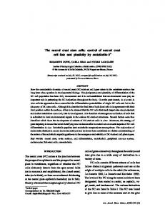
Fig. 1. A cleared, alizarin red S-stained head of a 18-day-old embryonic chick to show the ring of scleral ossicles encircling the orbit.
(Pratt, Larsen & Johnston, 1975; Pratt, Morriss & Johnston, 1976; Bolender, Seliger & Markwald, 1980; Newgreen & Thiery, 1980). Interaction with these environments is required for these cells to form cartilages, bones and connective tissues of the craniofacial region. Contact with epithelia and/or with epithelial products during migration is a prerequisite for differentiation of cartilage (Bee & Thorogood, 1980). Contact with epithelia within the mandibular arch after migration is a prerequisite for formation of membrane bones of the lower jaw (Tyler & Hall, 1977). Neural crest-derived cells only form scleral cartilage after undergoing a post-migratory interaction with extracellular products derived from the pigmented epithelium of the retina (Reinbold, 1968; Stewart & McCallion, 1975; Newsome, 1976). They only form scleral ossicles after interacting with specialized papillae within the scleral epithelium (scleral papillae) - each scleral papilla inducing a single scleral ossicle (Coulombre, Coulombre & Mehta, 1962; Palmoski & Goetinck, 1970; Johnson, 1973). The epithelia may be thin sheets of squamous epithelia as in the mandible, or they may be highly specialized scleral papillae which both grow away from the surface of the eye and penetrate into the underlying ectomesenchyme (Fyfe & Hall, 1981). Where does the specificity for epithelial morphology and epithelial-ectomesenchymal interactions reside? Heterotypic epithelia substituted for mandibular epithelium will allow mandibular membrane bones to form (Hall, 1978tf, 1981) indicating that the epithelium's osteogenic message is not highly site specific, although age of epithelium is important (Hall, 1978 a). Such heterotypic recombinations, in addition to identifying when the epithelia become inductively active, can also identify when the ectomesenchymal cells acquire the ability to respond to inductive influences emanating from the epithelia, and whether epithelial morphology is influenced by association with a particular ectomesenchyme. In this study I have utilized isolated epithelia and ectomesenchyme as well as heterotypic recombinations between scleral and mandibular epithelia and ectomesenchyme to determine: (1) timing of the induction of
Initiation of osteogenesis
111
scleral ossicles; (2) whether scleral ectomesenchyme can respond to mandibular epithelia by forming bone and vice versa; (3) whether ectomesenchyme or epithelium controls morphogenesis of scleral ossicles; (4) whether scleral papillae can form, or, if already present, be maintained in isolation from scleral ectomesenchyme; and (5) whether mandibular epithelium can be induced to form papillae in response to contact with scleral ectomesenchyme. MATERIALS AND METHODS
Incubation of eggs Fertile eggs of the common fowl, Gallus domesticus were obtained from Cook's Hatchery, Truro, Nova Scotia and incubated at 37 ± 0-5 °C and 55 ± 4 % R.H. in a forced draft Petersime Egg Incubator (Model k, Petersime Incubator Co., Gettysburg, Ohio, U.S.A.). Isolation of tissues Eggs were opened under sterile conditions and the embryos removed and staged according to the series of morphological stages (H.H. stages) described by Hamburger & Hamilton (1951).The embryos used in this study were between H.H. stages 22 and 36 (3|-10 days). Mandibular processes and/or portions of the eyes were removed under sterile conditions. Because the scleral papillae do not develop synchronously only papillae 11-13 (as defined by Coulombre et al. 1962) were used. Papilla number 12, the first to appear, arises at 7 days and is located over the temporal, long ciliary artery (Fig. 2). The epithelia and ectomesenchyme of sclera and mandibular processes were separated using trypsin and pancreatin as previously described (Hall, 1978 a), although some tissues were separated using a 0-5 % solution of ethylenediaminetetraacetic acid (EDTA) in calcium- and magnesium-free Tyrode's solution for one hour at 4 °C. The isolated mandibular or scleral ectomesenchyme was grafted or cultured either alone or after recombination with mandibular or scleral epithelia from the same or different aged embryos using techniques previously described (Hall, 1978 a). Chorioallantoic grafting Some isolated tissues and tissue recombinations were grafted onto the highly vascularized chorioallantoic membranes of host embryonic chicks using previously described techniques (Hall, 1978£)« Organ culture Millipore filters supporting tissues were placed onto stainless steel grids in 35 mm diameter Falcon plastic petri dishes containing 1-5 ml of BGJb supplemented with 15% foetal horse serum and 225 jug of ascorbic acid. Three
178
B. K. HALL
Fig. 2. Position of the 14 scleral papillae is shown on the eye of this H.H. stage-32 embryo. Papilla number 1 lies over the choroid figure (cf), number 12 lies over the temporal long ciliary artery (T). Each papilla identifies the future position occupied by a scleral ossicle (see Fig. 1). Reproduced with permission of the publisher from J.Morph (1981) 167, 201-209.
Table 1. The formation of cartilage and bone by chorioallantoic grafts of isolated mandibular and scleral ectomesenchyme Source of ectomesenchyme Mandibular Scleral (a) Enzyme isolated
(b) EDTA isolated
Number of grafts with:
Age H.H. stage
Days
22
3-5
15
0
15
30 35 36 35 36
6-5-7 9 10 9 10
8 18 6 8 8
0 0 4 0 6
8 18 7 8 8
Cartilage Bone
N
such culture assemblies were placed within a 100 mm diameter glass petri dish, and incubated for 2, 4 or 6 days at 37 °C in a humidified CO2 incubator (Forma Scientific, Model 3156) in an atmosphere of 5 % CO2 in air. The tissues rested at the medium: atmosphere interface, receiving nutrients by diffusion.
Initiation of osteogenesis
179
Table 2. The formation of bone in chorioallantoic grafts of homotypic, homochronic recombinations of mandibular and scleral ectomesenchyme and epithelium Source of Ectomesenchyme Epithelium
Age H.H. stage
Mandibular Mandibular Scleral Scleral
22 30 31 32 35 36
Days
Number of grafts with bone*
3-5 6-5-7 7 7-5 9 10
8/8 0/14 0/5 0/4 3/9 6/12
* Cartilage formed in all grafts.
005mm Fig. 3. A prominent rod of scleral cartilage (5C) and two scleral ossicles (0) formed from H.H. stage-36 scleral ectomesenchyme recombined with scleral epithelium and grafted to the chorioallantoic membrane.
Recovery of tissues and histology Grafts and cultures were fixed in neutral buffered formal saline, dehydrated in a graded series of ethanols, cleared in xylene, embedded in paraffin, serially sectioned at 6 fim and stained with a combination of haematoxylin (nuclear stain), alcian blue (for cartilage) and chlorantine fast red (for bone). RESULTS
The timing of scleral induction: evidence from isolated ectomesenchyme H.H. stage-22 mandibular ectomesenchyme when grafted alone always formed cartilage but never formed bone (Table 2). Scleral ectomesenchyme formed sheets of cartilage, whether the ectomesenchyme was taken from young
180
B. K. HALL
Table 3. The formation of cartilage and bone in chorioallantoic grafts of heterochronic recombinations of scleral epithelium and ectomesenchyme Age of epithelium
Age of ectomesenchyme A
r
H.H. stage
Days
H.H. stage
Days
Cartilage
Bone
AT
31 35
7 7
35 31
9 7
8 11
0 0
8 11
Table 4. The formation of bone in chorioallantoic grafts of heterochronic recombinations between scleral and mandibular epithelia and ectomesenchyme Mesenchyme Scleral H.H. stage
Epithelium 30 31 32 35 36
Mandibular H.H. stage 22 22 22
Mandibular H.H. stage 22 22 22 22 22 30 Scleral H.H. stage 32 35
Number (%) of grafts with bone 5/12 (42) (50) 2/4 (67) 4/6 (44) 4/9 (55) 5/9 3/7 (43) (83) 5/6 4/4 (100)
(H.H. stage 30) or from older (H.H. stage-36) embryos (Table 1). Newsome (1972) had previously shown that isolated scleral ectomesenchyme from embryos as young as H.H. stage 19 could chondrify so that the present result was not surprising. It did serve to confirm both the viability of the ectomesenchyme after dissection and enzyme treatment and the adequacy of the graft site for allowing chondrogenesis to occur. Osteogenesis was observed in grafts of isolated scleral ectomesenchyme obtained from H.H. stage-36 (10 day) embryos but not from younger embryos (Table 1). These results place the timing of the completion of the epithelialectomesenchymal interactions required for osteogenesis at H.H. stage 35 (9 days). In contrast, Coulombre et al. (1962) found that 50% of scleral ectomesenchyme isolated from H.H. stage-35 embryos formed bone when grafted to chorioallantoic membranes of host embryos, indicating an earlier age for Fig. 4,5. Scleral ossicles (O) formed by scleral ectomesenchyme from and H.H. stage30 (Fig. 4) and from an H.H. stage-36 embryo (Fig. 5) after recombination with mandibular epithelia from H.H. stage-22 embryos. Note the very sharp boundary of the bones and sparse number of cells (cf. Fig. 6). SC, scleral cartilage; w, millipore filter substrate. Fig. 6. A mandibular membrane bone (6) formed from H.H. stage-22 mandibular ectomesenchyme after recombination with scleral epithelium (e) from an H.H. stage-31 embryo. Note cellularity of the bone, sparsity of bone matrix, scalloped outline, presence of marrow and shape in comparison with scleral ossicles in Figs. 4 and 5.
Initiation of osteogenesis
# t
*»
i ,
181
182
B. K. HALL
Table 5. Scleral epithelia cultured alone or after recombination with scleral ectomesenchyme fail to form scleral papillae Age of embryo providing epithelium and/or ectomesenchyme H.H. stage (days) 28(6) 30(6-5-7) 32(7-5)
State of papillae at 0, 2, 4 and 6 days of in vitro cultivation* , * * 0 2 4 6 Isolated scleral epithelia + + +t -X -
-
N 12 11 14
Scleral epithelium + scleral ectomesenchyme 28 30 32
+ +f
+ -§
-
-
15 20 16
* + , papilla present; - , papilla absent. t papilla at state of maximal invasion into ectomesenchyme X §, 1/7 of \ and 2/6 of § possessed papillae
completion of the osteogenic scleral papilla - ectomesenchyme interaction. They used ectomesenchyme from the same region of the sclera as I did but performed the tissue separations using a chelating agent, EDTA, rather than the enzymatic digestion used in this study. I therefore used EDTA to separate H.H. stage-35 and -36 sclera but again found that no bone formed (Table 1). The timing of scleral induction - evidence from homotypic, homochronic tissue recombinations Recombination of mandibular ectomesenchyme with its own epithelium allowed osteogenesis to proceed in 100 % of the grafts (Table 2). This was not true for the recombinations involving scleral tissues. No bone formed unless the tissues were taken from embryos of at least H.H. stages 35 or 36 (9 or 10 days, Table 2, Fig. 3) and by 10 days the inductive interaction has already occurred (Table 1). To test for the possibility that the ectomesenchyme and/or the epithelium might not be active until H.H. stage-35 heterochronic recombinations were made between H. H. stage-31 and H.H. stage-35 scleral epithelia and ectomesenchyme. In neither case did bone form (Table 3). Specificity of the differentiation and morphogenesis of scleral ossicles - evidence from heterotypic, heterochronic tissue recombinations Can scleral ectomesenchyme from embryos of H.H. stages 30-36 respond to H.H. stage-22 mandibular epithelium by initiating osteogenesis? Scleral papillae are highly specialized structures (Murray, 1943; Fyfe, 1980; Fyfe & Hall, 1981) quite unlike mandibular epithelium. That all ages of scleral ectomesenchyme utilized (H.H. stages 30-36) responded to contact with mandibular epithelium
Initiation of osteogenesis
183
Fig. 7. Theflattened,squamous appearance of scleral epithelium taken from an H.H. stage-30 embryo and cultured in isolation for 48 h. Fig. 8. Mandibular epithelium (e) from an H.H. stage-22 embryo grafted in combination with scleral ectomesenchyme from an H.H. stage-30 embryo undergoes keratinization, as evidenced by the presence of keratohyaline granules (arrow) SC, scleral cartilage.
by forming bone (Table 4) indicated that the scleral papilla's inductive message was not unique and therefore probably not determined by its specialized morphology or behaviour. If it were, one would not expect scleral ectomesenchyme to respond to other epithelia. The percentage of the grafts which formed bone (Table 4) was independent of the age of scleral ectomesenchyme used, indicating no increasing responsiveness of scleral ectomesenchyme with time. The morphology of the bones formed was always that typical of scleral ossicles (Figs. 4, 5). In the second set of recombinations mandibular ectomesenchyme from H.H. stage-22 embryos was combined with scleral epithelia from embryos aged between H.H. stages 31 and 35. Each of these epithelia allowed mandibular ectomesenchyme to form bone (Table 4, Fig. 6). Epithelial inductive activity increased with age for the older the epithelium the greater the percentage of grafts which formed bone - 43 % with H.H. stage-31 epithelia, 83 % with H.H. stage 32 and 100 % with H.H. stage 35. The bone which formed from mandibular
184
B. K. HALL
Table 6. In vitro cultivation of mandibular epithelia with scleral ectomesenchyme fails to produce scleral papillae H.H. stage of embryo providing scleral ectomesenchyme*
Papilla present (+) or absent ( - )
State of epithelium at the end of the culture period
N
26 28 30 32
— -
Flattened squamous Keratinized Keratinized Keratinized
25 15 15 21
* ectomesenchyme was combined with mandibular epithelium from H.H. stage-22 embryos and cultured for up to six days.
ectomesenchyme was typically mandibular while that formed from scleral ectomesenchyme took the ossicular form typical of scleral ossicles (cf Figs. 4, 5 with Fig. 6). Origin and maintenance of scleral papillae The next experiment involved culturing scleral epithelia to determine whether scleral papillae could form, or, if already formed, be maintained, in the absence of scleral ectomesenchyme. The stages used corresponded to times preceding and following the appearance of scleral papilla number 12 (H.H. stages 28 and 30) and to maximal invasion of the papilla into the ectomesenchyme (H.H. stage 32 - see Fyfe & Hall, 1981 for a summary of the stages of scleral papilla development). Epithelia were cultured for 2, 4 or 6 days, lengths of time which corresponded to transition through the above stages. No papillae formed in vitro from scleral epithelia isolated from embryos before papilla formation had begun (H.H. stage 28, Table 5). The epithelium flattened out, remained mitotically active and spread along the Millipore filter substrates. Scleral epithelia from H.H. stage-30 embryos retained recognizable papillae for the first two days of culture. After that only a thin line of epithelial cells could be seen (Fig. 7). These papillae would have persisted in ovo until H.H. stage 36, i.e. for some 3£ days after H.H stage 30. Their behaviour in vitro was on the same time scale. Scleral epithelia isolated from H.H. stage-32 embryos all contained prominent scleral papillae at time zero. After two days in vitro only one of the seven epithelia still had a recognizable papilla (Table 5). No papillae were present after 4 or 6 days in vitro, a time course of disappearance which paralleled loss in ovo. Scleral epithelia from embryos of the same stages were then recombined with scleral ectomesenchyme and maintained under similar conditions in in vitro cultivation (Table 5). The results were the same as those obtained with the isolated scleral epithelia - scleral papillae did not form.
Initiation of osteogenesis
185
The next experiment was designed to determine whether papillae could be induced to form in mandibular epithelia cultured in contact with scleral ectomesenchyme. Scleral ectomesenchyme from embryos of H.H. stages 26-32 were combined with mandibular epithelia from H.H. stage-22 embryos and organ cultured for 2,4 or 6 days. Papillae did not form in any of the 76 cultures (Table 6). H.H. stage-26 scleral ectomesenchyme maintained mandibular epithelium as a squamous epithelium. However, epithelia in contact with older scleral ectomesenchyme keratinized (Fig. 8). H.H. stage-22 mandibular epithelia recombined with mandibular ectomesenchyme from embryos of H.H. stages 26-32 did not keratinize but epithelia cultured alone did. Thus, mandibular epithelium did not respond to scleral ectomesenchyme by forming papilla and older (H.H. stages 28-32) scleral ectomesenchyme was not able to substitute for mandibular ectomesenchyme and maintain mandibular epithelia in their normally unkeratinized state. DISCUSSION
The results may conveniently be discussed in relation to the five questions posed in the introduction. The object of the first set of experiments was to determine timing of the induction of scleral ectomesenchyme for scleral ossicle formation. Scleral ectomesenchyme from H.H. stage-36 (10 days of incubation) embryos could form bone in the absence of scleral papillae (Table 1). Ectomesenchyme from younger embryos could not, indicating that the inductive interaction continued until H.H. stage 36. The discrepancy in timing between this result and that of Coulombre et al. (1962) could not be resolved on the basis of the different methods of tissue separation (enzyme vs chelating agent) used in the two studies for I obtained similar results using either treatment (Table 1). Coulombre et al. did not provide any morphological stages for their embryos so their 9-day embryos may have been more advanced than the 9-day (H.H. stage-35) embryos used in this study, especially as their incubation temperature was higher than that used in this study (37-5 vs 37 °C). Temperature affects size of the embryonic blastoderm, surface area of the area vasculosa, time of appearance of various organs and tissues, rate of growth and attainment of particular morphological stages (Landauer, 1967; Romanoff, 1972). It seems reasonable to conclude that induction of scleral ossicle number 12 continues until H.H. stage 36. The next series of experiments, which involved recombining scleral ectomesenchyme with scleral epithelia of the same or different ages (Tables 2 and 3), confirmed that the inductive interaction was prolonged. Separation and recombination of the components at any age before H.H. stage 35 prevented initiation of osteogenesis (Table 2). This is in contrast to the epithelial-ectomesenchymal interaction which initiates mandibular osteogenesis, where separation and recombination at any age before completion of the induction is followed by bone formation (Hall, 1978 c, c). Failure of bone to form in hetero-
186
B. K. HALL
chronic recombinations where 'young' epithelium was combined with 'old' ectomesenchyme and vice versa (Table 3) also was consistent with the interaction being prolonged and continuous. The second objective was to determine whether scleral ectomesenchyme could respond to a 'foreign' (mandibular) epithelium and vice versa. As summarized in Table 4, scleral ectomesenchyme of any stage tested (H.H. stages 30-36) responded to mandibular epithelium by forming scleral ossicles. The scleral inductive interaction is therefore a permissive one (Wessells, 1977; Saxen, 1977; and Hall 1981) providing an environment necessary for initiation of differentiation. Because such interactions do not control determination of cell fate (a process which requires considerable specificity between the interacting tissues) several inducers may share the same ability of initiating cell differentiation. Mandibular epithelium does not have the same morphology or behaviour as do scleral papillae but possesses the same inductive activity. Therefore, osteogenic inductive ability of scleral papillae probably does not depend upon their specialized morphology. Fyfe & Hall (1981) emphasized that many of the striking changes in papilla development occur after completion of the induction of scleral ossicles. Strands of collagen have been described extending down into scleral ectomesenchyme beneath scleral papillae when induction is in progress (Murray, 1943; Coulombre et al. 1962; Puchkov, 1964; Van de Kamp, 1968). Collagen has recently been implicated as one of the osteogenic inductive components of mandibular epithelium (Bradamante & Hall, 1980) so that epithelially derived collagen may be a common denominator in these permissive osteogenic inductions. The second set of data in Table 4 indicate that scleral epithelium can act inductively on mandibular ectomesenchyme. This further supports the permissive nature of these interactions. That older (H.H. stage-35) scleral epithelia were more inductively active than epithelia from younger (H.H. stage-30) embryos coupled with the presence of inductive activity in H.H. stage-30 epithelia provided additional evidence that scleral epithelia retain inductive activity for a prolonged period. The third objective was to determine whether epithelia influenced the morphology of the bones formed. They did not, for morphology was always typical of the ectomesenchyme and not of the epithelium (cf. Figs 4 and 5 with Fig. 6). Morphology and histological organization of these two types of bone was strikingly different. We do not know where or how these differences originate. Both bones are derived from the neural crest. There could be clones of cranial neural crest cells which share the common feature of being determined for intramembraneous ossification but which differ in the type of bone which they form. Alternatively the differences may be imposed by the environments which these cells encounter during migration. In any event it is clear that epithelia at the site of differentiation play no role in determining the type of bone which will form. Mesenchymal control of morphogenesis is also seen in limb buds and in
Initiation of osteogenesis
187
teeth, for leg bud mesenchyme combined with wing bud epithelium produces a leg and vice versa (Cairns & Saunders, 1954; Z willing, 1955; Saunders, Cairns & Gasseling, 1957; Saunders, Gasseling & Gfeller, 1958) and molar tooth mesenchyme combined with incisor epithelium produces a molar tooth and vice versa (Kollar & Baird, 1969). The next question asked whether the presence of scleral ectomesenchyme was required for formation or maintenance of scleral papillae. Scleral epithelia taken from embryos which had not formed scleral papillae (H.H. stage 28) did not form papillae when organ cultured either alone of after recombination with scleral ectomesenchyme (Table 5). Nor did papilliform structures form in mandibular epithelia combined with scleral ectomesenchyme (Table 6). Furthermore the mandibular epithelium which does not keratinize when maintained with mandibular ectomesenchyme, but which does keratinize when maintained alone (Tyler & Hall, 1977), keratinized in the presence of scleral ectomesenchyme. We would be justified in concluding that scleral ectomesenchyme played no role in induction of papillae from scleral epithelia only if the epithelium was competent to respond under the conditions of in vitro cultivation used to maintain the recombined tissues. Mandibular epithelia are able to respond to mandibular ectomesenchyme under such conditions (see Table 2) but scleral epithelia may not be (Table 6). Scleral epithelia would not be responsive if formation of a scleral papilla was a response to the mechanical environment in the eye, an environment not mimicked in organ culture. The eye certainly exerts a mechanical influence which moulds the shape of the skull (Coulombre & Crelin, 1958; Silver, 1962) and spacing of papillae can be altered by modifying the environment within the eye (Coulombre & Coulombre, 1973). More significantly, the number of papillae which form can be reduced by slowing the growth of the eye, as occurs following intubation and removal of the vitreous humor. Coulombre et al. (1962) drained vitreous humor from the eyes of 4-day embryos. The number of scleral papillae which were present at 9 days of incubation (3-8) was very much below the normal number of 14, and correlated well with the diameter of the eye. Similarly, microphthalmic embryos have small eyes and fewer than normal scleral papillae. Reduced intraocular pressure in such embryos reduces tension on both the cornea and the sclera (Coulombre & Coulombre, 1958). A plausible proposal is that scleral epithelia cannot form scleral papillae unless they are under tension, a condition which did not exist in the present organ cultures. This would also explain why papillae do not regenerate after their removal (Coulombre et al. 1962). Any mechanical influence would have to be a localized consequence of the particular mode of growth of the eye for scleral papillae and scleral ossicles form in embryos totally paralysed by neuromuscular blocking agents such as decamethonium, curare or botulinum toxin (Murray & Drachman, 1969; Hall, unpublished observations). Whether the scleral ectomesenchyme plays an inductive role in papilla formation therefore cannot yet be determined and awaits experiments in which scleral ectomesen-
188
B. K. HALL
chyme can be recombined with epithelia under a variety of mechanical conditions. One future aim will be to devise such conditions, using approaches such as those developed by Leung, Glagor & Mathews (1976, 1977), Curtis and Seehar (1978) and Takeuchi (1979). The latter author has shown that migration of corneal epithelial cells from 8-day chick embryos can be promoted by application of tensile stresses to the epithelium. Migration of scleral epithelial cells towards a centre is a possible mechanism for formation of scleral papillae (Puchkov, 1964). Corneal ectomesenchyme also influences the migration of corneal epithelium (Takeuchi, 1972) and might exert its influence on the scleral epithelium by providing a prepattern as the dermis does in feather morphogenesis (Linsenmayer, 1972). Alternatively, mechanical factors may stimulate cell division within the epithelium as occurs in fibroblasts (Curtis & Seehar, 1978), smooth muscle (Leung et al. 1976, 1977), epidermis (MacKenzie, 1974), chondrocytes (Rodan, Mensi & Harvey, 1975) and periosteal progenitor cells (Hall, 1979). Autoradiographic analysis of sclera exposed to a pulse of [3H]thymidine shows a halo of labelled cells around the base of each papilla with little labelling within the papilla itself (Fyfe, 1980), an observation consistent with cell division (at the base) and migration (within the papilla) both playing a role in formation of papillae. Further studies on the roles of cell division, migration, biochemical factors and ectomesenchyme will all be required before the origin of scleral papillae is elucidated. That the induction of scleral ossicles is not based on a property unique to scleral papillae provides some consolation to those of us seeking a common developmental basis for the initiation of osteogenesis at various sites within the embryo. The supporting skeleton of the avian eye provides an accessible model system for studying the interplay between tissue interactions, biomechanical factors, growth, differentiation and morphogenesis. The Natural Sciences and Engineering Research Council of Canada (Grant A5056) and the Research Development Fund in the Sciences of Dalhousie University provided financial support for this study. I thank Sharon Brunt for expert technical assistance and Jim Hanken and Terry Mobbs for comments on the manuscript. REFERENCES BEE, J. & THOROGOOD, P.
V. (1980). The role of tissue interactions in the skeletogenic differentiation of avian neural crest cells. Devi Biol. 78, 47-62. BOLENDER, D. L., SELIGER, W. G. & MARKWALD, R. R. (1980). A histochemical analysis of polyanionic compounds found in the extracellular matrix encountered by migrating cephalic neural crest cells. Anat. Rec. 196, 401-412. BRADAMANTE, Z. & HALL, B. K. (1980). The role of epithelial collagen and proteoglycan in the initiation of osteogenesis by avian neural crest cells. Anat. Rec. 197, 305-315. CAIRNS, J. M. & SAUNDERS, J. W. Jr. (1954). The influence of embryonic mesoderm on the regional specification of epidermal derivatives in the chick. /. exp. Zool. 127, 221-248. COULOMBRE, A. J. (1965). The Eye. In Organogenesis (ed. R. deHaan & H. Ursprung), pp. 219-251. New York: Holt Rinehart and Winston. COULOMBRE, A. J. & COULOMBRE, J. L. (1958). The role of intraocular pressure in the development of the chick eye. IV. corneal curvature. A.M.A. Arch. Ophthalmol. 59, 502-506.
Initiation of osieogenesis
189
COULOMBRE, A. J. & COULOMBRE, J. L. (1973). The skeleton of the eye. III. Overlap of the
scleral ossicles of the domestic fowl. Devi Biol. 33, 257-267. COULOMBRE, A. J., COULOMBRE, J. L. & MEHTA, H. (1962). The skeleton of the eye. I. Con-
junctival papillae and scleral ossicles. Devi Biol. 5, 382-401. A. J. & CRELIN, E. S. (1958). The role of the developing eye in the morphogenesis of the avian skull. Amer. J. Phys. Anthrop. 16, 25-37. CURTIS, A. S. G. & SEEHAR, G. M. (1978). The control of cell division by tension or diffusion. Nature 274, 52-53. FYFE, D. MACG. (1980). The morphogenesis of scleral papillae and scleral ossicles in the eye of the chick embryo. M.Sc. Thesis, Dalhousie University, Halifax, N.S. 139 pp. FYFE, D. MACG. & HALL, B. K. (1981). A scanning electron microscopic study of the developing epithelial scleral papillae in the eye of the embryonic chick. /. Morph. 167, 201-209. HALL, B. K. (1978a). Initiation of osteogenesis by mandibular mesenchyme of the embryonic chick in response to mandibular and non-mandibular epithelia. Archs Oral Biol. 23, 1157-1161. HALL, B. K. (1978ft).Grafting organs and tissues to the chorioallantoic membrane of the embryonic chick. Tissue Culture Assoc. Manual 4, 881-884. HALL, B. K. (1978C). Developmental and Cellular Skeletal Biology. New York and London: Academic Press. HALL, B. K. (1979). Selective proliferation and accumulation of chondroprogenitor cells as the mode of action of biomechanical factors during secondary chondrogenesis. Teratology 20, 81-92. HALL, B. K. (1980). Chondrogenesis and osteogenesis in cranial neural crest cells. In Current Research Trends in Prenatal Craniofacial Development (ed. R. M. Pratt, &R. L. Christiansen) pp. 47-63. North Holland, New York: Elsevier. HALL, B. K. (1981). The induction of neural crest-derived cartilage and bone by embryonic epithelia: an analysis of the mode of action of an epithelial-mesenchymal interaction. /. Embryol. exp. Morph. (In the Press). HAMBURGER, V. & HAMILTON, H. L. (1951). A series of normal stages in development of the chick embryo. / . Morph. 88, 49-92. JOHNSON, L. G. (1973). Development of chick embryo conjunctival papillae and scleral ossicles after hydrocortisone treatment. Devi Biol. 30, 223-227. COULOMBRE,
JOHNSTON, M. C , NODEN, D. M., HAZELTON, R. D., COULOMBRE, J. L. & COULOMBRE, A. J.
(1979). Origins of avian ocular and periocular tissues. Expl Eye Res. 29, 27-45. E. J. & BAIRD, G. R. (1969). The influence of the dental papilla on the development of tooth shape in embryonic mouse tooth germs. / . Embryol. exp. Morph. 21, 137-148. LANDAUER, W. (1967). The hatchability of chicken eggs as influenced by environment and heredity. Monograph 1 (revised). Univ. Connecticut, Storrs, Agricultural Station, pp. 1-315. LELIEVRE, C. (1978). Participation of neural crest derived cells in the genesis of the skull in birds. J. Embryol exp. Morph. 47, 17-37. LEUNG, D. Y. M , GLAGOV, S. & MATHEWS, M. B. (1976). Cyclic stretching stimulates synthesis of matrix components by arterial smooth muscle cells in vitro. Science 191, 475477. LEUNG, D. Y. M., GLAGOV, S. & MATHEWS, M. B. (1977). A new in vitro system for studying cell response to mechanical stimulation. Different effects of cyclic stretching and agitation on smooth muscle cell biosynthesis. Expl Cell Res. 109, 285-298. LINSENMAYER, T. F. (1972). Control of integumentary patterns in the chick. Devi Biol. 27, 244-271. MACKENZIE, I. C. (1974). The effects of frictional stimulation on mouse ear epidermis. I. Cell proliferation. / . Invest. Dermatol. 62, 80-85. MURRAY, P. D. F. (1943). The development of the conjunctival papillae and of the scleral bones in the chick embryo. J. Anat. (Lond). 77, 225-240. MURRAY, P. D. F. & DRACHMAN, D. B. (1969). The role of movement in the development of joints and related structures: the head and neck in the chick embryo. /. Embryol. exp. Morph. 22, 349-371. KOLLAR,
7
EMB 66
190
B. K. HALL
D. & THIERY, J.-P. (1980). Fibronectin in early avian embryos: synthesis and distribution along the migration pathways of neural crest cells. Cell and Tissue Res. 211, 269-292. NEWSOME, D. A. (1972). Cartilage induction by retinal pigmented epithelium of chick embryos. Devi Biol. 27, 575-579. NEWSOME, D. A. (1976). In vitro stimulation of cartilage in embryonic chick neural crest cells by products of retinal pigmented epithelium. Devi Biol. 49, 496-507. NODEN, D. M. (1978). The control of avian cephalic neural crest cytodifferentiation. I. Skeletal and connective tissues. Devi Biol. 67, 296-312. PALMOSKI, M. J. & GOETINCK, P. F. (1970). An analysis of the development of conjunctival papillae and scleral ossicles in the eye of the scaleless mutant. /. exp. Zool. 174, 157-164. PRATT, R. M., LARSEN, M. A. & JOHNSTON, M. C. (1975). Migration of cranial neural crest cells in a cell-free hyaluronate-rich matrix. Devi Biol. 44, 298-305. PRATT, R. M., MORRISS, G. M. & JOHNSTON, M. C. (1976). The source, distribution and possible role of hyaluronate in the migration of chick neural crest cells. /. Gen. Physiol. 68, 15-16a. PUCHKOV, V. F. (1964). Mechanism by which the scleral papillae develop in the embryonic chick eye. Arkh. Anat. Gistol. Embriol. 46, 16-24. REINBOLD, R. (1968). Role du tapetum dans la differentiation de la sclerotique chez 1' embryon de poulet. /. Embryol. exp. Morph. 19, 43-47. RODAN, G. A., MENSI, T. & HARVEY, A. (1975). A quantitative method for the application of compressive forces to bone in tissue culture. Calcif. Tissue Res. 18, 125-132. NEWGREEN,
ROMANOFF, A. L. (1972). Pathogenesis of the Avian Embryo. An Analysis of Causes of Mal-
formations and Prenatal Death. New York: Wiley-Interscience. J. W., CAIRNS, J. M. & GASSELING, M. T. (1957). The role of the apical ridge of ectoderm in the differentiation of the morphological structure and inductive specificity of limb parts in the chick. /. Morph. 101, 57-87. SAUNDERS, J. W., GASSELING, M. T. & GFELLER, M. D. (1958). Interactions of ectoderm and mesoderm in the origin of axial relationships in the wing of the fowl. J. exp. Zool. 137, 39-74. SAXEN, L. (1977). Directive versus permissive induction: a working hypothesis. In Cell and Tissue Interactions (ed. J. W. Lash & M. M. Burger). New York: Raven Press. SILVER, P. H. S. (1962). In ovo experiments concerning the eye, the orbit and certain juxtaorbital structures in the chick embryo. /. Embryol. exp. Morph. 10, 423-450. STEWART, P. A. & MCCALLION, D. J. (1975). Establishment of the scleral cartilage in the chick. Devi Biol. 46, 383-389. TAKEUCHI, S. (1972). Wound healing of the cornea in the chick embryo. II. The stromal wound bed as a substratum for epithelial migration. /. Fac. Sci. Univ. Tokyo 12, 449-456. TAKEUCHI, S. (1979). Wound healing in the cornea of the chick embryo. IV. Promotion of the migratory activity of isolated corneal epithelium in culture by the application of tension. Devi Biol. 70, 232-240. TYLER, M. S. & HALL, B. K. (1977). Epithelial influences on skeleto^nesis in the mandible of the embryonic chick. Anat. Rec. 188, 229-240. VAN DE KAMP, M. (1968). Fine structural analysis of the conjunctival papillae in the chick embryo: a reassessment of their morphogenesis and developmental significance. /. exp. Zool. 169, 447-462. WESSELLS, N. K. (1977). Tissue Interactions and Development. Menlo Park, California: W. A. Benjamin. ZWILLING, E. (1955). Ectoderm-mesoderm relationships in the development of the chick embryo limb bud. /. exp. Zool. 128, 423-441. SAUNDERS,
(Received 3 April 1981, revised 8 June 1981)