Theoretical Analysis of Proton Therapy for Prostate Cancer. Buthainah .... Due to their relatively large mass protons are located, and have slight transverse side dispersion in tissues .... When the target is composed of more than one element, it is assumed that each component ... Atom name symbol Atomic number. Atomic ...
Theoretical Analysis of Proton Therapy for Prostate Cancer Buthainah Abdulmunem Ibrahim
Theoretical Analysis of Proton Therapy for Prostate Cancer Buthainah Abdulmunem Ibrahim Sciences College - Diyala University - Iraq Received 25 May 2016; Accepted 25 September 2016
Abstract The dependence of high proton energies has been investigated for proton therapy, which are governed by essential physical factors such as ion ranges and stopping powers. In addition, the volume of prostate is another variable factor varies according to human age and types of tumor. The aim of this theoretical study is about bombardment the prostate tumor by normal incidence protons from Medical Cyclotron at room temperature and energy ranges 60-250 MeV. It was studied by means of calculating Stopping Powers, 3D Ion ranges (longitudinal, lateral and radial) and the total target damage as a function of proton energies, which are calculated by TRIM computer code. The electronic stopping powers were calculated by Bethe-Bloch model, which have showed good coincidence with the TRIM results with 0.99 correlation factor. The ionization process is predominated through track of protons in tissues of the prostate, which yields a steep increase in total stopping power with penetration distance leading to the Bragg peak close to the end of range of the proton beams. The skewness and kurtosis are calculated, the negative sign of skewness for all spectra indicated that left tail (towards the surface of incidence protons) is larger than right (away from the surface) leading to reduce side effects to the neighboring normal tissues. The positive value of kurtosis means that the spectra are peaked. This indicates that the precise dose localization is delivered to the tissues. Proton therapy offers the benefit of specified dose localization and has favorable dose-depth distributions, compared with photon beam radiotherapy.
Vol: 13 No:2 , April 2017
146
P-ISSN: 2222-8373 E-ISSN: 2518-9255
Theoretical Analysis of Proton Therapy for Prostate Cancer Buthainah Abdulmunem Ibrahim
The angular distribution of protons with different energies are studied to reach the tumor in any position in prostate, it is noticed that at angle of 45 degree, there is equidimensional ion ranges. The total target damage increases with proton energies with values (0.58 - 1.7) KeV/ion for ranges energy (60 - 250) MeV respectively. In order to reach the tumor, the proton beam direction could be changed in definite steps or turned on and off very quickly.Volumes of the different shapes of prostates change according to age and kinds of the tumor, which are prolate ellipsoid, prolate spheroid, or spherical shape. This leads to select the precise proton energy to prevent the spread of radiation outside the prostate.This theoretical study can be considered as a tool and an indication to determine the precise proton energy required for prostate cancer treatment to minimize the side effects. Proton therapy is a promising technology as a result of the rapid development of nuclear accelerators. Keywords: proton Therapy – Cyclotron - Bragg Peak – Stopping Power – Ion Range Prostate Volume.
التحليل النظري لتقنية العالج البروتوني لسرطان البروستات بثينة عبد المنعم إبراهيم جامعة ديالى – بعقوبة – العراق- كلية العلوم- - قسم الفيزياء
الخالصة التي تحكمها عوامل فيزيائية أساسية مثل المدى االيوني،تم التحقيق في اعتماد طاقات ال بروتون العالية في العالج البروتوني ان هدف هذه الدراسة. باالضافة إلى ذلك حجم البروستات الذي يتغير تبعا إلى عمر االنسان ونوع الورم،وفدرة االيقاف النظرية هو قصف ورم البروستات بواسطة السقوط العمودي للبروتونات بواسطة معجل السايكلوترون الطبي بطاقات تتراوح وتمت الدراسة بحساب قدرات االيقاف والمدى االيوني ثالثي االبعاد.ف في درجة حرارة الغرفة.أ. م056 إلى06 من وحسبت قدرة االيقاف، القطري) والضرر الكلي للعينة كدالة لطاقة البروتونات باستخدام برنامج ترم، االفقي، (الطولي لقد هيمنت عملية.6.00 بلوخ وقد اظهرت النتائج توافقا جيدا مع برنامج ترم بعامل ارتباط-االلكترونية باستخدام نموذج بيث التأين اثناء مسار البروتونات في انسجة البروستات ونتج عن ذلك زيادة حادة في قدرة االيقاف الكلية مما أدى إلى تكون وقد تم حساب االلتواء والتفرطح حيث لوحظ االشارة السالبة لاللتواء.قمة براك بالقرب من نهاية مدى حزمة البروتونات
Vol: 13 No:2 , April 2017
147
P-ISSN: 2222-8373 E-ISSN: 2518-9255
Theoretical Analysis of Proton Therapy for Prostate Cancer Buthainah Abdulmunem Ibrahim
لجميع االطياف وهذا يعني ان الذيل االيسر للطيف (نحو سطح سقوط البروتونات) أكبر من الذيل االيمن (بعيدا عن السطح) مما يدل على قلة اآلثار الجانبية لألنسجة الطبيعية المجاورة للورم .وكذلك لوحظ القيمة الموجبة للتفرطح والذي يعني ان االطياف ذات قمم .ويدل ذلك على ايصال جرعة محددة في موقع النسيج .لذا فان العالج البروتوني يقدم فوائد اهمها الجرعة المحددة في موقع النسيج وتوزيع جرعة مناسب في عمق النسيج وهذا ما يفضله على العالج الفوتوني االشعاعي .وقد تم دراسة التوزيع الزاوي للبروتونات بطاقات مختلفة لغرض الوصول إلى المكان المحدد للورم في البروستات ولوحظ بتساوي االبعاد الثالثية للمدى االيوني عند سقوط البروتونات بزاوية 55درجة .وان الضرر الكلي للعينة ( ) 7.1- 6.50كيلو الكترون فولت /ايون يزداد عند الطاقات ( ) 056 – 06مليون الكترون فولت على التوالي..و لغرض الوصول الى الورم يجب ان يتغير اتجاه حزمة الب روتون في خطوات محددة أو تشغيلها وإيقافها بسرعة جدا.اشكالها اهليلجي ،كروي متطاول ،كروي ان معرفة حجم البروستات والتي تتغير حسب السن ونوع الورم وعادة تكون ،يؤدي إلى تحديد طاقة البروتونات المناسبة لمنع انتشار اإلشعاع خارج البروستات .ويمكن اعتبار هذه الدر اسة النظرية كأداة ومؤشرا لتحديد طاقة البروتون الدقيقة الالزمة لعالج سرطان البروستات للحد من اآلثار الجانبية .العالج البروتوني هي تقنية واعدة نتيجة للتطور السريع في المعجالت النووية. الكلمات المفتاحية :العالج البروتوني – السايكلوترون -قمة براك -قدرة االيقاف – المدى االيوني – حجم البروستات .
Background Robert R. Wilson suggested in 1946, that energetic protons might be an effective treatment method [1], whilst he had been embroiled in the design of the Harvard Cyclotron Laboratory (HCL) [2]. Therapy by this technology were performed up till now, and showed very highly uses and give excellent results. Therapy of proton uses ionized radiation. As protons are charged particles, a pencil proton beam can be exactly guided towards the tumor. Protons, heavy particles so they penetrate with minimal prevalence and they slow down comparatively fast when entering biological tissues. Due to their relatively large mass protons are located, and have slight transverse side dispersion in tissues; the beam is not widen frequently, stays focused on the tumor and transports low;dose to healthy tissues surrounded tumor. All protons of specified energy have certain range few protons penetrate behind that distance. Moreover, proton beams offer the advantage of
P-ISSN: 2222-8373 E-ISSN: 2518-9255
148
Vol: 13 No:2 , April 2017
Theoretical Analysis of Proton Therapy for Prostate Cancer Buthainah Abdulmunem Ibrahim
accurate dose localization and suitable dose-depth distributions, in comparison with photon radiotherapy in which neighboring tissues to tumor may receive similar dose and can be damaged. Proton beams have acute slope increment in energy deposition at Bragg peak [3] and transport few dose to the healthy tissues shortly after the Bragg peak location [4]. Accelerators are the main part used for proton therapy to produce protons for
energy
ranges (60 to 250 ) MeV, so that deep-seated tumors in prostate in any depth can be treated [5]. The proton beam causes damage of the DNA cells within the tumor [5]. Adjusting proton energy during therapy maximizes killing tumors or stop reproduction of the cancerous cells. The development of medical accelerator technology started to produce innovative designs and compact, high frequency, linear accelerators in order to reduce their costs, permitting more proton therapy centers to be accessible to as many hospitals as possible [6]. Notably, LIGHT (Linac for Image Guided Hadrons Therapy) is developed accelerator for proper treatment of cancer tissues with proton therapy [7]. In addition, it also brings innovations in design and manufacturing to reduce production costs [8]. The essential physical factors such as stopping powers and ion ranges depend on the protons energy and properties of prostate tissues. In addition, the volume of prostate is another variable factor, which varies according to human age and types of tumor. In this work, bombardment of the prostate tumor by normal incidence protons from Medical Cyclotron at room temperature and energy ranges 60-250 MeV. It was studied by means of calculating Stopping Powers, 3D Ion ranges (longitudinal, lateral and radial) and the total target damage as a function of proton energies, which are calculated by TRIM computer code [9] by using computer simulation for transport of protons in matter [10]. The electronic stopping powers were calculated by using Bethe-Bloch model, which have showed good agreement with the TRIM results with 0.99 correlation factor. The projected proton ranges (2.945 cm - 36.257 cm) with longitudinal straggles (0.124-1.511) cm and lateral straggles (0.076-0.853) cm increased with proton energies (60-250) MeV respectively, in contrary to the energy losses ( 11.28 - 4.079 ) MeV/cm and the stopping powers (10.84 – 3.923)KeV/(mg/cm2 ) decrease with increasing energy.The angular distribution of protons with different energies are studied to reach the tumor in any position in
Vol: 13 No:2 , April 2017
149
P-ISSN: 2222-8373 E-ISSN: 2518-9255
Theoretical Analysis of Proton Therapy for Prostate Cancer Buthainah Abdulmunem Ibrahim
prostate, it is noticed that at angle of 45 degree, there is equidimensional ion ranges. The total target damage increases with proton energies with values (0.58 - 1.7) KeV/ion for ranges energy (60 - 250) MeV respectively. In order to reach the tumor, the proton beam direction could be changed in definite steps or turned on and off very quickly. Many studies were done for calculating the volume of prostate, some of them show that, the common shapes of human prostate are: prolate ellipsoid or prolate spheroid [11] Objective The aim of the study is to reduce the side effects to healthy tissues of proton therapy for prostate cancer. The selection of specific energy and direction of protons have protected the healthy tissues from radiation. This is also achieved through precise calculation of prostate volume.
Theoretical Methods 1. Stopping power The ion is slowed down mainly by outer electrons of atoms of medium at high energy, and it moves nearly in a straight path. When the ion is slowed enough, the collision with the nucleus would be more probable. Finally, the nuclear stopping power was dominating the slowing down process [12] The total stopping powers are equal to the energy loss ( E ) per unit path length ( x), as shown by [13]. 𝑆(𝐸 ) = −𝑁𝑡
Vol: 13 No:2 , April 2017
𝑑𝐸 𝑑𝑥
150
(1)
P-ISSN: 2222-8373 E-ISSN: 2518-9255
Theoretical Analysis of Proton Therapy for Prostate Cancer Buthainah Abdulmunem Ibrahim
Where, 𝑑𝐸 𝑑𝑥
- Energy loss.
𝑁𝑡 - Atomic density (atom/𝑐𝑚3 ) The total stopping power depends on the energy and type of the incident radiation and on the properties of the substance through which it passes. Since the production of an ion pair requires specific amount of energy, the ionization density is proportional to the total stopping power. Generally total stopping powers are increased toward the end of ion ranges and reached a maximum, the Bragg peak soon before the energy drops to zero. The curve that describes the stopping power versus the target depth is called the Bragg curve which represents major practical importance for proton therapy. Electronic energy loss has calculated by using Bethe formula given by [13]: 𝑑𝐸
− 𝑑𝑥 ⌋ =
2𝜋𝑍1 2𝑒 2
𝑒
𝑀
𝑁𝑍2 (𝑚1 ) ln(
𝐸
𝑒
2𝑚𝑒 𝑣 2 𝐼𝑜
)
(2)
Where, E, 𝑀1 , 𝑣 – energy, mass and velocity of projectile respectively. N- Atomic density of target. me , e – mass and charge of the electron. Z1, Z2 - the atomic number of the projectile and the target. 𝐼𝑜 -ionizing potential as function of 𝑍2 , which is given [13] : 12 𝐼𝑜 = {
7
+ 𝑍 … … … … … . (𝑍2 < 13) 2
58.58
9.76 + 𝑍
2
Vol: 13 No:2 , April 2017
1.19
… … … … . . (𝑍2 ≥ 13)
151
(3)
P-ISSN: 2222-8373 E-ISSN: 2518-9255
Theoretical Analysis of Proton Therapy for Prostate Cancer Buthainah Abdulmunem Ibrahim
∴ The electronic stopping power is: 𝑆𝑒 (𝐸 ) = −
1 𝑑𝐸
⌋
(4)
𝑁 𝑑𝑥 𝑒
Nuclear stopping power based on ZBL (Ziegler, Biersack and Littmark ) interaction potential , which is given by [13]: 𝑉 (𝑟 ) =
1
𝑍1 𝑍2 𝑒 2
4𝜋𝜖𝑜
𝑟
∅(𝑟⁄𝑎)
(5)
Where, r - the distance between the projectile and the target ; 𝑎1 is the so-called screening parameter which is Given by: 𝑎1 =
0.8854 𝑎𝑜
(6)
𝑍1 0.23 +𝑍20.23
And the screening function is given by: ∅(𝑥 ) = 0.1818 𝑒 −3.2𝑥 + 0.5088 𝑒 −0.9423𝑥 + 0.2802 𝑒 −0.4029𝑥 + 0.02817𝑒 −0.2016𝑥
)1(
𝑟
Where, 𝑥 = 𝑎, and a0 is the Bohr atomic radius = 0.529 Å. Then, reduced nuclear stopping power is given by: 𝑆𝑛 (𝜖 ) = Where, 𝛾𝑜 =
𝑒 𝜋
𝑎2
𝛾 𝜀𝑜
𝑆𝑛 (𝐸 )
(8)
4𝑀1 𝑀2 (𝑀1 +𝑀2 )2
Vol: 13 No:2 , April 2017
152
P-ISSN: 2222-8373 E-ISSN: 2518-9255
Theoretical Analysis of Proton Therapy for Prostate Cancer Buthainah Abdulmunem Ibrahim
For practical calculation, the ZBL universal nuclear stopping power for an ion energy Eo, which is given By [13] : 𝑆𝑛 (𝐸𝑜 )[𝑒𝑉. 𝑐𝑚2 ] =
8.462×10⁻¹⁵ 𝑍1 𝑍2 𝑀1 (𝑀1 +𝑀2 )(𝑍1 0.23 +𝑍2 0.23 )
0.5 ln (1+1.1383𝜖)
𝑆𝑛 (𝜖 ) = {ln(𝜖)
𝑆𝑛 (𝜖 )
(9)
For 𝜖 ≤ 30
(𝜖+0.01321𝜖0.21226 +0.19593𝜖0.5 )
(10) For 𝜖 > 30
2𝜖
The reduced energy is given by: 𝜖=
32.53 𝑀2 𝐸𝑜
(11)
𝑍1 𝑍2 (𝑀1 +𝑀2 ) (𝑍10.23 +𝑍2 0.23 )
∴
𝑑𝐸
⌋ = 𝑁 𝑆𝑛 (𝐸)
𝑑𝑥 𝑛
(12) The total stopping power S (E) is given by [13]: 𝑆(𝐸 ) = 𝑆𝑒 (𝐸 ) + 𝑆𝑛 (𝐸)
(13)
When the target is composed of more than one element, it is assumed that each component contributes to stopping power, for compound 𝐴𝑥 𝐵𝑦 , where x + y =1, then the stopping power is given by the following equation : 𝑆𝐴𝐵 = 𝑥𝑆𝐴 + 𝑦𝑆𝐵
(14)
Where, 𝑆𝐴 𝑎𝑛𝑑 𝑆𝐵 are the stopping powers of each target atom A and B respectively [13]. Theoretically simulation methods by computer to calculate the motion of ions in the material, are now the predominant way of treating stopping powers [14].
Vol: 13 No:2 , April 2017
153
P-ISSN: 2222-8373 E-ISSN: 2518-9255
Theoretical Analysis of Proton Therapy for Prostate Cancer Buthainah Abdulmunem Ibrahim
2. Ion ranges The range, R of an energetic projectile with energy 𝐸𝑜 is determined by the rate of energy loss along the path of the projectile until stopping the projectile (𝐸𝑜 =0) [13]. 0
𝑅 = ∫𝐸
𝑜
𝑑𝐸 𝑑𝐸/𝑑𝑥
0
= ∫𝐸
𝑜
𝑑𝐸
(15)
𝑁𝑆(𝐸)
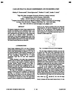
The general three dimensional ion ranges (Longitudinal, Lateral and Radial) are showed in figure (1).
Figure (1): three dimensional ion ranges Conventional methods used to compute ion ranges are based on the binary collision approximation (BCA) [10]. The well known BCA simulation program is, TRIM computer code based on the ZBL nuclear stopping and interatomic potential [9]. Volume of prostate One of the factors that reduce side effects is the precise measuring
of the prostate size to
maintain healthy cells from radiation, that’s why there are many studies about the prostate volume. Estimates of prostate volume
for various scanning modalities have been used.
Average gland sizes have calculated by the contoured ultrasound, ellipsoid ultrasound, and erMRI methods which are 33.99, 37.16, and 39.62 cc, respectively [15]. The relationship between age and human prostate size have been investigated in a population of men between the ages of 40 and 70 years to determine the normal prostate increase curve equation. The whole prostate volumes were measured by transrectal ultrasound (TRUS), these
Vol: 13 No:2 , April 2017
154
P-ISSN: 2222-8373 E-ISSN: 2518-9255
Theoretical Analysis of Proton Therapy for Prostate Cancer Buthainah Abdulmunem Ibrahim
equations and models can facilitate further studies about prostate growth and may enable early diagnosis of benign prostate hyperplasia (BPH) [16].The prostate volume has been estimated with transrectal ultrasound. Theoretically the volume is calculated by prolate ellipsoidal size formula [17] .The volume of the prostate was calculated by using TRUS. The height of the prostate is more precisely determined by using
transaxial than mid sagittal scanning
[18].Simple formula determined prostate volume (Prolate ellipsoid & prolate spheroid), based on prostate diameters, the results provide only marginally inferior to planimetry, which are preferable because they are simpler to perform in the clinic and are associated with less inconvenience for patient [11].
Results and Discussion The elemental compositions of human prostate (W&WJ were shown in table (1) ) [9]: Table (1): Prostate elemental compositions
Atom name
symbol
Atomic number
Atomic weight (amu)
Atomic percent
Hydrogen Carbon Nitrogen Oxygen Sodium Phosphorus Sulfur Potassium
H C N O Na P S K
1 6 7 8 11 15 16 19
1.008 12.011 14.007 15.999 22.99 30.974 32.066 39.098
64.32 4.57 1.10 29.87 0.05 0.02 0.04 0.03
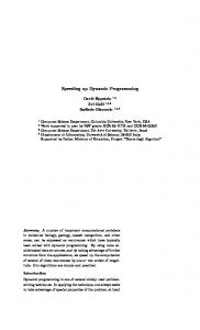
Stopping powers have calculated by using TRIM computer code for proton ions in prostate. The electronic stopping powers are calculated from equation (2) and by using TRIM computer code, they showed good agreement with correlation factor 0.99 as shown in
Vol: 13 No:2 , April 2017
155
figure (3).
P-ISSN: 2222-8373 E-ISSN: 2518-9255
Theoretical Analysis of Proton Therapy for Prostate Cancer Buthainah Abdulmunem Ibrahim
1.4
Trim calculations
Electronic stopping power(MeV/mm)
1.2
Our calculations
1 0.8 0.6
0.4 0.2
0 0
50
100
150 200 Energy(MeV)
250
300
Figure (3): electronic stopping power versus proton energy The skewness and kurtosis are calculated of the Bragg curves for vertical trajectory protons of different energies for bombarding
prostate as shown in figure(4 ), the negative sign of
skewness for all spectra indicate that left tail (towards the surface of incidence protons) is larger than right (away from the surface) leading to reduce side effects to the neighboring normal tissues. The positive value of kurtosis means that the spectra are peaked. This indicates that the precise dose localization deliver to the tissues.
Vol: 13 No:2 , April 2017
156
P-ISSN: 2222-8373 E-ISSN: 2518-9255
Theoretical Analysis of Proton Therapy for Prostate Cancer Buthainah Abdulmunem Ibrahim
60 MeV (depth =2.95cm)
100 MeV (depth = 7.37 cm)
160 MeV (depth = 16.8 cm)
200 MeV (depth = 24.8 cm)
250 MeV (depth = 36.3 cm) Figure (4): Bragg effect for different proton energies on prostate
The total target (prostate) damage increases as proton energy increases as shown in figure (5).
Vol: 13 No:2 , April 2017
157
P-ISSN: 2222-8373 E-ISSN: 2518-9255
Theoretical Analysis of Proton Therapy for Prostate Cancer
2 1.5 1
0.5
240
250
230
0
60 70 80 90 100 120 140 160 180 200 210 220
Total target damag(KeV/ion)
Buthainah Abdulmunem Ibrahim
Energy(MeV)
Figure (5): Total prostate damage versus proton energy The proton ranges are calculated for bombardment the prostate tumor by vertical incidence protons from Medical accelerator at room temperature and energy ranges (60-250) MeV. 3 D proton ranges (longitudinal, lateral and radial) are calculated with different proton energies, as shown in figure(6). Ion Ranges at Vertical Proton Incidence on Prostate Proton Range (mm)
400 Longitudinal Ranges(mm)
200
Lateral Ranges(mm) Radial Ranges(mm)
0 60
80 100 140 180 210 Energy (MeV)
230
250
Figure (6): Three dimensional ranges versus energy at vertical proton incidence At specific energy, the angular distributions of 3D proton ranges are taken for different angles (0 − 90)𝑜 with vertical on the prostate surface. The results show that equidimensional proton ranges at 45𝑜 as shown in figure (7). For other angles larger than zero, there are equidimensional lateral and radial ranges only, illustrated in figure (8).
Vol: 13 No:2 , April 2017
158
P-ISSN: 2222-8373 E-ISSN: 2518-9255
Theoretical Analysis of Proton Therapy for Prostate Cancer Buthainah Abdulmunem Ibrahim
Proton Range(mm)
Ion ranges at 45 degree proton incidence on prostate 300 200
Longitudinal Ranges Lateral Ranges
100 0 60
80 100 140 180 210 230 250 Energy(MeV)
Figure (7): Three dimensional ranges versus energy at proton incidence angle 45 o
400 300 Longitudinal Ranges
100
Lateral Ranges
0
Radial Ranges
Energy(MeV)
250
200
60 70 80 90 100 120 140 160 180 200 210 220 230 240
Proton Range(mm)
Proton Ranges at 60 degree Proton Incidence on Prostate
Figure (8): Three dimensional ranges versus energy at proton incidence angle 60o
Vol: 13 No:2 , April 2017
159
P-ISSN: 2222-8373 E-ISSN: 2518-9255
Theoretical Analysis of Proton Therapy for Prostate Cancer Buthainah Abdulmunem Ibrahim
Conclusions The conclusions of this study have indicated that, the ranges of proton ions in prostate tissues were increased with increasing proton energies. As well as, the total damage of the prostate increased due to increase the proton energy. Bragg effect means that, few side effects transferred to the neighboring vital tissues. High accuracy in calculating the size of the prostate have led to select the appropriate energy of proton beams and their directions according to the depth of tumors, which keep the radiation within the prostate volume to reduce the side effects to the healthy cells. The angular distribution of proton beams leads to decrease the longitudinal ranges (depths of tumor) with increasing the angle of proton incidence. On contrary, lateral and radial ion ranges (area of tumor) was increased, so that the protons direction could be changed at definite steps, in order to reach the tumors in any position. The vertical incidence of proton beams with high energy treat the distal depth tumors in prostate tissues, but the angular distribution of the proton beams treat the near depth tumors to the prostate surface. This theoretical study can be considered as a tool and an indication to select the precise proton energy required for prostate cancer treatment and to minimize the side effects. Proton therapy is a promised technology as a result of the fast development of particle accelerators.
Vol: 13 No:2 , April 2017
160
P-ISSN: 2222-8373 E-ISSN: 2518-9255
Theoretical Analysis of Proton Therapy for Prostate Cancer Buthainah Abdulmunem Ibrahim
References 1.
R. Wilson (1946), Radiological Use of Fast Protons, Radiology 47, 487- 491.
2.
L. E. Williams (2004)., A Brief History of Harvard University Cyclotrons, American Journal of Roentgenology 183(6), 1558 – 1558 (2004).
3.
H. Paganetti, T. Bortfeld (2005)., Proton Beam Radiotherapy—The State of the Art. in: New Technologies in Radiation Oncology (Medical Radiology Series),(Eds.) W. Schlegel, T. Bortfeld and A.-L. Grosu, Springer Verlag, Heidelberg, ISBN, 3-540.
4.
J. Metz (2002), Reduced normal tissue toxicity with proton therapy. Proton Info Web site: http://protoninfo. Com /Articles / University of Pennsylvania pdf. Published April, 28.
5.
O. Jäkel (2007), State of the art in hadron therapy. In NUCLEAR PHYSICS METHODS AND ACCELERATORS IN BIOLOGY AND MEDICINE: Fourth International Summer School on Nuclear Physics Methods and Accelerators in Biology and Medicine, 958(1), 70-77.
6.
E. Pedroni (2000), Latest developments in proton therapy. In Proceedings of EPAC, 240 – 244.
7.
U. Amaldi, R. Bonomi, S. Braccini, M. Crescenti, A. Degiovanni, M. Garlasché & R. Zennaro (2010), Accelerators for hadron therapy: from Lawrence cyclotrons to linacs. Nuclear Instruments and Methods in Physics Research Section A: Accelerators, Spectrometers, Detectors and Associated Equipment 620(2), 563-577.
8.
A. Lomax (1999), Intensity modulation methods for proton radiotherapy, Physics in medicine and biology 44(1), 185.
9.
J. F. Ziegler (1985), J. P. Biersack and U. Littmark, The Stopping and Range of Ions in Matter, New York, Pergamon.
10. M. T. Robinson, I. M. Torrens (1974), Computer simulation of atomic - displacement cascades in solids in the binary- collision approximation, Physical Review B 9 (12), 5008. 11. L. M. Eri, H. Thomassen, B. Brennhovd, & L.L. Håheim (2002), Accuracy and repeatability of prostate volume measurements by transrectal ultrasound. Prostate cancer and prostatic diseases 5(4), 273-278.
Vol: 13 No:2 , April 2017
161
P-ISSN: 2222-8373 E-ISSN: 2518-9255
Theoretical Analysis of Proton Therapy for Prostate Cancer Buthainah Abdulmunem Ibrahim
12. K. Nordlund, N. Runeberg & D. Sundholm (1997), Repulsive interatomic potentials calculated using Hartree-Fock and density-functional theory methods. Nuclear Instruments and Methods in Physics Research Section B: Beam Interactions with Materials and Atoms 132(1), 45-54. 13. M., J. Nastasi, W. Mayer, J.K. Hirvonen (2003), Ion –Solid Interaction: Fundamentals and Applications, Cambridge University Press. 14. J. P. Biersack, L. G. Haggmark (1980), Monte Carlo Computer Program for transport of energetic ions in amorphous targets. Nuclear Instruments and Methods 174(1), 257-269.
15. Y. R. Murciano-Goroff, L. D. Wolfsberger, A. Parekh, F. M Fennessy, K. Tuncali, P. F. Orio, & P. L. Nguyen (2014), Variability in MRI vs. ultrasound measures of prostate volume and its impact on treatment recommendations for favorable-risk prostate cancer patients: a case series, Radiation Oncology 9(1), 200. 16. S. J. Zhang, H. N. Qian, Y. Zhao, K. Sun, H. Q. Wang, G. Q. Liang & Z. Li (2013), Relationship between age and prostate size. Asian journal of andrology 15(1), 116. 17. T., T. Marchie, V. C. Onuora (2006), Determination of normal range of ultrasonic sizes of prostate in our local environment. West African Journal of Radiology 8(1), 54-64. 18.
S. B. Park, J. K. Kim, S. H. Choi, H. N. Noh, E. K. Ji & K. S. Cho (2000), Prostate volume measurement by TRUS using heights obtained by transaxial and midsagittal scanning: comparison with specimen volume following radical prostatectomy. Korean Journal of Radiology 1(2), 110-113.
Vol: 13 No:2 , April 2017
162
P-ISSN: 2222-8373 E-ISSN: 2518-9255