R esearch A rticle For reprint orders, please contact
[email protected]
Split calibration curve: an approach to avoid repeat analysis of the samples exceeding ULOQ Background: The current practice of using calibration curves with narrow concentration ranges during bioanalysis of new chemical entities has some limitations and is time consuming. In the present study we describe a split calibration curve approach, where sample dilution and repeat analysis can be avoided without compromising the quality and integrity of the data obtained. Results: A split calibration curve approach is employed to determine the drug concentration in plasma samples with accuracy and precision over a wide dynamic range of approximately 0.6 to 15,000 ng/ml for dapsone and approximately 1 to 25,000 ng/ml for cyclophosphamide and glipizide. A wide dynamic range of concentrations for these three compounds was used in the current study to construct split calibration curves and was successfully validated for sample analysis in a single run. Conclusion: Using this method, repeat analysis of samples can be avoided. This is useful for the bioanalysis of toxicokinetic studies with wide dose ranges and studies where the sample volume is limited. Quantification of drugs is very important in drug discovery and development research to understand the pharmacokinetic [1,2–7] and pharmacodynamic [3] relationships. Quantitative bioanalysis is generally carried out by LC–MS/MS [4–6] for drug discovery and development. Prediction of conventional calibration curve range is difficult for a new chemical entity (NCE) for which no prior data is available. In most cases, over a 5000-fold concentration range for constructing calibration curves is tricky, and does not fit in any of the well-known curve types. For example, quantification of the anti-influenza drug zanamivir in plasma utilizing high-throughput hydrophilic interaction LC–MS/MS using an approximately 50,000‑fold calibration curve range has been reported [8] . Our laboratory has reported a simple, rapid and universal assay to quantify five different antiretroviral drugs in rat plasma utilizing LC–MS/MS over a 2000-fold calibration curve range [9] . This kind of wide calibration curve range will not work in most cases. Thus, in normal practice, narrow range calibration curves (100- or 500fold) are selected and samples that exceed the upper limit of calibration curve are diluted in order that they fall into the calibration curve range and are reanalyzed. This process is very tedious, time consuming and expensive. In this article, a method has been proposed to split a wide dynamic range calibration curve
and analyze the samples in order to avoid the repeat analysis of the samples after dilution. There are few published methods for quantitation of dapsone with sample volume 0.1 ml and dynamic ranges from 0.5 to 100 ng/ml and 5 to 2000 ng/ml [10,11] . In the present study we show that dapsone, cyclophosphamide and glipizide can be analyzed using a split calibration approach without diluting and reanalyzing samples from ~0.6–14,900 ng/ml for dapsone, and ~1–25,000 ng/ml for cyclophosphamide and glipizide. The whole calibration curve range is split into two curves. Simultaneous determination of dapsone, cyclophosphamide and glipizide are carried out using meta-dapsone as an internal standard (IS). Dapsone and meta-dapsone are positional isomers with identical molecular mass and fragmentation patterns. Due to the similarity in the fragmentation patterns for these two positional isomers, chromatographic separation is essential to distinguish them. Plasma samples were extracted by protein precipitation. Chromatographic elution was accomplished on a C18 column interfaced with a triple-quadruple mass spectrometer. Pharmacokinetic (PK) parameters of dapsone, cyclophosphamide and glipizide were evaluated in Sprague Dawley (SD) rats using both a validated split calibration curve approach and a conventional single smaller calibration range approach.
Sudipta Basu*, Abdul Basit, Selvan Ravindran, Vandana B Patel, Subrahmanyam Vangala & Hitesh Patel
10.4155/BIO.12.219 © 2012 Future Science Ltd
Bioanalysis (2012) 4(19), 2375–2389
ISSN 1757-6180
Department of DMPK & Toxicology, Sai Advantium Pharma Ltd, Building 1, Plot 2, Chrysalis Enclave, International Biotech Park, Phase 2, Hinjewadi, Pune, 411057, India *Author for correspondence: Tel: +91 20 6674 3600 Fax: +91 20 6674 3645 Email:
[email protected]
2375
R esearch A rticle | Key Terms Conventional calibration curve: Narrow range
calibration curves (100- or 500-fold) where samples that exceed the upper limit of calibration curve are diluted in order that they fall into the calibration curve range and are reanalyzed.
Split calibration curve:
A wide calibration curve range that has been split into two calibration curves with a common calibration standard for both the curves. The common standard point serves as a ULOQ for the lower calibration curve and LLOQ for the higher calibration curve range.
Basu, Basit, Ravindran, Patel, Vangala & Patel
Experimental Chemicals & reagents Dapsone (4-aminophenyl sulfone, 97%), meta-dapsone (3-aminophenyl sulfone, 97%), cyclophosphamide (98%) and glipizide (98%) were purchased from Sigma-Aldrich (MO, USA). HPLC-grade acetonitrile and methanol were purchased from JT Baker (NJ, USA). Demineralized water was prepared in-house using millipore water system (USA). Absolute ethanol (99.9%) was purchased from Tedia Company Inc. (OH, USA). Tween-80 was purchased from Sigma-Aldrich (laborchemikalien, Switzerland). Other chemicals and buffers were of analytical grade and also purchased from Sigma-Aldrich. Animals
Healthy male SD rats (8–12 weeks old) weighing between 225 and 275 g were procured from the Advanced Centre for Treatment, Research and Education in Cancer (ACTR EC), Mumbai, India. One rat was housed in each polypropylene cage. Temperature and humidity were maintained at 22 ± 3°C and 40–70%, respectively, and illumination was controlled to give a sequence of 12-h light/dark cycle. Temperature and humidity were recorded by auto-controlled data logger system. All the animals were acclimatized to the experimental conditions for at least a week prior to dosing. All the animals were provided laboratory rodent diet (Nutrilab Rodent diet, Vetcare, The Netherlands) except for 14–16 h before treatment and 4 h post-dosing. Reverse osmosis water treated with UV light was provided ad libitum. LC–MS
instrumentation & analytical conditions Assay was performed on API 4000 Applied Biosystem MDS Sciex LC–MS/MS (Concord, Ontario, Canada) triple quadruple mass analyzer system with a turbo ion-spray ESI interface connected to a Shimadzu LC-20AD LC system (Shimadzu Corp., Japan). The instrument was operated in the MRM mode using the molecular parent and daughter ions. The optimum operating parameters of ESI interface in positive ion mode were: interface temperature 500°C, nebulizing gas 50 psi, drying gas 60 psi and ESI probe voltage 5000 V. Other voltages and MS parameters are reported in Table 1. The whole system was controlled by Analyst Classic 1.5® software (Applied Biosystem/MDS Sciex). 2376
Bioanalysis (2012) 4(19)
The chromatographic separation was carried out on a 150 × 4.6 mm, 5 µm, Zorbax Eclipse C18 column (Agilent Technologies, USA). Dapsone, cyclophosphamide, glipizide and IS were eluted isocratically using mobile phase consisting of acetonitrile/methanol/ammonium acetate (5 mM) (20/20/60, v/v/v) delivered at a flow rate of 0.8 ml/min. Metadapsone was used as an internal standard for dapsone, cyclophosphamide and glipizide. The column oven temperature was maintained at 40°C. The autosampler was kept at 4°C and 5 µl samples were injected. Preparation
of stock solution, calibration curves & QC samples Stock solutions (~1 mg/ml) were individually prepared in methanol and spiked together to obtain a mixture of working solution followed by serial dilution with methanol/water (50/50, v/v). All the stock and working solutions were stored at 2–8°C. Appropriate dilution of analytes were spiked into blank rat plasma containing K 2EDTA as anticoagulant, in order to get 13 different concentrations of calibration standards (CS) starting from ~0.6 to 15,000 ng/ml for dapsone (0.63, 1.25, 6.26, 31.29, 125.16, 312.90, 625.80, 1251.60, 3129.00, 6258.00, 8940.00, 11,920.00 and 14,900.00 ng/ml) in rat plasma. For cyclophosphamide the CS range was from ~1 to 25,000 ng/ml (1.06, 2.12, 10.60, 53.00, 212.01, 530.02, 1060.04, 2120.08, 5300.19, 10,600.39, 15,143.41, 20,191.21 and 25,239.01 ng/ml). Similarly, for glipizide the CS range was from ~1 to 25,000 ng/ml (1.09, 2.18, 12.12, 52.68, 210.74, 526.84, 1053.68, 2107.35, 5268.38, 10,536.75, 15,052.50, 20,070.00 and 25,087.50 ng/ml) in rat plasma. The whole calibration curve was split into two in case of split calibration curve approach. For dapsone, split calibration curve two ranges were selected as follows: the concentrations chosen for the lower calibration curve (LCC) range were from 0.63 to 625.80 ng/ml; and for the higher calibration curve (HCC) the range was from 625.80 to 14,900.00 ng/ml in rat plasma. For cyclophosphamide, the LCC range was from 1.06 to 1060.04 ng/ml and the HCC range was from 1060.04 to 25,239.01 ng/ml. Similarly, for glipizide the LCC range was from 1.09 to 1053.68 ng/ml and the HCC range was from 1053.68 to 25,087.50 ng/ml in rat plasma (Table 2) . QC samples were selected at eight different concentrations ranging from lower = L, medium A = MA, medium B = MB to high = H. For future science group
An approach to avoid repeat analysis of the samples exceeding ULOQ
| R esearch A rticle
Table 1. MS parameters for dapsone, cyclophosphamide, glipizide and meta-dapsone (internal standard). Drug
MW
MRM transition (m/z) Mode of detection
DP (V)
CE (V)
CXP (V)
Rt (min)
Dapsone Cyclophosphamide Glipizide Meta-dapsone
248.3 260.0 445.5 248.3
249.0→155.8 261.0→140.0 446.3→347.0 249.0→155.9
72 82 40 77
22.0 31.0 22.0 17.1
16.0 14.0 12.0 16.6
2.83 5.43 5.77 3.90
Positive Positive Positive Positive
All are voltage-dependent parameters of instrument. CE: Collision energy; CXP: Cell exit potential; DP: Declustering potential; MW: Molecular weight; Rt: Retention time.
dapsone, QC sample concentrations chosen were 2.52, 210.35, 420.70, 601.11, 2225.92, 4637.42, 8431.50 and 12,045.00 ng/ml, which were designated as LQC, MAQC, MBQC, HQC, higher LQC, higher MAQC, higher MBQC and higher HQC, respectively. QC sample concentrations of 2.52, 210.35, 420.70 and 601.11 ng/ml were chosen for the LCC. In the case of HCC range, the QC samples at concentrations 2225.92, 4637.42, 8431.50 and 12,045.00 ng/ml were selected. For cyclophosphamide, QC sample concentrations chosen were 4.18, 321.24, 642.47, 917.82, 3671.28, 7342.56, 13,350.11 and 18,802.97 ng/ml, which were designated as LQC, MAQC, MBQC, HQC, higher LQC, higher MAQC, higher MBQC and higher HQC, respectively. QC sample concentrations of 4.18, 321.24, 642.47 and 917.82 ng/ml were chosen for the LCC. In the case of HCC range, the QC samples at concentrations 3671.28, 7342.56, 13,350.11 and 18,802.97 ng/ml were selected. Similarly, for glipizide, QC sample concentrations chosen were 3.23, 359.44, 599.07, 921.65, 3544.80, 7385.00, 14,770.00 and 21,100.00 ng/ml that were designated as LQC, MAQC, MBQC, HQC, higher LQC, higher MAQC, higher MBQC and higher HQC, respectively. QC sample concentrations of 3.23, 359.44, 599.07 and 921.65 ng/ml were chosen for the LCC. For HCC range, the QC samples at concentrations 3544.80, 7385.00, 14,770.00 and 21,100.00 ng/ml were selected in rat plasma (Table 3) . Sample
preparation Aliquots (25 µl) of either study samples or calibration standards and QC samples were taken in labeled microcentrifuge tubes and mixed with 50 µl of internal standard (200 ng/ml in aceto nitrile [ACN]), except for blank where 50 µl of ACN was added. Extraction of drug was carried out by protein precipitation using 100 µl of ACN. Addition of ACN helped complete and better protein precipitation, and reduced the matrix effect. After centrifugation at 15,000 future science group
rpm for 10 min, the supernatant was transferred to a clean vial and was injected into the LC–MS/MS system. Drugs from the plasma PK study samples were also extracted with protein precipitation, as mentioned above. Validation
of the assay Method validation was carried out to evaluate method performance. All assay validation steps were carried out according to the 2001 version of the US FDA guidance for bioanalytical method validation [101] , however not extensively followed. Validation was carried out for both the calibration curve range, that is, LCC and HCC, using four different concentration range of QC samples (LQC, MAQC, MBQC and HQC) for each calibration curve (CC). Peak area ratio of the analyte to internal standard was utilized for the construction of calibration curve. Linearity was evaluated using a 1/X 2 (where X is the analyte concentration) weighted linear regression method. Concentration in unknown samples was calculated from the best-fit equation (y = mx + c), where y is the peak area ratio. The regression equation for the calibration curve was used to back-calculate the measured concentration at each QC level and unknown samples. The sensitivity of the analytical procedure was expressed as the LLOQ or the lowest concentration on the calibration curve that can be quantitatively determined with acceptable accuracy and precision (nominal ±20% and precision of 20%CV), and should be at least ten-times the response compared with that produced by the blank. The specificity of assay was determined by analysis of six lots of blank plasma from different animals. Noninterference from endogenous or exogenous material observed at the retention time in each analyte channel and in the internal standard was ensured. Injection carryover experiments were carried out by injecting ULOQ level concentration (both neat and extracted), followed by injecting extracted matrix blank (MB) and mobile phase (MP; 20 parts of acetonitrile, 20 parts of methanol www.future-science.com
2377
2378
0.9994
0.9961
and 60 parts of ammonium acetate [5mM]), and observed for any carryover. The following sequence was used to check the injection carry over: MP→Neat ULOQ→MP→MP→Neat ULOQ→MP→MP→MB→Extracted ULOQ →MB→MB→Extracted ULOQ→MB →MP. The accuracy and precision were assessed by determining QC samples with a minimum of four concentration levels from three different precision and accuracy validation batches. The QC samples at each level were prepared for six replicates together with calibration samples. Matrix effect and recovery were assessed, as described in detail by Matuszewski et al. [12] , by comparing the peak areas of the analyte standards, spiked before and after extraction in six different lots of plasma at four different concentration levels (LQC, MAQC, MBQC, HQC and also with higher LQC, higher MAQC, higher MBQC and higher HQC). The stock solution stability was determined by placing the stock solution mixture and internal standard at room temperature for 10 h and at 2–8°C for 10 days. The freeze–thaw, shortterm benchtop autosampler and long-term stability studies were evaluated. For freeze–thaw stability, QC plasma samples were subjected to five cycles from -70°C to room temperature. Short-term benchtop stability was performed by placing samples on the benchtop at room temperature for 10 h. The autosampler stability was assessed by placing processed QC samples in an autosampler at 4°C for 32 h, and long-term stability was evaluated by freezing QC samples at -70°C for a month, then comparing the concentrations with freshly processed calibration curve and QCs.
0.9950
0.9948
4.68 4.98 4.64 2.36 2.58 5.84 3.74 5.49 6.04 6.92 3.66 2.33 4.73 6.67 1.82 1.70 1.17 5.60 2.00 3.54 3.66
103.70 94.13 102.88 101.87 98.81 106.57 94.90 97.80 107.70 94.34 100.87 99.74 109.89 95.68 100.92 97.25 105.39 101.31 98.85 103.75 91.31
0.9951
625.80 1251.60 3129.00 6258.00 8940.00 11,920.00 14,900.00 1060.04 2120.08 5300.19 10,600.39 15,143.41 20,191.21 25,239.01 1053.68 2107.35 5268.38 10,536.75 15,052.50 20,070.00 25,087.50
615.21 1279.30 3214.50 6340.42 8908.39 11,636.78 14,539.23 1031.95 2227.60 5502.41 10,678.14 15,183.56 20,539.81 23,726.47 1032.50 2246.88 5271.99 10,550.35 15,072.40 20,085.99 24,469.28
3.31 3.98 3.68 1.09 0.97 1.24 4.42 3.53 6.60 1.24 1.06 0.61 4.14 1.65 1.27 2.55 1.34 0.06 0.30 0.19 2.28
98.31 102.21 102.73 101.32 99.65 97.62 97.58 97.35 105.07 103.82 100.73 100.27 101.73 94.01 97.99 106.62 100.07 100.13 100.13 100.08 97.54
0.9989
Basu, Basit, Ravindran, Patel, Vangala & Patel
Bioanalysis (2012) 4(19)
n = 3.
0.63 1.25 6.26 31.29 125.16 312.90 625.80 Cyclophosphamide 1.06 2.12 10.60 53.00 212.01 530.02 1060.04 Glipizide 1.09 2.18 12.12 52.68 210.74 526.84 1053.68
0.65 1.18 6.44 31.88 123.67 333.45 593.90 1.04 2.28 10.00 53.46 211.46 582.44 1014.28 1.10 2.12 12.77 53.37 208.32 546.60 962.12
PK
Dapsone
Mean Mean r accuracy (%) Mean %CV Nominal conc. Mean found (ng/ml) conc. (ng/ml) Mean Mean r accuracy (%) Nominal conc. Mean found Mean (ng/ml) conc. (ng/ml) %CV
Lower calibration curve Drug
Table 2. Linearity data for lower and higher calibration curves (linear regression 1/X 2 weighting).
Higher calibration curve
R esearch A rticle |
study PK of dapsone, cyclophosphamide and glipizide were studied following a single 5 mg/kg intravenous (iv.) and oral (p.o.) dose of dapsone, single 30 mg/kg intraperitoneal (ip.) dose of cyclophosphamide and single 1 mg/kg iv. and 5 mg/kg p.o. dose of glipizide in male SD rats (250 ± 20 g). For dapsone, drug was administered as solution (prepared in 5% w/w ethanol and 5% w/w Tween-80 in water) through tail vein and oral gavages for iv. and p.o. dosing, respectively. For cyclophosphamide, drug was administered as solution (prepared in sterile normal saline (0.9% w/v NaCl) in water) by injecting into the intraperitoneal cavity. Similarly, for glipizide, drug was administered as solution and suspension (iv. prepared in 5% future science group
future science group
Level
2.52 210.35 420.70 601.11 4.18 321.24 642.47 917.82 3.23 359.44 599.07 921.65
(ng/ml) 3.66 3.93 5.78 4.26 5.96 4.45 5.54 4.03 3.99 3.70 3.67 6.03
97.36 107.43 106.39 101.06 100.07 102.54 102.76 104.15 96.70 102.62 106.39 106.69
2.44 221.62 454.11 622.16 4.24 334.68 653.60 938.64 3.28 374.49 620.99 975.12
4.51 4.20 5.06 4.54 6.16 3.99 4.90 4.20 3.96 3.54 4.38 5.84
Mean %CV
96.86 105.36 107.94 103.50 101.36 104.18 101.73 102.27 101.53 104.19 103.66 105.80
Mean accuracy (%)
Inter-batch
2225.92 4637.42 8431.50 12,045.00 3671.28 7342.56 13,350.11 18,802.97 3544.80 7385.00 14,770.00 21,100.00
www.future-science.com
Level
2.52 210.35 420.70 601.11 4.18 321.24 642.47 917.82 3.23 359.44 599.07 921.65
3421 26,8581 51,4380 73,2937 2503 18,9882 36,6604 54,3683 2860 71,294 13,3119 19,3134
3317 26,0742 50,1808 71,7212 2314 17,6837 34,3707 50,0294 2735 68,921 12,2193 18,8412
3161 24,5910 46,1916 66,8954 2174 16,5149 32,0821 46,5209 2542 65,183 11,3418 17,7530
Spiked Mean peak area QC Set 1 Set 2 Set 3 conc. (ng/ml) 95.30 94.31 92.05 93.27 93.95 93.39 93.34 92.99 92.96 94.58 92.82 94.22
5.34 4.45 3.31 7.70 3.78 3.41 2.64 7.36 2.00 4.02 2.44 9.72
96.95 97.08 97.56 97.85 92.44 93.13 93.75 92.02 95.65 96.67 91.79 97.55
2.28 4.01 2.36 1.42 3.82 4.42 3.50 3.37 2.66 2.88 5.17 4.61
Mean %CV Mean %CV ME† of RE‡ % of RE % ME
Lower calibration curve
2225.92 4637.42 8431.50 12045.00 3671.28 7342.56 13,350.11 18,802.97 3544.80 7385.00 14770.00 21100.00
2,755,726 5,732,686 10,696,939 15,266,514 1,910,289 3,907,588 7,341,829 10,259,414 679186 1455898 2863185 4217257
Set 2
2,377,791 5,190,351 9,429,331 13,741,225 1,708,578 3,465,801 6,638,335 9,509,252 61,3514 1,368,723 2,495,440 3,806,752
Set 3
Mean peak area
2,980,440 6,222,552 11,998,635 16,782,251 1,994,472 4,203,261 7,9144,57 11,449,041 70,7507 154,7772 318,7919 442,7825
Spiked higher QC Set 1 conc. (ng/ml)
110.85 106.86 104.73 108.46 101.69 102.49 103.98 106.47 106.77 106.11 102.37 103.43
Mean accuracy (%)
86.29 90.54 88.15 90.01 89.44 88.69 90.42 92.69 90.33 94.01 87.16 90.27
3.26 7.44 3.48 1.43 3.46 7.46 4.59 2.09 4.31 7.70 4.54 3.16
92.46 92.13 89.15 90.97 95.78 92.97 92.76 89.61 96.00 94.06 89.81 95.24
5.12 2.18 5.62 5.69 8.82 2.63 2.48 2.29 1.67 6.31 4.50 11.32
Mean %CV Mean %CV ME† of RE‡ % of RE % ME
2.86 4.95 5.68 5.54 3.56 4.20 5.20 2.83 4.86 6.85 6.89 5.27
Mean %CV
Inter-batch
2467.39 4955.59 8830.04 13,064.37 3733.17 7525.28 13,881.55 20,020.04 3784.83 7836.03 15,120.66 21,823.67
Higher calibration curve
110.63 109.68 107.52 111.67 103.01 103.62 103.91 107.69 108.96 104.07 102.94 103.95
Mean accuracy (%)
n = 6, total three batches. † Matrix effect expressed as the ratio of the mean peak area of analyte spiked post extraction (set 2) to the mean peak area of the same analyte standards (set 1) multiplied by 100. ‡ Recovery calculated as the ratio of the mean peak area of an analyte spiked before extraction (set 3) to the mean peak area of an analyte spiked post extraction (set 2) multiplied by 100.
LQC MAQC MBQC HQC Cyclophos- LQC phamide MAQC MBQC HQC Glipizide LQC MAQC MBQC HQC
Dapsone
Drug
3.45 5.02 3.83 4.49 3.37 5.31 5.50 2.87 4.95 6.33 5.43 5.78
Mean %CV
Mean found conc. (ng/ml)
Higher calibration curve Intra-batch
2462.51 5086.12 9065.43 13,450.19 3781.68 7608.01 13,871.70 20,249.07 3862.51 7685.47 15,204.79 21,932.66
(ng/ml)
Spiked higher QC Mean conc. found (ng/ml) conc.
Table 4. Matrix effect and recovery of dapsone, cyclophosphamide and glipizide in rat plasma.
2.45 225.99 447.56 607.50 4.18 329.40 660.22 955.87 3.12 368.86 637.33 983.33
Mean found conc. (ng/ml)
Lower calibration curve
Spiked Intra-batch QC Mean Mean Mean conc. found %CV accuracy (ng/ml) conc. (%)
n = 6, total three batches.
LQC MAQC MBQC HQC Cyclophos- LQC phamide MAQC MBQC HQC Glipizide LQC MAQC MBQC HQC
Dapsone
Drug
Table 3. Accuracy and precision of QC samples with lower and higher calibration curve (linear regression 1/X 2 weighting).
An approach to avoid repeat analysis of the samples exceeding ULOQ
| R esearch A rticle
2379
R esearch A rticle |
Basu, Basit, Ravindran, Patel, Vangala & Patel
DMSO, 20% cremophore in normal saline, p.o. prepared in 0.1% Tween‑80, 0.5% NaCl in water) through tail vein and oral gavages for iv. and p.o. dosing, respectively. Blood samples (~200 µl) were collected from the retro-orbital plexus into labeled tubes, containing 10 µl of K 2EDTA solution (20%) as anticoagulant. For dapsone, blood samples were collected at 0.08, 0.25, 0.5, 1, 2, 4, 6, 8 and 24 h post-dose. For cyclophosphamide, blood samples were collected at 0.08, 0.15, 0.25, 0.5, 1, 2, 4, 6, 8 and 24 h post-dose. For glipizide iv. the time points were 0.08, 0.25, 0.5, 1, 2, 4, 8 and 24 h postdose; and for p.o. the time points were 0.25, 0.5, 1, 2, 4, 6, 8 and 24 h post-dose. Pre-dose samples were collected from different groups of untreated animals. Plasma was harvested from the blood by centrifugation (Kubota 3500, Japan) at 4000 rpm for 10 min at 4°C and stored in deep-freezer whose temperature is below -70°C (Thermo Scientific, USA) until analysis. Plasma samples were quantified by LC–MS/MS. The pharmacokinetic parameters such as Tmax, Cmax, AUC and T½ in rat plasma were determined from the concentration– time data using noncompartmental analysis (WinNonlin Enterprise version 5.2, Pharsight Corporation, USA).
A
B 36
2.82
34 32 30 28
Intensity (cps)
24 22 20 18 16
2.29
14 12
1.97 1.02 1.55
10 0.170.80 8 0.47 6
4.17
4.81 4.30
3.05 3.574.05
6.28 5.81
Intensity (cps)
26
6.46
5.14 5.59
4 2 0 0.5 1.0 1.5 2.0 2.5 3.0 3.5 4.0 4.5 5.0 5.5 6.0 6.5 Time (min)
1450 1400 1350 1300 1250 1200 1150 1100 1050 1000 950 900 850 800 750 700 650 600 550 500 450 400 350 300 250 200 150 100 50 0
Results & discussion Method development During tuning in the mass spectrometer (Q1 scan) with flow injection analysis (FIA) using single standard solution, it was observed that all the analytes and internal standard were ionized in positive polarity ESI. MRM scan mode was used for quantitation of all analytes. Compound-dependent parameters were optimized to achieve highest intensity for each fragment. Detailed parameters are summarized in Table 1. It was difficult to find a compound that could ideally mirror all three analytes to serve as a good internal standard. Several compounds were investigated to find a suitable internal standard, and finally meta-dapsone was found to be the most appropriate one for the present study. There was no significant effect of internal standard on analytes recovery, sensitivity or ion suppression. The results of method validation using meta-dapsone as the internal standard were acceptable in this study based on FDA guidelines. Various bonded-phase columns, including ODS (C18), were evaluated in an attempt to get better separation of analytes in a short runtime. Among the various bonded phases, the Zorbax Eclipse C18 (150 mm × 4.6 mm, 5µm) provided good chromatography
4.42
3.05 3.86
1.56 0.5 1.0 1.5 2.0 2.5 3.0 3.5 4.0 4.5 5.0 5.5 Time (min)
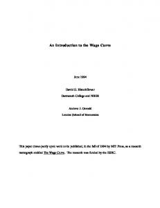
Figure 1. Representative LC–MS chromatograms of (A) plasma blank and (B) LLOQ for dapsone.
2380
Bioanalysis (2012) 4(19)
future science group
An approach to avoid repeat analysis of the samples exceeding ULOQ and separation. The above mentioned column provided better separation of analytes, especially for dapsone and meta-dapsone, as they are positional isomers. Care was taken to adjust dwell times and delays between ion transitions so that there were at least ten to 12 data points for each monitored peak. Failure to meet this requirement may result in inadequate peak integration and considerably poorer precision [13] . A dwell time of 50 ms for each MS/MS transition was used. Construction
of calibration curve & QC samples The entire range of calibration standards were analyzed using pre-optimized chromatographic and MS conditions. For split calibration curve the whole range of calibration standard was divided into two sets. All the calibration curves were best fitted by linear regression with 1/X 2 (1/concentration × concentration) as weighing factor. Eight QC samples were selected for two sets of calibration curves. LQC, MAQC, MBQC and HQC were selected for LCC whereas higher LQC, higher MAQC, higher MBQC and higher HQC were selected for HCC. The method was challenged extensively by analyzing six more replicates of the QC samples (higher LQC, higher MAQC,
| R esearch A rticle
higher MBQC and higher HQC) against LCC range after diluting 20-times using blank rat plasma. Selectivity,
sensitivity (LLOQ) & carryover The selectivity of the method towards endogenous plasma matrix was evaluated in six different lots of animal plasma by analyzing blank and spiked samples at LLOQ levels. No significant direct interference in the blank plasma traces was observed from the endogenous substance in drug-free plasma at the retention time of the analytes and internal standard. Typical chromatograms of extracted blank and drug free rat plasma spiked with analyte at LLOQ levels are illustrated in Figures 1–3. In the current study, LLOQ of 0.63, 1.06 and 1.09 ng/ml have been selected for dapsone, cyclophosphamide and glipizide, respectively. LLOQ concentrations were selected based on method development and the in-house study. With the latest triple-quadruple instrumentation a much lower LLOQ can be obtained. Rinsing solution was optimized to avoid carryover and consisted of acetonitrile/methanol/DMSO (30/40/30, v/v/v) with 0.1% formic acid. No carryover was observed using this mixture.
A
B 24 6.14 23 22 2.39 4.15 21 20 3.15 19 3.75 18 17 5.49 16 3.36 0.44 15 2.64 2.77 1.64 14 5.77 6.63 1.31 13 2.17 0.86 12 4.68 4.83 11 0.17 10 6.34 9 8 7 6 5 4 3 2 1 0 0.5 1.0 1.5 2.0 2.5 3.0 3.5 4.0 4.5 5.0 5.5 6.0 6.5
5.45
68 64 60 56 52 48 Intensity (cps)
Intensity (cps)
5.39
44 40 36 32 28 24 3.30 4.52 0.91 16 0.440.75 1.46 2.20 2.86 1.35 12 3.814.35 4.87 2.29 8 1.60 2.57 4 20
5.85 6.01 6.68
0
Time (min)
0.5 1.0 1.5 2.0 2.5 3.0 3.5 4.0 4.5 5.0 5.5 6.0 6.5 Time (min)
Figure 2. Representative LC–MS chromatograms of (A) plasma blank and (B) LLOQ for cyclophosphamide.
future science group
www.future-science.com
2381
R esearch A rticle |
Basu, Basit, Ravindran, Patel, Vangala & Patel B
88 84 80 76 72 68 64 60 56 52 48 44 40 36 32 28 24 20 16 12 8 4 0
2.61
5.76
4.39 2.01 0.64 0.30 0.80 4.08 1.32 2.76 3.05 5.39 1.07 3.52 3.83 4.84 1.67
6.60 6.44
0.5 0.1 0.5 2.0 2.5 3.0 3.5 4.0 4.5 5.0 6.0 6.5 7.0 Time (min)
Intensity (cps)
Intensity (cps)
A
5.72 280 270 260 250 240 230 220 210 200 190 180 170 160 150 140 130 120 110 100 90 80 70 4.11 0.51 1.06 60 2.49 2.97 3.62 6.53 6.66 50 0.21 0.90 1.42 4.50 4.67 5.50 3.17 2.36 6.08 40 4.89 2.04 30 20 10 0 0.5 0.1 0.5 2.0 2.5 3.0 3.5 4.0 4.5 5.0 6.0 6.5 7.0 Time (min)
Figure 3. Representative LC–MS chromatograms of (A) plasma blank and (B) LLOQ for glipizide.
Linearity
A weighted linear regression of the peak area ratio versus concentrations was performed for dapsone, cyclophosphamide and glipizide. The whole calibration curve range for all three analytes did not fit in a linear regression. The observed peak area ratios were linear over the concentration range of 0.63–625.80 ng/ml for dapsone in rat plasma using 1/X 2 regression. The higher calibration curve standards (625.80 to 14,900.00 ng/ml) were also fitted to a linear equation with 1/X 2 regression, but as a whole did not fit in a linear regression. For cyclophosphamide, the LCC range that fitted in a linear regression was 1.06 to 1060.04 ng/ml and the HCC range was 1060.04 to 25,239.01 ng/ml in rat plasma. Similarly, for glipizide the LCC range selected was from 1.09 to 1053.68 ng/ml and the HCC range was from 1053.68 to 25,087.50 ng/ml in rat plasma, selected with 1/X 2 regression (Table 2) . A linear regression y = mx + c was used for quantitation, where y is the peak area ratio of analyte to internal standard, m is the slope of calibration curve, c is the intercept, x is the analyte concentration (ng/ml). For dapsone, the mean slope and intercept values for LCC were 0.9645 and 3.8597, respectively, and for HCC 0.9725 and 114.7953, respectively. For cyclophosphamide the mean slope and intercept values for 2382
Bioanalysis (2012) 4(19)
LCC were 0.9765 and 7.1595, respectively, and for HCC 0.9616 and 327.9867, respectively. Similarly, for glipizide the slope and intercept values for LCC were 0.9286 and 8.5380, respectively, and for HCC 0.9815 and 145.6990, respectively. Precision
& accuracy The intra- and inter-batch precision and accuracy were evaluated by assaying the QC samples (Table 3) . In this assay, for QC samples analyzed under LCC, the intra- and inter-batch precision was 7.62% or less, the accuracy ranges from ~95 to 111%, at low, medium and high QC (LQC, MAQC, MBQC and HQC, repsectively) levels. For the QC samples analyzed under HCC, the intra- and inter-batch precision was 9.82% or less for undiluted and 5.50% or less for diluted QC samples, and the accuracy ranges from ~96 to 111% for undiluted and from ~94 to 111% for diluted QC samples, at LQC, MAQC and HQC levels. These results demonstrate that the values were within the acceptable range and the method was sufficiently accurate and precise. Matrix
effect & recovery Matrix effect was evaluated by comparing peak area of analyte and internal standards in blank plasma samples spiked after the sample preparation with those obtained by direct injection future science group
Name of the stability experiment
future science group
www.future-science.com
n = 6.
Benchtop (10 h at room temperature) stability Autosampler (32 h at 4ºC) stability Freeze–thaw (five cycles) stability Long-term plasma (30 days at -70ºC) stability Cyclophos- Benchtop (10 h at room phamide temperature) stability Autosampler (32 h at 4ºC) stability Freeze–thaw (five cycles) stability Long-term plasma (30 days at -70ºC) stability Glipizide Benchtop (10 h at room temperature) stability Autosampler (32 h at 4ºC) stability Freeze–thaw (five cycles) stability Long-term plasma (30 days at -70ºC) stability
Dapsone
Drug
LQC HQC LQC HQC LQC HQC LQC HQC LQC HQC LQC HQC LQC HQC LQC HQC LQC HQC LQC HQC LQC HQC LQC HQC
Level
2.61 622.45 2.63 638.83 2.66 642.33 2.73 659.56 4.58 948.11 3.89 958.60 4.36 906.09 4.57 899.43 3.22 874.25 3.02 898.19 2.98 868.41 3.54 952.06
6.61 7.22 8.30 7.17 5.52 3.79 5.19 1.95 6.80 3.01 1.48 4.90 13.05 4.65 7.19 1.52 4.48 3.02 2.79 13.02 10.39 4.05 6.80 3.01
103.54 103.55 104.33 106.28 105.71 3.79 108.42 109.72 109.59 103.30 93.18 104.44 104.34 4.65 109.37 98.00 99.82 94.86 93.55 97.46 92.26 94.22 109.59 103.30
2225.92 12,045.00 2225.92 12,045.00 2225.92 12,045.00 2225.92 12,045.00 3671.28 18,802.97 3671.28 18,802.97 3671.28 18,802.97 3671.28 18,802.97 3544.80 21,100.00 3544.80 21,100.00 3544.80 21,100.00 3544.80 21,100.00
2368.34 12,375.64 2297.63 12,634.00 2343.30 12,022.52 2460.12 12,725.54 3848.79 19,151.77 3420.78 19,638.45 3853.74 19,524.38 3504.91 17,319.10 3329.22 21,261.06 3114.82 18,424.52 3206.09 18,007.80 3716.19 21,491.41
9.53 8.91 6.38 6.16 3.44 10.53 4.06 4.18 2.87 2.60 1.48 4.90 0.49 2.17 3.86 3.13 2.10 6.40 2.35 2.17 1.14 2.65 2.87 2.60
106.40 102.75 103.22 104.89 105.27 99.81 110.52 105.65 104.84 101.86 93.18 104.44 104.97 103.84 95.47 92.11 93.92 100.76 87.87 87.32 90.45 85.35 104.84 101.86
Spiked higher Mean found Mean Mean QC conc. (ng/ml) conc. (ng/ml) %CV accuracy (%)
Spiked QC Mean found Mean Mean conc. (ng/ml) conc. (ng/ml) %CV accuracy (%) 2.52 601.11 2.52 601.11 2.52 601.11 2.52 601.11 4.18 917.82 4.18 917.82 4.18 917.82 4.18 917.82 3.23 921.65 3.23 921.65 3.23 921.65 3.23 921.65
Higher calibration curve
Lower calibration curve
Table 5. Stability assay results of dapsone, cyclophosphamide and glipizide in rat plasma.
An approach to avoid repeat analysis of the samples exceeding ULOQ
| R esearch A rticle
2383
R esearch A rticle |
Basu, Basit, Ravindran, Patel, Vangala & Patel
Table 6. Pharmacokinetic parameters of dapsone following single intravenous and oral administration (5 mg/kg) to male Sprague Dawley rats (n = 3) analyzed under conventional single calibration curve and split calibration curve. Single calibration curve
Split calibration curve
Route
iv.
p.o.
iv.
p.o.
Dose (mg/kg) Formulation † Cmax (ng/ml)
5 Solution 2748.931 ± 265.951 –
5 Solution 2279.933 ± 255.461 0.500
5 Solution 2748.59 ± 269.768 –
5 Solution 2259.467 ± 89.358 0.667 ± 0.289
T1/2 (h)
17,926.129 ± 1854.778 21,793.266 ± 3157.259 10.534 ± 1.386
18,958.948 ± 1133.814 20,779.76 ± 995.606 –
17,457.999 ± 2457.447 21,154.239 ± 3417.320 10.593 ± 1.707
17,269.469 ± 510.377 19,214.257 ± 468.634 –
MRTlast (h)
6.841 ± 0.325
6.384 ± 0.280
6.818 ± 0.427
6.34 ± 0.448
– –
4.016 ± 0.710 2.965 ± 0.408
– –
Tmax (h) AUClast (ng/ml*h) AUCinf (ng/ml*h)
Clearance (ml/min/kg) 3.882 ± 0.604 Vss (l/kg) 2.871 ± 0.125
C0 back-extrapolated concentration of iv. profile. AUCinf: Area under the curve from 0 h to infinity; AUClast: Area under the plasma concentration–time curve from 0 h to the last quantifiable concentration; Cmax: Peak concentration; iv.: Intravenous; MRT: Mean residence time; p.o.: Per oral; T1/2: Terminal half-life; Tmax: Time to peak concentration; Vss: Steady state volume of distribution. †
of neat standards. Though the multiple cleanup procedures or stable-isotope-labeled internal standards were not employed in sample preparation, sufficient chromatographic retention on the analytical column was achieved. As a result, the method almost showed no matrix effect from biological material. Acceptable matrix effect with mean peak area of 92.05–95.30% for LCC and 86.29–94.01% for HCC, obtained from post-extracted samples to standards, and recovery with that of 91.79–97.85% for LCC and 89.15–96.00% for HCC, obtained from
standards spiked before and after extraction in six lots of rat plasma. Data of matrix effect and recovery are given in Table 4. Stability
Stock solution of analytes and internal standard were stable at room temperature for 10 h and at 2–8°C for a 10 days. For the short-term benchtop stability, the QC plasma samples were kept at room temperature for 10 h and extracted as per the extraction procedure, with the results indicating reliable stability behaviors under
Table 7. Pharmacokinetic parameters of cyclophosphamide following single intraperitoneal administration (30 mg/kg) to male Sprague Dawley rats (n = 3) analyzed under conventional single calibration curve and split calibration curve. Single calibration curve
Split calibration curve
ip. 30 Solution 19,021.848 ± 731.709 0.103 ± 0.040
ip. 30 Solution 20,602.373 ± 2219.330 0.103 ± 0.040
T1/2 (h)
9053.929 ± 294.999 9054.639 ± 295.139 1.991 ± 0.101
8872.284 ± 359.271 8873.467 ± 359.033 2.103 ± 0.024
MRTlast (h)
0.401 ± 0.035
0.424 ± 0.047
Route Dose (mg/kg) Formulation † Cmax (ng/ml) Tmax (h) AUClast (0–24h) (ng/ml*h) AUClast (0-inf) (ng/ml*h)
AUClast: Area under the plasma concentration–time curve from 0 h to the last quantifiable concentration; Cmax: Peak concentration; ip.: Intraperitoneal; MRT: Mean residence time; T1/2: Terminal half-life; Tmax: Time to peak concentration.
2384
Bioanalysis (2012) 4(19)
future science group
An approach to avoid repeat analysis of the samples exceeding ULOQ
| R esearch A rticle
Table 8. Pharmacokinetic parameters of glipizide following single intravenous (1 mg/kg) and oral (5 mg/kg) administration to male Sprague Dawley rats (n = 3) analyzed under conventional single calibration curve and split calibration curve. Single calibration curve Route Dose (mg/kg) Formulation
Split calibration curve
iv. 1 Solution
p.o. 5 Suspension
iv. 1 Solution
p.o. 5 Suspension
26,780.859 ± 6343.647 –
18,081.147 ± 728.021
27,253.136 ± 6825.172
18,967.32 ± 1671.929
1.00 ± 0.866
–
0.667 ± 0.289
AUClast (ng/ml*h)
68,058.079 ± 1513.042
14,6470.288 ± 7888.919
67,749.659 ± 1855.209
15,7302.602 ± 8894.819
AUCinf (ng/ml*h)
68,601.460 ± 1617.311
14,6470.288 ± 7888.919
68138.582 ± 2160.509
15,7302.602 ± 8894.819
T1/2 (h)
3.531 ± 0.792
2.26 ± 0.072
3.277 ± 0.446
2.335 ± 0.166
MRTlast (h)
5.17 ± 0.353
5.388 ± 0.219
5.221 ± 0.368
5.443 ± 0.181
Clearance (ml/min/kg) Vss (l/kg)
0.243 ± 0.006 0.078 ± 0.005
– –
0.245 ± 0.008 0.079 ± 0.006
– –
†
Cmax (ng/ml)
Tmax (h)
C0 back-extrapolated concentration of iv. profile. AUCinf: Area under the curve from 0 h to infinity; AUClast: Area under the plasma concentration–time curve from 0 h to the last quantifiable concentration; Cmax: Peak concentration; iv.: Intravenous; MRT: Mean residence time; p.o.: Per oral; T1/2: Terminal half-life; Tmax: Time to peak concentration; Vss: Steady state volume of distribution. †
experimental conditions of the regular analytical procedure. The stability of QC samples kept in the autosampler for 32 h was also assessed. The result indicated that the solution of analytes and internal standard can remain in the autosampler for at least 32 h without showing any significant loss in quantified values, indicating that samples should be processed within this period of time. Analytes in rat plasma were stable for a minimum of five freeze–thaw cycles. Different stability experiments in plasma, and the values for the precision, %CV and accuracy are shown in the Table 5. Incurred sample ana lysis was performed (results not shown), with no significant changes observed, that is, the deviations in concentration was within 15% of the first analysis values. PK
parameters The individual PK parameters were compared using a validated split calibration curve approach and a conventional smaller calibration range approach. In the split calibration curve approach, PK study samples were analyzed without any dilutions whereas in conventional smaller calibration range approach (dapsone: 0.63–625.80 ng/ml; cyclophosphamide: 1.06– 1060.04 ng/ml; and glipizide: 1.09–1053.68 ng/ ml) samples that exceed the ULOQ were diluted in order to fall within the calibration curve range and were reanalyzed using suitable dilution factor. For comparison, same study samples were processed twice (by split calibration curve and conventional method) to avoid variability. PK future science group
parameters of iv., ip. and p.o. samples, and comparisons of PK parameters between two methods, are tabulated in Tables 6–8. Split calibration curve to avoid repeat analysis of sample exceeding ULOQ
The analysis of drugs or analytes over a wide range of concentrations became cumbersome using the conventional narrow calibration curve method. In general, it was observed that due to higher fold in calibration curve, the curve does not fit in any of the well-known curve types. This may be due to a wide range in the curve. However, if the same calibration curve splits into two calibration curves with a common standard point, it fits in the well-known curve type. When the entire range (for dapsone: 0.63–14900.00 ng/ml; cyclophosphamide: 1.06–25,239.01 ng/ml; and glipizide: 1.09–25087.50 ng/ml) were analyzed, the calibration curve range did not fit in the linear as well as the quadratic regression. The instrumental response were found linear up to certain levels (dapsone: 0.63–625.80 ng/ml; cyclophosphamide: 1.06–1060.04 ng/ml; and glipizide: 1.09–1053.68 ng/ml) with 1/X 2 as the weighing factor, but non linear towards further higher standard points. In the first part of our work we generated data for a conventional approach of analysis by diluting the QC samples whose concentration was appropriately higher than the upper limit of LCC so that they fell within the range. Undiluted QC samples in the range of LCC were also analyzed www.future-science.com
2385
2386
Basu, Basit, Ravindran, Patel, Vangala & Patel to confirm precision and accuracy of the method at lower range. Linearity, precision and accuracy of the method were proved by analyzing three precision and accuracy batches. Linearity of the method expressed in terms of precision and accuracy of the method at lower calibration range is summarized in terms of %CV and mean accuracy, respectively, in Table 3. In the second part of our work we validated using a split calibration curve instead of the conventional method described above. The whole range of calibration standards and QC samples were evenly processed. The whole range was spliced into smaller ranges, that is, for dapsone: 0.63–625.80 ng/ml for LCC and 625.80– 14900.00 ng/ml for HCC. Similarly, for cyclophosphamide, the LCC range was 1.06–1060.04 ng/ml and HCC was 1060.04–25,239.01 ng/ml. For glipizide, the LCC range was selected as 1.09– 1053.68 ng/ml and for HCC 1053.68 –25,087.50 ng/ml in rat plasma. The HCC range was best fit by linear equation with 1/X 2 as weighting factor. The higher QCs were analyzed undiluted under HCC. The precision and accuracy of HQC after analysis without dilution under HCC was compared with data generated by conventional method. The differences exhibited were very minimal and within acceptable range (Table 9) .
2184.08 4484.13 9317.00 13,410.13 3474.73 7261.53 14,754.23 20,814.82 3444.29 7258.32 14,717.56 21,123.16 110.85 106.86 104.73 108.46 101.69 102.49 103.98 106.47 106.77 106.11 102.37 103.43 2.86 4.95 5.68 5.54 3.56 4.20 5.20 2.83 4.86 6.85 6.89 5.27 2467.39 4955.59 8830.04 13,064.37 3733.17 7525.28 13,881.55 20,020.04 3784.83 7836.03 15,120.66 21,823.67
Application of split calibration curve in PK study
n = 6, total three batches. HCC: Higher calibration curve; LCC: Lower calibration curve.
2462.51 5086.12 9065.43 13,450.19 3781.68 7608.01 13,871.70 20,249.07 3862.51 7685.47 15,204.79 21,932.66 2225.92 4637.42 8431.50 12,045.00 3671.28 7342.56 13,350.11 18,802.97 3544.80 7385.00 14,770.00 21,100.00 LQC MAQC MBQC HQC Cyclophos- LQC phamide MAQC MBQC HQC Glipizide LQC MAQC MBQC HQC Dapsone
(ng/ml)
3.45 5.02 3.83 4.49 3.37 5.31 5.50 2.87 4.95 6.33 5.43 5.78
110.63 109.68 107.52 111.67 103.01 103.62 103.91 107.69 108.96 104.07 102.94 103.95
Mean accuracy (%) Mean %CV Mean found conc. (ng/ml)
2225.92 4637.42 8431.50 12,045.00 3671.28 7342.56 13,350.11 18,802.97 3544.80 7385.00 14,770.00 21,100.00
Mean found conc. (ng/ml)
1.92 3.00 1.73 3.77 1.48 3.25 7.01 4.37 5.40 6.06 2.64 4.24
Mean %CV
98.12 96.69 110.50 111.33 94.65 98.90 110.52 110.70 97.16 98.28 99.64 100.11
2190.05 4518.21 9245.39 13,180.37 3432.90 7278.18 14,183.11 20,683.55 3414.45 7210.14 14,645.25 21,103.31
1.85 4.04 2.80 6.34 2.01 4.07 8.73 7.19 5.25 6.27 3.23 3.87
98.39 97.43 109.65 109.43 93.51 99.12 106.24 110.00 96.32 97.63 99.16 100.02
Mean Mean %CV accuracy (%) Mean accuracy (%)
Mean found conc. (ng/ml)
Inter-batch Intra-batch
Spiked Intra-batch higher Mean Mean Mean QC conc. found %CV accuracy (ng/ml) conc. (%)
Inter-batch
Spiked higher QC conc. (ng/ml)
Higher QCs diluted and analyzed under LCC Higher QCs undiluted and analyzed under HCC Level Drug
Table 9. Comparative data of precision and accuracy of undiluted higher QC samples analyzed with higher calibration curve and diluted higher QC samples analyzed with lower calibration curve.
R esearch A rticle |
Bioanalysis (2012) 4(19)
The validated method was used for analysis of PK study samples. Study samples were processed twice and analyzed against conventional and split calibration methods. For conventional method, samples that fall above calibration curve range were diluted appropriately using blank rat plasma and analyzed against suitable dilution factor. For split calibration curve, sample analysis was carried out over wide calibration curve range without diluting the samples. Concentrations that fell under LCC were quantified against LCC. Sample concentrations that fell above LCC ULOQ (dapsone: 625.80 ng/ ml; cyclophosphamide: 1060.04 ng/ml; glipizide: 1053.68 ng/ml) were analyzed against HCC. Similar concentration levels were achieved using both approaches, with negligible differences. The comparative PK parameters after analysis with conventional method and split calibration curve approach are tabulated in Tables 6–8. Mean plasma concentration–time profile of dapsone, cyclophosphamide and glipizide in SD rats after analyzing with conventional method and split calibration curve approach are future science group
An approach to avoid repeat analysis of the samples exceeding ULOQ 3000
Matrix: plasma; route: iv.; dose: 5 mg/kg; dapsone single calibration curve Matrix: plasma; route: iv.; dose: 5 mg/kg; dapsone split calibration curve Matrix: plasma; route: p.o.; dose: 5 mg/kg; dapsone single calibration curve Matrix: plasma; route: p.o.; dose: 5 mg/kg; dapsone split calibration curve
2500 Mean plasma conc. (ng/ml)
| R esearch A rticle
2000
1500
1000
500
0 0
4
12
8
16
20
24
Time (h)
Figure 4. Mean plasma concentration–time profile of dapsone following single intravenous and oral administration of 5 mg/kg in Sprague Dawley rats, analyzed under conventional single calibration curve and split calibration curve. iv.: Intravenous; p.o.: Per oral.
represented in Figures 4–6, respectively. The outcomes of PK parameters from both the methods are very similar. Conclusion The split calibration curve approach is advantageous to analyze samples with wide concentration
ranges, often encountered in bioanalysis of earlystage drug discovery. Apart from analyzing samples, this approach is useful to ensure the quality of data generated in terms of accuracy and precision. Dilution and repeat analysis of samples that exceed the ULOQ can be avoided using this method. This method is also useful for the
Mean plasma conc. (ng/ml)
25,000 Matrix: plasma; route: ip.; dose: 30 mg/kg; single calibration curve Matrix: plasma; route: ip.; dose: 30 mg/kg; split calibration curve
20,000
15,000
10,000
5000
0 0
4
8
12
16
20
24
Time (h)
Figure 5. Mean plasma concentration–time profile of cyclophosphamide following single intraperitoneal administration of 30 mg/kg in Sprague Dawley rats, analyzed under conventional single calibration curve and split calibration curve. ip.: Intraperitoneal.
future science group
www.future-science.com
2387
R esearch A rticle |
Basu, Basit, Ravindran, Patel, Vangala & Patel 25,000
Mean plasma conc. (ng/ml)
Matrix: plasma; dose: 1 mg/kg; route: iv.; glipizide single calibration curve 20,000
Matrix: plasma; dose: 1 mg/kg; route: iv.; glipizide split calibration curve
15,000
Matrix: plasma; dose: 5 mg/kg; route: p.o.; glipizide single calibration curve Matrix: plasma; dose: 5 mg/kg; route: p.o.; glipizide split calibration curve
10,000
5000
0 0
4
8
12 Time (h)
16
20
24
Figure 6. Mean plasma concentration–time profile of glipizide following single intravenous (1 mg/kg) and oral (5 mg/kg) administration in Sprague Dawley rats, analyzed under conventional single calibration curve and split calibration curve. iv.: Intravenous; p.o.: Per oral.
bioanalysis of samples where sample volume is very limited for repeat analysis. Future perspective Quantification of drugs is necessary for PK assessment, which is integral to the drug-discovery and development processes. Application of a split calibration curve to quantify drugs in PK studies can be extended to pharmacodynamic, metabolic and toxicokinetic studies. A split calibration curve can also be used to quantify lipids, peptides, metabolites, impurities and environmental pollutants. Quantification of drugs along with metabolites is a time-consuming laborious process, where the split calibration curve approach will be very useful. Similarly, in cassette dosing where many drugs have to be analyzed in a shorter period of time without any error, the split calibration approach will be advantageous. It will also be useful where the sample volume is small, such as cerebro spinal fluid samples or samples collected from neonates. Samples obtained from different types of biological matrices, such as plasma, urine and microsomes, may present various challenges to quantify the necessary component in the matrix. Herein we report a split calibration curve 2388
Bioanalysis (2012) 4(19)
approach that can quantify the NCE or drugs in complex biological matrices, so as to save time, effort and costs of bioanalysis. Thus, application of linear fit for low calibration curve range and linear for higher calibration range makes the split calibration curve approach rapid and efficient, while being accurate. Acknowledgements The authors are thankful to P Patole and R Sahu for valuable discussions and critical review.
Financial & competing interests disclosure The authors thank the Sai Advantium Pharma Limited for financial support. The authors have no other relevant affiliations or financial involvement with any organization or entity with a financial interest in or financial conflict with the subject matter or materials discussed in the manuscript apart from those disclosed. No writing assistance was utilized in the production of this manuscript.
Ethical conduct of research The authors state that they have obtained appropriate institutional review board approval or have followed the principles outlined in the Declaration of Helsinki for all human or animal experimental investigations. In addition, for investigations involving human subjects, informed consent has been obtained from the participants involved. future science group
An approach to avoid repeat analysis of the samples exceeding ULOQ
| R esearch A rticle
Executive summary Split calibration curve to avoid repeat analysis of samples exceeding ULOQ
In this article, a method has been proposed to split a wide dynamic range in calibration curve and analyze the samples in order to avoid the repeat analysis of the samples after dilution.
Due to higher fold in calibration curve, the curve does not fit in any of the well-known curve types. This may be due to wide range in the curve. However, if the same calibration curve is split into two calibration curves with a common standard point, it fits in the wellknown curve type.
Application of split calibration curve in pharmacokinetic study
Three drugs (analytes) were validated using the split calibration curve approach and their pharmacokinetics parameters compared with conventional methods of bioanalysis, and similar results were observed.
This approach may be more useful for discovery bioanalysis rather than regulated bioanalysis.
This method is also useful for the bioanalysis of exploratory toxicokinetic studies where different dosages starting from low to very high were used, and will also be useful where the sample volume is small, such as cerebrospinal fluid samples or the samples collected from neonates.
References
in serum/plasma using a monolithic column. Ther. Drug Monit. 32(1), 102–106 (2010).
Papers of special note have been highlighted as: n of interest 1
n
2
3
4
5
Palm C, Bjork, O, Bjorkholm M, Eksborg S. Quantification of doxorubicin in plasma – a comparative study of capillary and various blood sampling. Anti-Cancer Drugs 12 (10), 859–864 (2001). Interesting article about quantification of doxorubicin by capillary blood sampling in pediatric patients. Kim SJ, Koo TS, Ha DJ et al. Liquid chromatography–tandem mass spectrometry for quantification of lacosamide, an antiepileptic drug, in rat plasma and its application to pharmacokinetic study. Biomed. Chromatogr. 7, 305–712 (2011). Malizia AL, Gunn RN, Wilson SJ et al. Benzodiazepine site pharmacokinetic/ pharmacodynamic quantification in man: direct measurement of drug occupancy and effects on the human brain in vivo. Neuropharmacology 35 (9–10), 1483–1491 (1996). Kostiainen R, Kotiaho T, Kuuranne T, Auriola S. Liquid chromatography/ atmospheric pressure ionization–mass spectrometry in drug metabolism studies. J. Mass Spectrom. 38, 357–372 (2003). Heideloff C, Bunch DR, Wang S. A novel HPLC method for quantification of 10 antiepileptic drugs or metabolites
future science group
6
Barfield M, Spooner N, Lad R, Parry S, Fowles S. Application of dried blood spots combined with HPLC–MS/MS for the quantification of acetaminophen in toxicokinetic studies. J. Chromatogr. B 870 (1), 32–37 (2008).
7
Krishna MM, Purna CM, Ramy C, Nirmala D, Dheeraj G. Toxicokinetics: an important tool in new drug development. Intl J. Pharm. Bio. Sci. 1(3), 319–327 (2011).
8
Lindegargh N, Hanpithakpong W, Kamanikom B et al. Quantification of the anti-influenza drug zanamivir in plasma using high-through put HILIC–MS/MS. Bioanalysis 3(2), 157–165 (2011).
n
9
n
10
One of the better methods to quantify the polar drug Zanamivir® in biological matrices. Basu S, Sahu R, Velamuri R et al. Simultaneous determination of five antiretroviral drugs in rat plasma by liquid chromatography–tandem mass spectrometry. J. Pharm. Res. 4(10), 3738–3742 (2011). This new method offers a platform to analyze several drugs in a wide concentration range using less sample processing volume. Helton DR, Osborne DW, Pierson SK, Buonarati MH, Bethem RA. Pharmacokinetic
www.future-science.com
profiles in rats after intravenous, oral, dermal administration of dapsone. Drug Metab. Dispos. 28(8), 925–929 (2000). n
11
Quantitative pharmacokinetic profile favors the dermal administration of dapsone over intravenous and oral routes to reduce side effects. Chen Y, Junga H, Jiang X, Naidong W. Simultaneous determination of theophylline, tolbutamide, mephenytoin, debrisoquin, and dapsone in human plasma using high-speed gradient liquid chromatography/tandem mass spectrometry on a silica-based monolithic column. J. Sep. Sci. 26 (17), 1509–1519 (2003).
12 Matuszewski BK, Constanzer ML, Chavez-
Eng CM. Strategies for the assessment of matrix effect in quantitative bioanalytical methods based on HPLC–MS/MS. Anal. Chem. 75, 3019–3030 (2003). 13 Andrew V, Christopher A, Lillian T, Steven
JS. Simple rapid method for quantification of antiretrovirals by liquid chromatography– tandem mass spectrometry. Clin. Biochem. 35, 99–103 (2002).
Website 101 US FDA. Guidance for Industry,
Bioanalytical Method Validation. www.fda.gov/cder/guidance/4252fnl.pdf
2389