STATISTICAL RECOGNITION OF BREATHING BY MS KINECT DEPTH SENSOR. Martin Schätzb, Fabio Centonzeb,c, Jirı Kuchynkad, Ondrej ËTupab,.
STATISTICAL RECOGNITION OF BREATHING BY MS KINECT DEPTH SENSOR b ˇ Martin Sch¨atzb , Fabio Centonzeb,c , Jiˇr´ı Kuchyˇnkad , Ondˇrej Tupa , a,d e a,b Oldˇrich Vyˇsata , Oana Geman , Aleˇs Proch´azka a
b
Czech Institute of Informatics, Robotics and Cybernetics, Czech Technical University, Prague, CZ University of Chemistry and Technology in Prague, Dept of Computing and Control Engineering, CZ c Politecnico di Milano, Department of Mechanical Engineering, Italy d Charles University, Department of Neurology, University Hospital in Hradec Kralove, CZ e Stefan cel Mare University of Suceava, Department of Health and Human Development, Romania ABSTRACT
Measuring of breathing with contact methods, like respiratory belts, is very uncomfortable for patients and in case of complex sleep analysis, cables from different sensors can substantially affect the quality of the sleep. This paper presents the contactless measuring of breathing using the MS Kinect depth sensor, and it compares the results obtained with records of breathing observed by the flowmetry. The methodological part of the paper is devoted to spectral analysis of data acquired, feature extraction, and their Bayesian classification. The proposed classifier is able to distinguish the Sleep and Wake classes with the accuracy of 100% (crossvalidation: 0) for given data. The achived accuracy of classification into 3 classes (Sleep, Falling Asleep and Wake) is 97% (cross-validation: 0.0248) in the given case. Index Terms— MS Kinect, range image processing, breathing analysis, signal processing 1. INTRODUCTION Range Imaging methods are based upon specific computational technologies forming matrices with their elements carrying information about the distance of the corresponding image component from the sensor (also known as the depth map). The beginning of these methods can be dated back to 1904 when radar was discovered [1]. But the first range imaging sensor was used by the Mars rover in 1977 [2] only. The technology detecting the time of flight is also used in the MS Kinect v2 depth sensor, or in the LIDAR (LIght Detection And Ranging) systems. The breathing rate during all stages of sleep is significantly more rapid and shallow comparing to that during the awake stage and the most significant reduction occurs during the REM sleep [3]. There are no significant differences between the sexes either in ventilation (corrected for body surface area) or in end-tidal concentrations during any stage of sleep. There are no significant differences in the levels c 978-1-4673-8457-5/15/$31.00 2015 IEEE
of ventilation in non-REM sleep, but there can be significant fall in the REM sleep. This reduction in ventilation exists because of a breathing pattern which is considerably shallower in all stages of sleep than during wakefulness [3]. The respiratory rate and the respiratory dynamics or the variation of the breathing pattern provide useful information for diagnostics or therapeutic [4]. Breathing rate is one of the most important vital signs [5]. There already exist different ways how to measure breathing with contactless methods, one of them is using radio frequency. It behaves essentially like a radar system [6, 7, 4]. A common form of this system uses Doppler radar, which is based on the frequency shift of a signal caused by the relative velocities of a transmitter and receiver. The transmitted signal reflected from the chest will change in frequency according to the movement of the chest. Another method uses Eddy current to exploit changes in torso conductance during respiration, that are caused by varying quantities of air, blood and fluids. It provides possibility to measure volume changes of air in the lungs [8]. This paper focuses on using the depth sensor from the MS Kinect v2 for contactless measuring of breathing. 2. METHODS Depth maps acquired by the MS Kinect v2 depth sensor were recorded to follow chest movement, to analyze breathing and to detect its disorders. The range of this sensor is officially 4.5m [9], but it is able to measure depth up to 8m with the sampling rate up to 30 frames per second [9]. The resulting matrix has 512x424 elements [9, 10], and precision of depth measurement was increased in comparison with the MS Kinect v1. The depth sensor of the MS Kinect v2 is not affected by outside light and errors from it are not present in depth maps. But single rough errors randomly distributed still exist and they can substantially affect the captured scene. The method of reducing of these errors is similar to processing of the salt-and-pepper noise in standard images and the 2D median filter was used for the depth map processing as well.
Flowmetry
Kinect v1
5 4.5
.
4.5
4
.
4
3.5 Frequency (Hz)
For processing of depth maps there were considered two methods, both dealing with the mean of depth over the selected area of M rows and N columns. The first method computes the mean value of selected area in the depth map 1 XX y(t) = DMt (i, j) (1) MN i j where the DMt is the depth map, acquired in time t, and i, j are indexes of area of interest. The second method evaluates the mean of differences of two consecutive depth maps 1 XX y(t) = (DMt (i, j) − DMt−1 (i, j)) (2) MN i j where DMt is depth map, acquired in time t, and i, j are indexes of area of interest. Resulting signal has the mean value zero and the reduced amplitude in comparison with the original signal. Fig. 1 presents a selected part of data acquired by MS Kinect showing the time evolution of the average distance of the chest from the MS Kinect used for analysis.
5
Max
.
2.5
.
2 . 1.5
.
.
3 .
2.5 2
.
1.5 .
1
0.5
Min 10
20
30 40 Time (s)
.
1
.
0.5 0
Max
3.5
.
3
Frequency (Hz)
2.1. Data Acquisition
0
50
. Min 10
20
30 40 Time (s)
50
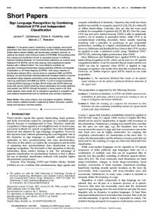
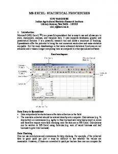
Fig. 2. The comparison of spectrograms evaluated from independent data; (a) acquired by the contact method and (b) obtained by the the MS Kinect.
It is visible that both spectrograms have most energy between 0.1 and 0.5 Hz, corresponding to breathing rate of 10 to 30 breaths per minute.
Processed record divided in parts (2)
Difference of Depth Map [mm]
0.3 Whole record Wake Sleep FA
0.2
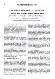
2.2. Spectral Analysis and Feature Extraction The record presented in Fig.1 presents partitioning of the signal into three sections. In fact two of sections are during sleep, just divided in two parts because recorded person moved during sleep. Spectral analysis of these parts (Fig. 3) reveals that there is difference in frequency of breathing while being awake and sleep.
0.1 0 −0.1 −0.2 −0.3 0
200
400
600 Time [s]
800
1000
1200 (a) Frequency analysis of record during wakefulness
Fig. 1. The time evolution of the average distance of the chest from MS Kinect used for feature extraction and classification. As the MS Kinect depth sensor is not precise enough with its error increasing with the distance of the measured object, the output of any depth map processing will be affected by noise components. Typical respiratory rate for a healthy adult at rest is 12-20 breaths per minute [4, 11, 12, 13], which corresponds to 0.2–0.33 Hz. This fact can be used for the selection of the cut-off frequency of the low-pass FIR filter proposed to filter out noise and to preserve the slow periodic signal of breathing. The best option is to design a Savitzky-Golay filter of the second order, because it is able to preserve steep changes in the signal while smoothing it. The cut-off frequency of 0.7 Hz (42 breaths per minute) was chosen. Comparison of results obtained by a contact method and by the MS Kinect is presented in Figure 2. Data recorded by the depth sensor of the MS Kinect were filtered and results of their time-frequency analysis were compared with data acquired by the contact method of the standard polysomnography which is used for recording of breathing during the sleep. Spectrogram was used to compare both records confirming that important frequencies of breathing are preserved.
120
(b) Frequency analysis of record during sleep
25
100
20
80 15 60 10 40 5
20
0
0.1 0.2 0.3 0.4 0.5 0.6 0.7 0.8 0.9 Frequency [Hz]
1
0
0.1 0.2 0.3 0.4 0.5 0.6 0.7 0.8 0.9 Frequency [Hz]
1
Fig. 3. Frequency analysis of selected parts of the record showing frequency componets (a) during the wake stage and (b) during the sleep. Taking into account all changes of processed signal the most important differences of selected parts of signal include (a) the change of frequency and (b) the change of the amplitude. During wakefulness the dominant frequency is around 0.15 Hz and the height of amplitude is 0.13 while during the sleep the dominant frequency is around 0.25 Hz with amplitude height of 0.5. The frequencies cannot be directly transferred into breaths per second, because of unstable sampling frequency changing from 8 Hz to 30 Hz, with its mean of 18 Hz.
Table 2. D ISTRIBUTION OF FEATURES (2) Class Count Percent
2.3. Bayesian Classification Bayesian classification is based on the Bayes’ theorem, which is stated by relation: P (B|A) =
P (A|B)P (B) P (A)
(3)
where P (A) and P (B) are independent probabilities, P (A|B) is conditional probability (probability A given B is true) and P (B|A) is conditional probability (probability of B given A is true). The goal of classification is to find an estimate of class cˆk of an unknown instance x, expressed as: cˆk = maxc1 ,c2 ,...,cM (P (ck , x)) then probability of x being in class {ck }M k=1 is P (x|ck )P (ck ) P (ck |x) = PM k=1 P (x|ck )P (ck )
(4)
It is then possible to find fundamental characteristics of each attribute xj , including mean µck ,xj and variance σc2k ,xj for each class ck , assuming Gaussian distribution of their values and independence of their features. Gaussian distribution of each attribute xj and class ck is then defined by relation: � (x − µ 2� 1 j ck ,xj ) P (xj |ck ) = q exp − 2σc2k ,xj 2πσc2k ,xj It is possible to find out how likely to see observation x for class ck by relation: P (x|ck ) =
R Y
Wake Sleep FA
P (xj |ck ) 4. CONCLUSION
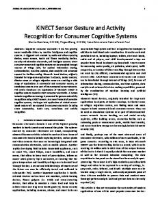
Fig. 1 presents data processed and classified into three classes (two for sleep). Classes are Wake, Sleep1 and Sleep2. The distribution of selected features including the signal amplitude and dominant frequency is presented in Table 1. The classifier can be then defined to distinguish between the stage of wakefulness and sleep. Classes for the second classification include Wake, Falling Asleep (FA) and Sleep. The selected parts of processed recordings prepared for feature extraction are presented in Fig. 1, with distribution of features in Table 2. The classifier Table 1. D ISTRIBUTION OF FEATURES (1) Class Count Percent 43.06% 48.15% 8.80%
Classification (2)
Classification (1) 0.25
Sleep Wake
0.25
Sleep Wake FA
0.2 Amplitude
3. RESULTS
This paper shows possibility of measuring breathing with MS Kinect’s depth sensor, and shows that it is good alternative for other already published articles about measurement of respiration rate with various non contact methods.
Amplitude
where R is number of features.
186 208 38
35.43% 39.62% 24.95%
defined for these features shows possibility to classify also some states in between of wakefulness and the sleep. As is shown in Fig. 2, the depth map can be processed with introduced methods using Eqs (1) and (2) in a way that brings out the breathing rate out of noise, errors of depth sensor, and movement of recorded person. The whole process is done in three steps, denoising of depth maps (using the 2D median filter), processing of depth maps, and the low-pass filtering of resulting signal (FIR filter). The analysis of record during sleep revealed changes in frequency of breathing rate (Fig. 3) and amplitude of signal, the breathing rate during sleep is more rapid and shallow than during when person is awake (as it should be [3]). This change of breathing in processed signal shows that used method is sensitive enough to be useful. This established property of record was also used to train Bayesian classifiers, using dominant frequency of signal and maximal change in amplitude as features. The first classifier was trained on two classes (Sleep and Wake), and has 100% accuracy (Fig 4) with cross-validation of 0, calculated by leave one out method. The second classifier was trained on three classes (Sleep, Falling Asleep and Wake), with purpose to test possibilities of Bayesian classification for this kind of problem, and has 97% accuracy (Fig 5) with cross-validation of 0.0248, calculated by leave one out method.
J=1
Wake Sleep1 Sleep2
186 208 131
0.2 0.15 0.1
0.15
0.1
0.15
0.2 0.25 Dominant frequency
Fig. 4. Classification (1)
0.15
0.2 0.25 Dominant frequency
Fig. 5. Classification (2)
The whole process consists of denoising images with the 2D median filter, computing difference of chest position using the difference of depth maps and post-processing using FIR filter for final denoising, and on part of the recorded data are trained Bayesian classifiers. The first classifier uses amplitude and dominant frequency to distinguish from breathing between when person is awake and when person is asleep with 100%. This also shows that the whole processing is sensitive enough to produce results that can be used as part of more complex analysis of sleep. It is assumed that the following research will be devoted to integration of MS Kinect observations into the whole polysomnography and data acquisition in the sleeping laboratory where the analysis of breathing is used for more complex diagnostical purposes. The data fusion will form the fundamental part of this study. Acknowledgements: All procedures were approved by the local Ethics committee. The project was supported by grant MH CZ DRO (UHHK, 00179906).
5. REFERENCES [1] C. Hulsmeyer, “Hertzian wave projecting and receiving apparatus adapted to indicate or give warning of the presence of a metallic body, such as a ship or a train,” in the line of projection of such waves. [patent]. U.K., vol. 13, pp. 170, 1904. [2] R. A. Lewis and A. R. Johnston, “A scanning laser rangefinder for a robotic vehicle,” in Proceedings 5th International Joint Conference on Artificial Intelligence, 1977, pp. 762–768. [3] N. J. Douglas, D. P. White, Ch. K. Pickett, J. V. Weil, and W. Clifford, “Respiration during sleep,” pp. 840– 844, 1982. [4] Y. S. Lee, P. N. Pathirana, Senior Member, and Ch. L. Steinfort, “Respiration Rate and Breathing Patterns from Doppler Radar Measurements,” , no. December, pp. 8–10, 2014. [5] J. F. Fieselmann, M. S. Hendryx, C. M. Helms, and D. S. Wakefield, “Respiratory rate predicts cardiopulmonary arrest for internal medicine inpatients,” Journal of general internal medicine, vol. 8, no. 7, pp. 354–360, 1993. [6] O. Kaltiokallio, H. Yiˇgitler, R. J¨antti, and N. Patwari, “Non-invasive respiration rate monitoring using a single COTS TX-RX pair,” IPSN 2014 - Proceedings of the 13th International Symposium on Information Processing in Sensor Networks (Part of CPS Week), pp. 59–69, 2014.
[7] T. Taheri and A. S. Anna, “Non-Invasive Breathing Rate Detection Using a Very Low Power Ultra-wideband Radar,” pp. 78–83, 2014. [8] A. Richer and A. Adler, “Eddy current based flexible sensor for contactless measurement of breathing,” in Instrumentation and Measurement Technology Conference, May 2005, pp. 257–260. [9] “Kinect for Windows features,” http://www. microsoft.com/en-us/kinectforwindows/ meetkinect/features.aspx, 2015. [10] R. Smeenk, “Kinect V1 and Kinect V2 fields of view compared,” http://smeenk.com/ kinect-field-of-view-comparison/, 2014. [11] K. E. Barrett, S. M. Barman, S. Boitano, and H. Brooks, “Ganong’s Review of Medical Physiology, 24th Edition,” . [12] Ch. Patel, “Breathing,” in The Complete Guide to Stress Management, pp. 141–161. Springer US, 1991. [13] W. Lindh, M. Pooler, C. Tamparo, and B. Dahl, Delmar’s Comprehensive Medical Assisting: Administrative and Clinical Competencies, Cengage Learning, 2009. [14] M. Sch¨atz, “Statistical Recognition and Spatial Monitoring of Breathing Using Data Acquired by the MS KINECT,” M.S. thesis, ICT Prague, 2015. [15] A. Prochazka, M. Schatz, O. Tupa, M. Yadollahi, O. Vysata, and M. Walls, “The MS kinect image and depth sensors use for gait features detection,” in 2014 IEEE International Conference on Image Processing (ICIP). Oct. 2014, pp. 2271–2274, IEEE. [16] Ch. W. Wang, A. Hunter, N. Gravill, and S. Matusiewicz, “Unconstrained video monitoring of breathing behavior and application to diagnosis of sleep apnea,” IEEE Transactions on Biomedical Engineering, vol. 61, no. 2, pp. 396–404, 2014. [17] S. Ostadabbas, Ch. Bulach, D. N. Ku, L. J. Anderson, and M. Ghovanloo, “A passive quantitative measurement of airway resistance using depth data,” in 2014 36th Annual International Conference of the IEEE Engineering in Medicine and Biology Society. Aug. 2014, pp. 5743–5747, IEEE. [18] S. V. Vaseghi, Advanced Digital Signal Processing and Noise Reduction, John Wiley & Sons Ltd, West Sussex, 2006. [19] G. Benchetrit, “Breathing pattern in humans: Diversity and individu- ality,” Respiration Physiology, vol. 122, pp. 123–9, 2000.