LETTERS PUBLISHED ONLINE: 29 MARCH 2009 | DOI: 10.1038/NMAT2421
Stepwise surface encoding for high-throughput assembly of nanoclusters Mathew M. Maye1 *, Dmytro Nykypanchuk1 , Marine Cuisinier1 , Daniel van der Lelie2 and Oleg Gang1† Self-assembly offers a promising method to organize functional nanoscale objects into two-dimensional (2D) and 3D superstructures for exploiting their collective effects1–3 . On the other hand, many unique phenomena emerge after arranging a few nanoscale objects into clusters, the so-called artificial molecules4–10 . The strategy of using biomolecular linkers between nanoparticles has proven especially useful for construction of such nanoclusters4–6,11–16 . However, conventional solution-based reactions typically yield a broad population of multimers or isomers of clusters; furthermore, the efficiency of fabrication is often limited4–6,11–16 . Here, we describe a novel high-throughput method for designing and fabricating clusters using DNA-encoded nanoparticles assembled on a solid support in a stepwise manner. This method efficiently imparts particles with anisotropy during their assembly and disassembly at a surface, generating remarkably high yields of well-defined dimer clusters and Janus (two-faced) nanoparticles. The method is scalable and modular, assuring large quantities of clusters of designated sizes and compositions. The ability of biomolecules grafted onto a surface to specifically recognize molecules with corresponding affinity is promoting the development of a broad range of biodetection platforms16 . Recently, the suitability of this interaction was demonstrated in assembling designed nanoclusters of nanoparticles, wherein recognition properties are encoded through biofunctionalization4,5,11–15 . Here, we construct a designed cluster through sequential bio-recognition and immobilization events. Resembling the synthesis of biopolymers17 or DNA constructs on solid supports18 , this strategy begins with the recognition between an encoded support and a nano-object (such as a nanoparticle) that continues at each step between the object and extra biomolecular linkers or nano-objects. We implemented this concept to more easily fabricate nanoclusters using DNA-encoded nanoparticles of various sizes and compositions. The programmability and structural plasticity of DNA, critical for such a process, was recently demonstrated as a powerful molecular tool for organizing nano-objects as one-dimensional (1D) groupings5,11,15 , onto 2D scaffolds12–14 or into 3D crystalline assemblies4,19–21 . Figure 1 schematically represents our approach, illustrating the initial encoding (Fig. 1a) and fabrication of either dimer nanoclusters (Fig. 1b) or Janus particles and clusters (Fig. 1c). The protocol for stepwise assembly begins with surface encoding, which we achieved by grafting A0 single-stranded (ss) DNA (see Supplementary Table S1) onto the surface of micrometresized (1–4 µm) magnetic-bead supports, denoted as A0 -s. In the first assembly step (step 1), model nano-objects of 11 nm gold nanoparticles, functionalized22 with an equal surface fraction of two types of ssDNA, A and B (particle AB-p), are added to the solution containing the A0 -s. In this process, the A strands from AB-p
hybridize to A0 -s through a 15-base-pair (bp) A–A0 recognition, and the unreacted B encodes the attachment of the next component at the particle–solution interface. An equimolar regime of A and B on a particle (∼60ssDNA/AB-p), adequately facilitates moderate binding to the support’s surface while preserving the availability of B to act as the next binding site22 . We monitored the hybridization of AB-p onto A0 -s, reflecting the assembly of particles on the support, by ultraviolet–visible spectrophotometry (UV–vis) measurements of the gold surface plasmon resonance band in the supernatant, after the magnetic support with assembled particles was separated by a magnetic field. When assembly was complete (typically 98% of AB-p, Supplementary Fig. S7), we followed this separation procedure by removing the supernatant, and rinsing the material multiple times with the buffer (see the Methods section). In step 2, a 33-base (b) linker ssDNA, B0 C 0 − l, is added. The recognition of the B0 C 0 − l and the AB-p, through a 15-bp B0 B hybridization, directs the assembly of this linker primarily at the particle–solution interface; meanwhile, hybridization is suppressed sterically at the particle–support interface (AA0 region). The linker’s C 0 terminus thus provides an 18-b encoding at the top hemisphere, thereby transforming the AB-p with its initial isotropic A and B distribution over the particle’s surface into a anisotropic particle, AB-p-B0 C 0 (Fig. 1a) immobilized on the surface. From these initial assembly steps (Fig. 1a), we subsequently explored two alternative strategies for fabricating nanoclusters; both have advantages in terms of fidelity or use in further assembly due to their difference in steric effects. In the first strategy (Fig. 1b), we produced dimer nanoclusters by assembling them at the surface, and then releasing them. In contrast, in the second strategy (Fig. 1c), we used the initially encoded particles, which on release are considered as Janus nanoparticles, to generate multivalent Janus nanoclusters through an extra step of conventional solution-based assembly. To fabricate dimer nanoclusters (Fig. 1b, step 3a), we introduced a second nanoparticle of similar size to the immobilized AB-p-B0 C 0 . For example, a C-functionalized particle (C-p) specifically recognizes only the C 0 -terminated regions of the AB-p-B0 C 0 by a 15-bp C 0 C hybridization, the assembly of which we followed either using UV–vis or visually by loss of colour. In principle, the number of C-p particles bound to AB-p-B0 C 0 can be regulated by varying the number of linkers, B0 C 0 -l, attached to each AB-p. The resulting cluster architecture, however, will also depend on the binding constraints near the surface, connectivity of multiple linkers between particles and steric repulsions caused by interactions with non-complementary DNA, either between similar particles or a particle and a surface. We found that for similarly sized particles AB-p and C-p, the conditions of equimolar particle concentration and of an average three B0 C 0 -l linkers per AB-p favour a predominantly single-particle binding, as illustrated below
1 Center
for Functional Nanomaterials, 2 Biology Department, Brookhaven National Laboratory, Upton, New York 11973, USA. *Present address: Department of Chemistry, Syracuse University, Syracuse, New York 13244 USA. † e-mail:
[email protected]. NATURE MATERIALS | ADVANCE ONLINE PUBLICATION | www.nature.com/naturematerials © 2009 Macmillan Publishers Limited. All rights reserved.
1
NATURE MATERIALS DOI: 10.1038/NMAT2421
LETTERS a
AB–p +
B’ +
A’¬s
C’
C’
A’ B C’ (2)
(1)
(3a or 3b)
Rinse
b
(3a)
C¬p
c
(3b) + A”
+
C’ C’ +
Separate
(4b)
C’ Janus particle AB¬p¬B’C’
+
C¬p
Rinse (4a)
+A”
(5a)
+ C”
+
Janus cluster
Dimer
Separate AB¬p¬B’C’¬C¬p
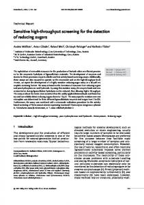
Figure 1 | The assembly and encoding steps in fabricating symmetric dimer nanoclusters or asymmetric Janus particles and clusters. a, Surface encoding begins by grafting A0 -ssDNA strands at paramagnetic bead supports (A0 -s), followed by the immobilization of A- and B-ssDNA-functionalized particles (AB-p) through 15-bp A0 A hybridization (step 1). After a purification step (rinse), facilitated by magnetic separation of the A0 -s, the B0 C0 -l ssDNA linker is added, which recognizes only the AB-p through 15-bp BB0 hybridization (step 2). b, Dimer nanoclusters can then be fabricated by the assembly of C-ssDNA-functionalized particles (C-p) through 18-bp C0 C hybridization (step 3a). Next, the assembled dimers are released from the support by adding A00 fuel strands that recognize the A0 -s through 23-bp A0 A00 hybridization, thus replacing the A0 A linkages. Finally, any free C0 recognition sites are passivated by adding C00 ssDNA (step 5a). c, Alternatively, the immobilized particles from steps 1 and 2 (a) are released from the A0 -s (step 3b), thus forming Janus particles. The particles’ asymmetric structure and multivalent binding can be verified by adding C-p, wherein assembly is realized in solution through 18-bp C0 C hybridization, resulting in Janus nanoclusters.
(Fig. 2a,b). At the same time, the ability to control the number of linkers enabled us to assemble complex clusters when permitted by differences in the particles’ sizes, as described below for the second approach (steps 3b and 4b, Fig. 1c). We released the two-particle dimer nanoclusters (AB-p-B0 C 0 -C-p) from the A0 -s support (step 4a) by substituting the A0 A double-stranded (ds) DNA hybridization with the energetically more favourable hybridization of A0 A00 , wherein A00 is the so-called fuel strand (see Supplementary Table S1, see the Methods section)23 . Specifically, A00 binds to A0 through the formation of a thermodynamically more stable 23-bp dsDNA, replacing the 15-bp AA0 linkage between AB-p and the support, and thus, liberating the dimers. Control experiments with non-complementary nanoparticles (D-p), linkers (B0 F -l) or fuel strands (G) revealed less than 1% nonspecific adsorption over 12 h, or lack of assembly or release, respectively, during the ∼24 h of our assembly process. Finally, adding a 15-b C 00 effectively quenched any free C 0 recognition, so suppressing the aggregation of dimers after their release. 2
We sampled the assembled and released dimer nanoclusters by transmission electron microscopy (TEM) without any further purification. Figure 2a, a typical TEM micrograph, demonstrates the remarkably successful fabrication of two-particle morphology, that is, dimer nanoclusters. A representative statistical analysis (Fig. 2b) revealed a morphological yield of ∼73% dimers, ∼26% monomers and only ∼1% clusters with more than two particles. In addition to these high yields and lack of larger assemblies, the micrographs demonstrated that this approach engenders high concentrations of dimer products, scalable from nanomolar to micromolar concentrations, an outcome challenging to achieve with existing methods. Similar results were obtained at multiple concentrations, TEM grid locations and in replicate trials wherein dimer yields were consistently between 70–83% (see Supplementary Fig. S1). We probed the assembled dimer structures in situ after release using dynamic light scattering (DLS) and UV–vis. Figure 2c shows a representative set of DLS profiles characterizing the numberaveraged hydrodynamic diameter (Dh ) population for isolated C-p and assembled dimers (gold core size ∼11 nm). The dimer nanoclusters possessed a Dh ≈ 37 nm, in contrast to Dh ≈ 26 nm for isolated C-p (core size + DNA shell), agreeing well with an estimate of 37.4 nm for an ellipsoid of two DNA-capped particles linked by B0 C 0 -l (see Supplementary Information)24 . Moreover, we validated, by gel electrophoresis, the increase in size of the dimers compared with that of unassembled monomers (see Supplementary Fig. S4), as demonstrated by a decrease in their electrophoretic mobility, similar to that reported by others for DNA-conjugated nanoparticle groupings5,11,15 . In the second approach (step 3b, Fig. 1c), we fabricated nanoparticles with strongly anisotropic binding motifs, that is, Janus particles7,8,10,25,26 , for assembling asymmetric Janus clusters26 . The anisotropic interactions of nanoscale objects offer unique opportunities for forming novel materials25 , such as 1D plasmonic structures. Analogous to the methods detailed above (Fig. 1a), a first layer of large gold AB-p (50 nm) was assembled at A0 -s (step 1), followed by anisotropic encoding with B0 C 0 -l (step 2). The Janus particles, AB-p-B0 C 0 , were released from the surface using the A00 strand (step 3b). AB-p-B0 C 0 particles possess distinguishably different recognition sites on both hemispheres, wherein the number of C 0 recognition sites is regulated by adjusting the ratio between the linkers and particles while they are at the solid support. For instance, after hybridizing these Janus particles with a ∼5× ratio of B0 C 0 -l, and subsequently releasing them, we probed their binding characteristics by assessing their reaction with a ∼5× molar ratio of a smaller complementary C-p. For the range of particle sizes and the number of linkers used, we can neglect the effect of steric repulsion between bound particles (C-p) on the availability of recognition sites. The TEM image in Fig. 2d reveals the assembly of Janus clusters containing C-p particles on the B0 C 0 -l-functionalized hemisphere of the Janus particles, AB-p-B0 C 0 . A relatively high population of these Janus clusters consisted of ∼5 small C-p per large AB-p, attributed to their nearly pentavalent anisotropic binding property (see Supplementary Fig. S2). The optical properties of nanoclusters, such as dimers, recently attracted significant interest because of their potential applications, and value as model systems for elucidating particle–particle surfaceplasmon coupling, surface-enhanced Raman spectroscopy, singlemolecule detection27–29 and plasmonic quenching of fluorescence30 . To examine the characteristics of their surface plasmon resonance band, we exploited our modular strategy to produce dimers (steps 1–5a) containing particles of various sizes (11, 20, 50 and 75 nm) and compositions (gold and silver). For instance, dimers of ∼75 nm gold nanoparticles exhibited strong surface plasmon resonance band coupling, as demonstrated by a redshift from λ0 = 546 nm (isolated 75 nm particles) to λ ≈ 556 nm (Fig. 3a). This finding contrasts with the minimal surface plasmon resonance bandshift
NATURE MATERIALS | ADVANCE ONLINE PUBLICATION | www.nature.com/naturematerials © 2009 Macmillan Publishers Limited. All rights reserved.
NATURE MATERIALS DOI: 10.1038/NMAT2421
LETTERS
a
Population (%)
b
100
50
0
c Number (%)
50 nm
1 2 3¬10 >10 Particles/cluster (n)
20 10 0
10
100
Dh
d
100 nm
25 nm
25 nm
Figure 2 | Dimer and Janus morphologies. a, A representative TEM micrograph for nanoparticle dimers assembled and released through the encoded solid-support approach, sampled without further purification or size selection. b, Statistical analysis of the TEM micrograph, revealing ∼73% dimers, ∼26% monomers and only 1% structures with three or more particles (n = 1,670 particles). c, The DLS-measured Dh population for C-p monomers (black) and assembled dimers (red). d, Representative TEM micrographs sampled after assembly of 50 nm AB-p Janus particles with a ∼5× ratio of 11 nm C-p.
50 nm
0.01
0
Dh
100
50 nm
0 550 λ (nm)
650
10 0
0.5
450
20
10
50 nm
50 nm
0
0.5 d/D
1.0
Nor. Abs
10
Number (%)
20
0
Nor. Abs
c
b
Δλ/Δ0
Number (%)
a 30
Dh
100
25 nm
0.8 0 400
500 600 λ (nm)
Figure 3 | Dimer optical characteristics. a, A corresponding set of DLS (top panel) and UV–vis (bottom panel) results for isolated 75 nm C-p (black) and assembled dimers (red). b, Comparison of the observed surface plasmon resonance bandshift (1λ/λ0 ), and the ratio of interparticle distance to particle diameter (d/D) (left panel). The exponential decay was fitted by y = C1 exp(−(x/τ )) + y0 , where τ = 0.14 ± 0.028, C1 = 0.06 ± 0.005. TEM micrographs for each dimer system sampled from each solution (right panel). c, A corresponding set of DLS (top panel) and UV–vis (bottom panel) results for 27 nm silver AB-p (black), 11 nm gold C-p monomers (green) and their assembled hetero-dimer (red), with a TEM micrograph sampled from the solution.
(1–2 nm) for dimers containing small particles, 11 nm and 20 nm diameter, but is consistent with an intermediate redshift detected for gold dimers with ∼50 nm diameters (see Supplementary Fig. S3). The enhanced surface plasmon resonance band coupling for larger particles (50, 75 nm) reflects an increase in the particle’s diameter (D), such that it exceeds the interparticle surface-tosurface distance (d). Here, d, is defined by the particles’ DNA shells (AB-, C-) and linkers (B0 C 0 -l). For each of these systems, d is constant, as defined by identical DNA constructs, and was ∼12 nm, measured by in situ small-angle X-ray scattering (see Supplementary Fig. S6). Figure 3b summarizes the relationship between the surface plasmon resonance shift (1λ/λ0 ) and the ratio of interparticle distance to core size (d/D); it follows the recently described plasmon-ruler equation; 1λ/λ ≈ C1 exp(−(d/D)/τ )+y0 , where C1 is a constant and τ is the decay constant29 . The observed τ for our dimer systems, 0.14±0.03, is slightly lower than the in-plane polarized measurements of gold nanodiscs on silicon, fabricated by electron beam lithography29 .
This approach can also be applied for fabrication of hetero-material clusters. Figure 3c illustrates the results of optical (UV–vis) and structural (DLS and TEM) measurements of silver–gold hetero-dimers. They consist of AB-capped ∼27 nm silver particles, and 11 nm gold particles, C-p. On their assembly and release, following the identical method to that used for gold dimers (steps 1–5a), the assembled hetero-dimers exhibited a 3 nm redshifted silver surface plasmon resonance band (∼405 nm) and a gold particle surface plasmon resonance band (∼525 nm). These results are consistent with dimers in solution, as was confirmed by light scattering and TEM (Fig. 3c). In summary, we demonstrated the high-throughput fabrication of both symmetric and asymmetric nanoclusters through a stepwise surface-encoding protocol. The modularity of our approach, based on the programmability of DNA interactions, facilitates the incorporation of isotropic or anisotropic nano-objects of different sizes or composition. We consider that as the number of available DNA-based geometries and motifs grows, along with the ability
NATURE MATERIALS | ADVANCE ONLINE PUBLICATION | www.nature.com/naturematerials © 2009 Macmillan Publishers Limited. All rights reserved.
3
NATURE MATERIALS DOI: 10.1038/NMAT2421
LETTERS to functionalize biotic and abiotic nano-objects with DNA, this approach will afford a convenient platform for the rational assembly of designed nano-architectures with tailored functions.
Methods In a typical dimer-fabrication experiment, we quantified a solution of AB-p ([AB-p] = 10 nM, 11.1 ± 1.2 nm gold core) in 0.1 M PBS buffer (300–1,000 ml, 10 mM phosphate buffer, pH = 7.4, 0.1 M NaCl) using UV–vis. Next, we added ∼75 µl of A0 -s ([A0 -s] ∼ 0.2 mg ml−1 ) and stirred the mixture at room temperature for 3–6 h (step 1). Meanwhile, we monitored the concentration of bound AB-p by UV–vis, which quantified the particles’ concentration by measuring the extinction of the gold particles’ surface plasmon resonance band. In this process, we separated the AB-p bound to the A0 -s support by a magnetic field (rare-earth magnet, see Supplementary Fig. S5), and then measured the support-free supernatant (see Supplementary Fig. S7). When the sample was assembled completely, it was purified by collecting the AB-p bound to A0 -s with an applied magnetic field, removing the supernatant and then rinsing the sample at least three times with buffer. This step adequately removed any unbound or inert AB-p, or free DNA. Next (step 2), we added the ssDNA linker B0 C 0 -l at a 3× molar ratio to the measured concentration of bound AB-p ([B0 C 0 -l]/[AB-p] = 3), and allowed them to hybridize (through B to B0 recognition) for 6–12 h, after which the system was purified as described above. The hybridization of B0 C 0 -l occurs only at the top of the particles owing to geometrical constraints and the entropic barrier for hybridization with the B strand in the AA’ region, so generating anisotropically functionalized particles22 . In the next step (step 3a), C-p was added at a 1:1 molar ratio with the bound concentration of AB-p, and allowed to assemble for 6–12 h through C to C 0 hybridization over 6–12 h. Again, we followed the progression of assembly of the second layer through UV–vis. After its completion, the bound sample was separated and purified. The final product was re-dispersed in fresh buffer and a 1000× molar excess of A00 ‘fuel’ strand was added (step 4a). The fuel strand preferentially binds to A0 -s because of the higher stability of the A0 A00 hybridization (23 bp) versus the A0 A particle recognition (15 bp). Consequently, the bound dimers were liberated after stirring for 3–6 h at room temperature. Ultraviolet–visible spectrophotometry was used to monitor this disassembly process, which revealed an increase in the extinction of the surface plasmon resonance band. After this release finished, we added C 00 in a 1:1 molar ratio to the assembled B0 C 0 -l to passivate any free C 0 recognition sites in the system (step 5a). The released dimers were separated from A0 -s using a magnetic field, and stored at 4 ◦ C at desired concentrations before sampling for TEM, DLS and UV–vis studies. In tests requiring the removal of excess A00 fuel strands from solution, an excess of A0 -s was added. In addition, excess A00 or C 00 was removed with a molecular weight cutoff filter, again monitoring the process through UV–vis. We achieved the assembly and release of Janus particle monomers in an identical manner, except for particle disassembly through A00 after step 2 (that is, step 3b). The released Janus particles were quantified by UV–vis, combined with the desired ratio of C-p (step 4b) and left to assemble free in solution for at least 12 h before sampling for TEM, DLS and UV–vis observations. Several control experiments confirmed the selective step-by-step assembly or disassembly based on DNA recognition. For instance, a non-complementary DNA-capped gold particle (D-p) showed less than ∼1% nonspecific absorption (quantified by UV–vis) to the A0 -s support after mixing for 12 h. Furthermore, the second layer of particles did not assemble when non-complementary linking strands (B0 F -l) were used, and using a non-complementary fuel strand (G) indefinitely prevented the particles’ disassembly from the A0 -s support.
Received 23 July 2008; accepted 24 February 2009; published online 29 March 2009
References 1. Lee, J., Hernandez, P., Govorov, A. O. & Kotov, N. A. Exciton-plasmon interactions in molecular spring assemblies of nanowires and wavelength-based protein detection. Nature Mater. 6, 291–295 (2007). 2. Redl, F. X., Cho, K. S., Murray, C. B. & O’Brien, S. Three-dimensional binary superlattices of magnetic nanocrystals and semiconductor quantum dots. Nature 423, 968–971 (2003). 3. Csaki, A. et al. A parallel approach for subwavelength molecular surgery using gene-specific positioned metal nanoparticles as laser light antennas. Nano Lett. 7, 247–253 (2007). 4. Mirkin, C. A., Letsinger, R. L., Mucic, R. C. & Storhoff, J. J. A DNA-based method for rationally assembling nanoparticles into macroscopic materials. Nature 382, 607–609 (1996). 5. Alivisatos, A. P. et al. Organization of ‘nanocrystal molecules’ using DNA. Nature 382, 609–611 (1996). 6. Bidault, S., de Abajo, F. J. G. & Polman, A. Plasmon-based nanolenses assembled on a well-defined DNA template. J. Am. Chem. Soc. 130, 2750–2751 (2008).
4
7. Sardar, R. & Shumaker-Parry, J. S. Asymmetrically functionalized gold nanoparticles organized in one-dimensional chains. Nano Lett. 8, 731–736 (2008). 8. Sung, K. M., Mosley, D. W., Peelle, B. R., Zhang, S. G. & Jacobson, J. M. Synthesis of monofunctionalized gold nanoparticles by Fmoc solid-phase reactions. J. Am. Chem. Soc. 126, 5064–5065 (2004). 9. Aldaye, F. A. & Sleiman, H. F. Dynamic DNA templates for discrete gold nanoparticle assemblies: Control of geometry, modularity, write/erase and structural switching. J. Am. Chem. Soc. 129, 4130–4131 (2007). 10. DeVries, G. A. et al. Divalent metal nanoparticles. Science 315, 358–361 (2007). 11. Fu, A. et al. Discrete nanostructures of quantum dots/Au with DNA. J. Am. Chem. Soc. 126, 10832–10833 (2004). 12. Zhang, J. P., Liu, Y., Ke, Y. G. & Yan, H. Periodic square-like gold nanoparticle arrays templated by self-assembled 2D DNA nanogrids on a surface. Nano Lett. 6, 248–251 (2006). 13. Deng, Z. X., Tian, Y., Lee, S. H., Ribbe, A. E. & Mao, C. D. DNA-encoded self-assembly of gold nanoparticles into one-dimensional arrays. Angew. Chem. Int. Ed. 44, 3582–3585 (2005). 14. Pinto, Y. Y. et al. Sequence-encoded self-assembly of multiple-nanocomponent arrays by 2D DNA scaffolding. Nano Lett. 5, 2399–2402 (2005). 15. Claridge, S. A., Liang, H. Y. W., Basu, S. R., Fréchet, J. M. J. & Alivisatos, A. P. Isolation of discrete nanoparticle–DNA conjugates for plasmonic applications. Nano Lett. 8, 1202–1206 (2008). 16. Katz, E. & Willner, I. Integrated nanoparticle-biomolecule hybrid systems: Synthesis, properties, and applications. Angew. Chem. Int. Ed. 43, 6042–6108 (2004). 17. Merrifield, R. B. Solid phase peptide synthesis. 1: Synthesis of a tetrapeptide. J. Am. Chem. Soc. 85, 2149–2154 (1963). 18. Zhang, Y. & Seeman, N. C. Construction of a DNA-truncated octahedron. J. Am. Chem. Soc. 116, 1661–1669 (1994). 19. Nykypanchuk, D., Maye, M. M., van der Lelie, D. & Gang, O. DNA-guided crystallization of colloidal nanoparticles. Nature 451, 549–552 (2008). 20. Park, S. Y. et al. DNA-programmable nanoparticle crystallization. Nature 451, 553–556 (2008). 21. Xiong, H. M., van der Lelie, D. & Gang, O. DNA linker-mediated crystallization of nanocolloids. J. Am. Chem. Soc. 130, 2442–2443 (2008). 22. Maye, M. M., Nykypanchuk, D., van der Lelie, D. & Gang, O. DNA-regulated micro- and nanoparticle assembly. Small 3, 1678–1682 (2007). 23. Yurke, B., Turberfield, A. J., Mills, A. P., Simmel, F. C. & Neumann, J. L. Nature 406, 605–608 (2000). 24. Brenner, H. Rheology of a dilute suspension of axisymmetric Brownian particles. Inter. J. Multiphase Flow 1, 195–341 (1974). 25. Xu, X. Y., Rosi, N. L., Wang, Y. H., Huo, F. W. & Mirkin, C. A. Asymmetric functionalization of gold nanoparticles with oligonucleotides. J. Am. Chem. Soc. 128, 9286–9287 (2006). 26. Perro, A., Reculusa, S., Ravaine, S., Bourgeat-Lami, E. B. & Duguet, E. Design and synthesis of Janus micro- and nanoparticles. J. Mater. Chem. 15, 3745–3760 (2005). 27. Liu, G. L. et al. A nanoplasmonic molecular ruler for measuring nuclease activity and DNA footprinting. Nature Nanotech. 1, 47–52 (2006). 28. Sonnichsen, C., Reinhard, B. M., Liphardt, J. & Alivisatos, A. P. A molecular ruler based on plasmon coupling of single gold and silver nanoparticles. Nature Biotech. 6, 741–745 (2005). 29. Jain, P. K., Huang, W. Y. & El-Sayed, M. A. On the universal scaling behavior of the distance decay of plasmon coupling in metal nanoparticle pairs: A plasmon ruler equation. Nano Lett. 7, 2080–2088 (2007). 30. Gueroui, Z. & Libchaber, A. Single-molecule measurements of gold-quenched quantum dots. Phys. Rev. Lett. 93, 166108 (2004).
Acknowledgements Research was supported by the US DOE Office of Science and Office of Basic Energy Sciences under contract No. DE-AC-02-98CH10866. M.M.M. acknowledges a Goldhaber Distinguished Fellowship at BNL sponsored by Brookhaven Science Associates.
Author contributions M.M.M., D.N., D. vdL. and O.G. contributed to the design of the experiment and manuscript preparation. M.M.M. and M.C. carried out the experiments. M.M.M., D.N. and O.G analysed data. O.G. directed the research.
Additional information Supplementary Information accompanies this paper on www.nature.com/naturematerials. Reprints and permissions information is available online at http://npg.nature.com/ reprintsandpermissions. Correspondence and requests for materials should be addressed to O.G.
NATURE MATERIALS | ADVANCE ONLINE PUBLICATION | www.nature.com/naturematerials © 2009 Macmillan Publishers Limited. All rights reserved.