Arthritis Care & Research Vol. 65, No. 7, July 2013, pp 1177–1182 DOI 10.1002/acr.21958 © 2013, American College of Rheumatology
ORIGINAL ARTICLE
Sternoclavicular Joint Involvement in Rheumatoid Arthritis: Clinical and Ultrasound Findings of a Neglected Joint ˜ A,3 SAU ´ L LEO ´ N-HERNA ´ NDEZ,3 PEDRO RODRI´GUEZ-HENRI´QUEZ,1 CARLA SOLANO,2 ANGE´LICA PEN 3 4 3 ´ NDEZ-DI´AZ, MARWIN GUTIE´RREZ, AND CARLOS PINEDA CRISTINA HERNA
Objective. To describe the prevalence of sternoclavicular (SC) joint involvement and the relationship between clinical and ultrasound (US) findings in patients with rheumatoid arthritis (RA). Methods. One hundred three consecutive patients with RA and 103 age- and sex-matched healthy individuals were enrolled. Clinical evaluation and blinded US examinations of the SC joint were performed bilaterally in both groups. The presence of gray-scale synovitis, osteophytes, erosions, and intraarticular power Doppler (PD) was recorded. Interobserver agreement was calculated. Results. A total of 412 SC joints were evaluated: 206 from patients with RA and 206 from healthy controls. In the RA group, 39 joints (19%) were found to be clinically involved (pain/swelling), in contrast to only 4 (1.9%) in the control group (P ⴝ 0.0001). In the RA group, US abnormalities were recorded in 89 SC joints (43%) compared with 36 (17%) in the healthy control group (P ⴝ 0.0001), comprising osteophytes in 59 (29%) versus 25 (12%; P ⴝ 0.0001), synovitis in 31 (15%) versus 5 (2%; P ⴝ 0.0001), erosions in 23 (11%) versus none (P ⴝ 0.0001), and intraarticular PD in 5 (2%) versus none (P ⴝ 0.03). Furthermore, a correlation between the presence of US synovitis (P < 0.001) and intraarticular PD (P < 0.0001) with a higher Disease Activity Score in 28 joints (DAS28) was found. Conclusion. In patients with RA, US detected a higher number of involved SC joints than with clinical assessment. Our results indicate that both gray-scale and PD US findings were more prevalent in patients with RA than in healthy controls. US synovitis and synovial hyperperfusion correlated with the DAS28, suggesting that SC joints actively participate in the systemic inflammatory process of RA.
INTRODUCTION Rheumatoid arthritis (RA) is a chronic inflammatory rheumatic disease with systemic features of which the main targets are the synovial joints and periarticular tissues. It 1 Pedro Rodrı´guez-Henrı´quez, MD: Hospital General “Dr. Manuel Gea Gonza´lez,” Col. Seccio´n XVI, Tlalpan, 14080 Me´xico, DF, Mexico; 2Carla Solano, MD: Hospital Nacional Rosales and Instituto Salvadoren˜o del Seguro Social, San Salvador, El Salvador; 3Ange´lica Pen˜a, MD, Sau´l Leo´nHerna´ndez, MD, MSc, Cristina Herna´ndez-Dı´az, MD, Carlos Pineda, MD, MSc: Instituto Nacional de Rehabilitacio´n, Col. Arenal de Guadalupe, Tlalpan, 14389 Me´xico, DF, Mexico; 4 Marwin Gutie´rrez, MD: Universita` Politecnica delle Marche, Ancona I-60035, Italy. Dr. Gutie´rrez has received consultant fees, speaking fees, and/or honoraria (less than $10,000 each) from Abbott Laboratories, UCB Pharma, Bristol-Myers Squibb, and Esaote. Address correspondence to Carlos Pineda, MD, MSc, Calzada Mexico-Xochimilco No. 289, Col. Arenal de Guadalupe, Tlalpan, 14389 Me´xico, DF, Mexico. E-mail:
[email protected]. Submitted for publication July 16, 2012; accepted in revised form January 8, 2013.
can lead to both physical disability and joint destruction (1). The sternoclavicular (SC) joint is a true diarthrodial joint that can be involved during the course of RA; however, its clinical implications appear to continue to be underestimated by the rheumatology community (2,3). In daily clinical practice, conventional radiography is considered the standard imaging technique for assessing SC joint involvement (4). Unfortunately, it may represent a problem in the diagnostic evaluation because it provides little information regarding soft tissue involvement. Moreover, it is less sensitive than other imaging techniques for assessment of bony abnormalities. Computed tomography (CT) is considered the gold standard for detection of bone erosions at different joint levels, including the SC joint (4). Its major disadvantage is related to ionizing radiation. The value of magnetic resonance imaging (MRI) is remarkable because it allows identification of bone marrow edema, extraarticular abnormalities, disc and cartilage lesions, and synovial membrane involvement. However, its use is frequently limited due to its high cost. Currently, ultrasound (US) is a widely accepted imaging technique in both clinical practice and in rheumatology 1177
1178
Rodrı´guez-Henrı´quez et al
Significance & Innovations ●
Ultrasound revealed a high prevalence of sternoclavicular joint involvement in patients with rheumatoid arthritis.
●
Ultrasound was demonstrated to be more sensitive than clinical examination for detecting sternoclavicular joint abnormalities.
●
The sternoclavicular joint actively participates in the systemic inflammatory process of rheumatoid arthritis, as do other peripheral synovial joints.
●
The present study provides evidence that ultrasound is a good alternative diagnostic tool in sternoclavicular joint involvement in patients with rheumatoid arthritis.
research to visualize joints and soft tissues. To date, there is a consistent body of evidence supporting its validity, reliability, and feasibility in the assessment of chronic inflammatory arthritis and its higher sensitivity than clinical examination in the diagnosis of synovitis, enthesitis, and tenosynovitis in these patients (5–11). Moreover, its multiplanar scanning capability allows for accurate assessment of areas not satisfactorily evaluated by conventional radiography (12). Despite this, there continues to be a clear paucity of studies regarding the potential role of US in the assessment of the SC joint in RA (13,14). Therefore, the aims of the present study were to describe the prevalence of SC joint involvement and to investigate the relationship between clinical and US findings in patients with RA.
SUBJECTS AND METHODS Patients. The study was conducted on 103 consecutive patients with a definite diagnosis of RA according to the 1987 American College of Rheumatology criteria (15) and on 103 age- and sex-matched healthy controls. Exclusion criteria comprised the following: age ⬍18 years; steroid injection for the SC joint in the 4 weeks prior to the beginning of the study; and a history of injury, surgery, or an infectious disease process involving the SC joint or the anterior chest wall or the presence of a concomitant chronic inflammatory disease (i.e., psoriatic arthritis, calcium pyrophosphate deposition disease, gout). All patients were seen at the Rheumatology Outpatient Clinic of the Instituto Nacional de Rehabilitacio´n (Mexico City, Mexico). The study was conducted according to the Declaration of Helsinki and local regulations. The Instituto Nacional de Rehabilitacio´n Review Board for Human Research approved the study protocol and written informed consent was obtained from both the patients and healthy controls. Clinical assessment. An experienced rheumatologist (CS) who did not participate in the US examinations clin-
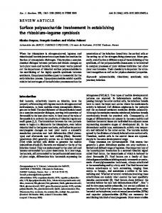
Figure 1. Scanning technique of the sternoclavicular joint in a healthy subject. A, Transducer placement. B, Longitudinal scan showing a normal sternoclavicular joint. C ⫽ clavicle; S ⫽ sternum.
ically evaluated all patients and healthy controls. The following data were recorded for each patient: age, sex, disease duration, and history of SC joint pain and/or swelling. Moreover, RA disease activity was evaluated using the Disease Activity Score in 28 joints (DAS28) (16). Both the patients and healthy controls were specifically asked about the current or past presence of signs or symptoms reminiscent of SC joint involvement (i.e., pain and/or swelling). The presence of SC joint pain and/or swelling and crepitus was clinically evaluated bilaterally by inspection, direct palpation (with the patient sitting in a neutral position with a 90° backrest), and during active movement (abduction of the glenohumeral joint ⬎60°). These clinical maneuvers allowed us to categorize the SC joint as clinically involved or not. Both data (history of pain and/or swelling and the presence of abnormal SC joint findings on physical examination) allowed us to consider the clinically affected SC joint. US assessment. US examinations of all study participants were performed by an experienced rheumatologist (⬎6 years) in musculoskeletal US (PR-H) who was blinded to the clinical data. The patients and healthy controls underwent US examinations at random and were asked not to talk about their clinical condition with the sonographer. All US examinations were performed using a MyLab25 (Esaote Biomedica) device equipped with a 6 –18-MHz broadband, linear array transducer. For each patient and healthy control, US examinations included longitudinal and transverse scans of the SC joint performed bilaterally, according to a previously described scanning technique (12,17). Briefly, the transducer was placed parallel and perpendicular to the long axis of the medial clavicle. The SC joint was identified moving the probe medially. The cortical surfaces of the medial clavicle and sternum were identified with the hypoechoic joint space between them. Figure 1 shows the probe position and the respective normal US image. Each SC joint was initially scanned in gray scale to detect morphostructural changes and subsequently with the power Doppler (PD) technique to detect intraarticular synovial blood perfusion. Blood flow assessment was carried out with settings standardized as follows: pulse repetition frequency 750 Hz, Doppler frequency between 6 and 8 MHz, and wall filter 4. PD gain was adjusted to avoid random noise visualization. Synovial PD signal was scored on the basis of the following semiquantitative scoring: 0 ⫽ absent (no synovial flow), 1 ⫽ mild (ⱕ3 PD signals within
Ultrasound Assessment of the Sternoclavicular Joint the synovial area), 2 ⫽ moderate (⬎3 PD signals in less than one-half of the synovial area), and 3 ⫽ marked (signals in more than one-half of the synovial area) (9). For US elementary lesions, the Outcome Measures in Rheumatology preliminary definitions were adopted (18). The following abnormalities were recorded in both groups: synovitis, osteophytes, and erosions. Synovitis was defined as the presence of either synovial hypertrophy and/or joint effusion. Synovial effusion was defined as abnormal hypo- or anechoic (relative to subdermal fat, but sometimes may be iso- or hyperechoic) intraarticular material that is displaceable and compressible, but that does not exhibit a Doppler signal. Synovial hypertrophy was defined as abnormal hypoechoic (relative to subdermal fat, but sometimes may be iso- or hyperechoic) intraarticular tissue that is nondisplaceable and poorly compressible, and that may exhibit a Doppler signal. Bone erosion was defined as an intraarticular discontinuity of the bone surface that is visible in 2 perpendicular planes. Additionally, osteophytes were defined as a step-up bony prominence at the end of the normal bone contour or at the margin of the joint, with or without an acoustic shadow. In order to assess interobserver agreement, a second investigator with 3 years of experience in musculoskeletal US carried out sonographic examinations in 30 patients. Both examiners were instructed to perform the same US scanning technique and to adopt the same US interpretation scheme. Statistical analysis. Statistical analysis was performed using SPSS software, version 13. Standard descriptive results were expressed as the mean or median, SD, and 95% confidence intervals (95% CIs) for the median. Frequencies (%) were used to describe categorical data and were compared using the chi-square test. Mean ⫾ SD was used for continuous variables and differences were assessed by unpaired t-tests. Fisher’s exact test was used to compare SC joint clinical and US variables between the groups. A statistical significance value was set at less than 0.05. The interobserver agreement was calculated using an unweighted kappa test. A kappa value of 0 – 0.20 was considered poor, 0.21– 0.40 was considered fair, 0.41– 0.60 was considered moderate, 0.61– 0.80 was considered good, and 0.81–1.00 was considered excellent.
1179
Table 1. Demographic and medical history of the study population*
Characteristics
RA patients (n ⴝ 103)
Healthy controls (n ⴝ 103)
P
Women/men, no. Age, years Disease duration, years DAS28
90/13 50.9 ⫾ 13.5 13.7 ⫾ 9.1 3.5 ⫾ 1.1
83/20 49.4 ⫾ 10.9 N/A N/A
0.18 0.39 N/A N/A
* Values are the mean ⫾ SD unless otherwise indicated. RA ⫽ rheumatoid arthritis; N/A ⫽ not applicable; DAS28 ⫽ Disease Activity Score in 28 joints.
cantly higher SC joint involvement in patients with RA than in healthy controls (P ⬍ 0.0001). For the interobserver agreement, 2 investigators examined 60 SC joints of 30 patients. Kappa values showed good to excellent agreement for synovitis, osteophytes, and bone erosions ( ⫽ 0.86, 0.65, and 0.75, respectively). Prevalence of US findings. As shown in Table 2, in the RA group, the most frequent US finding was osteophytes, detected in 59 (29%) of 206 SC joints. Synovitis and erosions were found in 31 (15%) and 23 SC joints (11%), respectively. Five SC joints (2%) were found to be positive for intraarticular PD signal. Three SC joints displayed a moderate PD signal and 2 displayed a marked PD signal. All positive PD cases also demonstrated gray-scale signs of synovitis (Figure 2). In no cases was sonographic evidence of bilateral synovitis or bony erosions found. In contrast, the control group displayed a lower frequency of synovitis (5 [2%] of 206 SC joints; P ⫽ 0.0001) and absence of erosions (P ⫽ 0.0001) and intraarticular PD signal (P ⫽ 0.03). Osteophytes were detected in 25 (12%) of 206 SC joints (P ⫽ 0.0001). Relationship between US findings and history of SC joint involvement. Table 3 shows the relationship between US elementary lesions and a clinical history of SC joint pain or swelling. The risk of synovitis was 4 times more likely for patients with SC joint pain or swelling in contrast to patients without these symptoms (odds ratio [OR] 4.1, 95% CI 1.6 –10.0; P ⫽ 0.01). In addition, erosions were found more frequently in patients with RA with a
RESULTS The demographic and medical history of the patients with RA and healthy controls is shown in Table 1. A total of 412 SC joints were evaluated clinically and sonographically from 103 patients with RA and 103 age- and sex-matched healthy controls. In the group of patients with RA, 39 (19%) of 206 SC joints were considered clinically involved, in contrast to only 4 (1.9%) in the control group (P ⫽ 0.0001). All 4 controls referred pain during joint palpation. At least 1 US elementary lesion suggestive of SC joint involvement was recorded in 89 SC joints (43%) in the RA group compared with 36 SC joints (17%) in the healthy control group; therefore, US assessment showed signifi-
Table 2. Ultrasonographic findings of sternoclavicular joints*
Elementary lesion Synovitis Erosions Osteophytes Synovial hyperperfusion (PD)
RA patients (n ⴝ 206)
Healthy controls (n ⴝ 206)
P
31 (15) 23 (11) 59 (29) 5 (2)
5 (2) 0 (0.0) 25 (12) 0 (0.0)
0.0001 0.0001 0.0001 0.03
* Values are the number (percentage). RA ⫽ rheumatoid arthritis; PD ⫽ power Doppler.
1180
Rodrı´guez-Henrı´quez et al
Table 4. Correlation between ultrasound findings and the DAS28*
Synovial hyperperfusion (PD) Positive Negative Synovitis Yes No Figure 2. Longitudinal scan of a sternoclavicular joint of a patient with rheumatoid arthritis shows moderate joint cavity widening with the presence of synovial hypertrophy, irregularities of bone profile, and a marked intraarticular power Doppler signal.
N
Mean ⴞ SD DAS28
5 98
5.3 ⫾ 1.2 3.4 ⫾ 1.0
31 72
4.1 ⫾ 1.1 3.3 ⫾ 1.0
P 0.0001
0.001
* DAS28 ⫽ Disease Activity Score in 28 joints; PD ⫽ power Doppler.
DISCUSSION history of SC joint pain and/or swelling (P ⫽ 0.03) in contrast to patients without SC joint symptoms. We did not find a correlation between intraarticular PD and a history of SC joint symptoms (P ⫽ 0.27). When analyzing the subgroup of patients with RA displaying erosions (n ⫽ 23), there were 12 (52%) with concomitant synovial hypertrophy in contrast to those lacking erosions (n ⫽ 80), among whom only 18 (23%) showed synovial hypertrophy (OR 3.5, 95% CI 1.3–9.2; P ⫽ 0.01).
The SC joint is often overlooked during routine rheumatologic clinical examinations despite its synovial membrane lining and biomechanical properties as part of the shoulder joint complex. This joint should be carefully examined because it is clinically involved in nearly onethird of the most common arthritides (2). Involvement of the SC joint in RA has been reported from as few as 1% to as many as 41% in different series (2,3), while our study disclosed at least 1 US elementary lesion in up to 43% of SC joints in patients with RA, highlighting the relevance of ultrasonographic assessments of these joints in daily rheumatologic practice. To the best of our knowledge, this is the first US study providing evidence about a higher number of involved SC joints than in clinical assessment in patients with RA. Moreover, our results indicate that both gray-scale and PD findings were more prevalent in patients with RA than in age- and sex-matched healthy controls. The precise role of US in the assessment of the SC joint in patients with RA has yet to be established firmly in rheumatologic practice. To date, only a few investigators have concentrated their attention on this joint (4,17).
RA disease activity and US findings. On average, patients with RA had a moderate DAS28 (mean ⫾ SD 3.5 ⫾ 1.1). The DAS28 was higher in those with SC joint synovial hypertrophy than in those not exhibiting this US elementary lesion (mean ⫾ SD 4.1 ⫾ 1.1 versus 3.3 ⫾ 1.0; P ⫽ 0.001). The highest DAS28 (⬎5.2) was found in patients who displayed intraarticular PD in contrast to patients without intraarticular PD (mean ⫾ SD 5.3 ⫾ 1.2 versus 3.4 ⫾ 1.0; P ⫽ 0.0001). Table 4 shows a correlation between the presence of US synovitis and intraarticular PD and the DAS28 score.
Table 3. Correlation between ultrasound findings and history of SC joint pain and/or swelling in the rheumatoid arthritis group* History of SC joint pain/swelling
Synovitis Presence Absence Intraarticular PD signal Presence Absence Erosions Presence Absence Osteophytes Presence Absence
Presence
Absence
Total (n ⴝ 206)
19 2
12 173
31 175
3 0
2 201
5 201
13 0
10 183
23 183
22 0
37 147
59 147
P
OR (95% CI)
0.01
4.1 (1.6–10.0)
0.27
2.5 (0.4–16.1)
0.03
2.7 (1.0–6.9)
0.52
0.9 (0.4–2.1)
* SC ⫽ sternoclavicular; OR ⫽ odds ratio; 95% CI ⫽ 95% confidence interval; PD ⫽ power Doppler.
Ultrasound Assessment of the Sternoclavicular Joint Further analysis of our results allows us to formulate additional considerations: first, the correlation between the presence of B-mode synovitis and synovial hyperperfusion with a higher DAS28 score in some patients suggests that SC joints actively participate in the inflammatory process of RA; second, from the clinical point of view, accurate detection of synovitis is important in both the diagnosis and outcome. Our US study demonstrated that the risk of having SC joint synovitis and erosions was greater for patients with a history of pain and/or swelling than for patients without these findings. The SC joint is often underestimated during the clinical examination; it is noteworthy that at least one-third of patients with RA exhibit crepitus and tenderness of the SC joint (2). Therefore, the SC joint should be systematically examined both clinically and sonographically. Third, synovial hypertrophy was present more frequently in patients with erosive abnormalities, confirming the well-established concept that its presence is an independent risk factor for the development of erosive arthropathy (19). Furthermore, the presence of hypervascular synovitis is a stronger predictor for radiologic damage and US erosions (7,9,19 –21). Finally, the high prevalence of osteophytes in patients with RA with respect to healthy controls (29% versus 12%) could be the expression of the simultaneous presence of inflammatory and degenerative changes, such as in data reported for distal interphalangeal joints in patients with RA (22,23). Another possible explanation of this phenomenon is that unlike the majority of synovial joints, SC joint articular cartilage is composed of fibrocartilage rather than hyaline cartilage, which renders the joint more susceptible to degenerative changes (24,25). On the other hand, SC joint degenerative changes become increasingly common with older age (26,27). In fact, Kier et al (28) radiographically examined 55 cadaveric SC joints, showing that moderate or severe degenerative changes were uncommon in specimens of individuals ages ⬍40 years, but were present in more than one-half of specimens of persons ages ⱖ60 years. The mean ⫾ SD age of our patients with RA was 50.9 ⫾ 13.5 years versus 49.4 ⫾ 10.9 years in the healthy controls; this supports the notion that SC joint changes are likely a result of RA with secondary osteoarthritis. We are aware that our study has several limitations: first, a lack of assessment of the SC joints by a second imaging technique (MRI or CT); second, the absence of a longitudinal followup prevents us from knowing the predictive value of US in the development of SC joint abnormalities; and third, the fact that a dichotomous assessment (presence/absence) of gray-scale US findings was used. In this way, very small and focal irregularities had the same value as prominent and multiple osteophytes and/or erosions. In conclusion, the present study provides US evidence that reveals a higher prevalence of SC joint involvement in patients with RA than in age- and sex-matched healthy controls. US is more sensitive than clinical examination for detecting SC joint involvement in RA. The correlation among US synovitis, intraarticular PD, and the DAS28 suggests that SC joints actively participate in the systemic inflammatory process of RA. The precise role of US in the assessment of the SC joint in patients with RA is yet to be established firmly in rheumatologic practice.
1181 AUTHOR CONTRIBUTIONS All authors were involved in drafting the article or revising it critically for important intellectual content, and all authors approved the final version to be published. Dr. Pineda had full access to all of the data in the study and takes responsibility for the integrity of the data and the accuracy of the data analysis. Study conception and design. Rodrı´guez-Henrı´quez, Pen˜a, Pineda. Acquisition of data. Solano, Herna´ndez-Dı´az. Analysis and interpretation of data. Leo´n-Herna´ndez, Gutie´rrez.
REFERENCES 1. Gordon AG, Hastings DE. Clinical features of rheumatoid arthritis. In: Hochberg MC, Silman AJ, Smolen JS, Weinblatt ME, Weisman MH, editors. Rheumatology. 3rd ed. Vol 1. London: Mosby-Wolfe; 2003. p. 765– 80. 2. Yood RA, Goldenberg DL. Sternoclavicular joint arthritis [review]. Arthritis Rheum 1980;23:232–9. 3. Laine VA, Vainio KJ, Pekanmaki K. Shoulder affections in rheumatoid arthritis. Ann Rheum Dis 1954;13:157– 60. 4. Guglielmi G, Cascavilla A, Scalzo G, Salaffi F, Grassi W. Imaging of sternocostoclavicular joint in spondyloarthropathies and other rheumatic conditions. Clin Exp Rheumatol 2009;27:402– 8. 5. D’Agostino MA, Said-Nahal R, Hacquard-Bouder C, Brasseur JL, Dougados M, Breban M. Assessment of peripheral enthesitis in the spondylarthropathies by ultrasonography combined with power Doppler: a cross-sectional study. Arthritis Rheum 2003;48:523–33. 6. Alarcon GS, Lopez-Ben R, Moreland LW. High-resolution ultrasound for the study of target joints in rheumatoid arthritis [letter]. Arthritis Rheum 2002;46:1969 –70. 7. Salaffi F, Ciapetti A, Gasparini S, Carotti M, Filippucci E, Grassi W. A clinical prediction rule combining routine assessment and power Doppler ultrasonography for predicting progression to rheumatoid arthritis from early-onset undifferentiated arthritis. Clin Exp Rheumatol 2010;28:686 –94. 8. Dougados M, Jousse-Joulin S, Mistretta F. Evaluation of several ultrasonography scoring systems for synovitis and comparison to clinical examination: results from a prospective multicentre study of rheumatoid arthritis. Ann Rheum Dis 2010;69:828 –33. 9. Naredo E, Collado P, Cruz A, Palop MJ, Cabero F, Richi P, et al. Longitudinal power Doppler ultrasonographic assessment of joint inflammatory activity in early rheumatoid arthritis: predictive value in disease activity and radiologic progression. Arthritis Rheum 2007;57:116 –24. 10. Wakefield RJ, Gibbon WW, Conaghan PG, O’Connor P, McGonagle D, Pease C, et al. The value of sonography in the detection of bone erosions in patients with rheumatoid arthritis: a comparison with conventional radiography. Arthritis Rheum 2000;43:2762–70. 11. Filippucci E, Gabba A, Di Geso L, Girolimetti R, Salaffi F, Grassi W. Hand tendon involvement in rheumatoid arthritis: an ultrasound study. Semin Arthritis Rheum 2012;41:752– 60. 12. Ferri M, Finlay K, Popowich T, Jurrians E, Friedman L. Sonographic examination of acromioclavicular and sternoclavicular joints. J Clin Ultrasound 2005;33:345–55. 13. Robinson CM, Jenkins PJ, Markham, PE, Beggs I. Disorders of the sternoclavicular joint. J Bone Joint Surg 2008;90:685–96. 14. Andonopoulos AP, Meimaris N, Yiannopoulos G, Pastromas V, Dimopoulos P. Large synovial cysts originating from the sternoclavicular joints in a patient with rheumatoid arthritis [letter]. Ann Rheum Dis 2003;62:1119 –20. 15. Arnett FC, Edworthy SM, Bloch DA, McShane DJ, Fries JF, Cooper NS, et al. The American Rheumatism Association 1987 revised criteria for the classification of rheumatoid arthritis. Arthritis Rheum 1988;31:315–24. 16. Prevoo ML, van ’t Hof MA, Kuper HH, van Leeuwen MA, van de Putte LB, van Riel PL. Modified disease activity scores that include twenty-eight–joint counts: development and valida-
1182
17.
18.
19.
20.
21.
tion in a prospective longitudinal study of patients with rheumatoid arthritis. Arthritis Rheum 1995;38:44 – 8. Delle Sedie A, Riente L, Iagnocco A, Filippucci E, Meenagh G, Valesini G, et al. Ultrasound imaging for the rheumatologist. VI. Ultrasonography of the elbow, sacroiliac, parasternal and temporomandibular joints. Clin Exp Rheumatol 2006;24:617– 21. Wakefield RJ, Balint PV, Szkudlarek M, Filippucci E, Backhaus M, D’Agostino MA, et al, for the OMERACT 7 Special Interest Group. Musculoskeletal ultrasound including definitions for ultrasonographic pathology. J Rheumatol 2005;32:2485–7. McGonagle D, Conaghan PG, O’Connor P, Gibbon W, Green M, Wakefield R, et al. The relationship between synovitis and bone changes in early untreated rheumatoid arthritis: a controlled magnetic resonance imaging study. Arthritis Rheum 1999;42:1706 –11. Pascual-Ramos V, Contreras-Yanez I, Cabiedes-Contreras J, Rull-Gabayet M, Villa AR, Vazquez-Lamadrid J, et al. Hypervascular synovitis and American College of Rheumatology classification criteria as predictors of radiographic damage in early rheumatoid arthritis. Ultrasound Q 2009;25:31– 8. Karim Z, Wakefield RJ, Quinn M, Conahan PG, Brown AK, Veale DJ, et al. Validation and reproducibility of ultrasonog-
Rodrı´guez-Henrı´quez et al
22. 23. 24. 25. 26. 27. 28.
raphy in the detection of synovitis in the knee: a comparison with arthroscopy and clinical examination. Arthritis Rheum 2004;50:387–94. Jacob J, Sartoris D, Kursunoglu S, Pate D, Pineda CJ, Braun RM, et al. Distal interphalangeal joint involvement in rheumatoid arthritis. Arthritis Rheum 1986;29:10 –5. Abbott GT, Bucknall RC, Whitehouse GH. Osteoarthritis associated with distal interphalangeal joint involvement in rheumatoid arthritis. Skeletal Radiol 1991;20:495–7. Klein MA, Spreitzer AM, Miro PA, Carrera GF. MR imaging of the abnormal sternoclavicular joint: a pictorial essay. Clin Imaging 1997;21:138 – 43. Klein MA, Miro PA, Spreitzer AM, Carrera GF. MR imaging of the normal sternoclavicular joint: spectrum of findings. Am J Roentgenol 1995;165:391–3. Arlet J, Ficat P. Osteo-arthritis of the sterno-clavicular joint. Ann Rheum Dis 1958;17:97–100. Silberberg M, Frank EL, Jarrett SR, Silberberg R. Aging and osteoarthritis of the human sternoclavicular joint. Am J Pathol 1959;35:851– 65. Kier R, Wayne SL, Apple J, Martinez S. Osteoarthritis of the sternoclavicular joint: radiographic features and pathologic correlation. Invest Radiol 1986;21:227–30.