PROTOCOL
© 2006 Nature Publishing Group http://www.nature.com/naturemethods
PUBLISHED IN ASSOCIATION WITH COLD SPRING HARBOR LABORATORY PRESS
Strategies for protein coexpression in Escherichia coli Niraj H Tolia & Leemor Joshua-Tor Keck Structural Biology Laboratory, Cold Spring Harbor Laboratory, 1 Bungtown Road, Cold Spring Harbor, New York 11724, USA. Correspondence should be addressed to N.H.T. (
[email protected]).
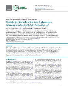
E. coli is a convenient host for heterologous protein expression. Its advantages include high levels of heterologous gene expression and scalability of experiments, low cost, fast growth, a lack of posttranslational modification and an ability to express labeled (isotope or seleno-methionine) proteins. However, heterologous gene expression in E. coli can lead to the production of insoluble and/or nonfunctional target proteins. This is often due to the absence of cofactors or post-translational modifications required for function, stability or folding. Coexpression of multiple genes in E. coli, such as the members of a stable multiprotein complex1 or a protein with a chaperone2,3, can in many cases alleviate these problems. Coexpression involves the transformation of E. coli with several plasmids that have compatible origins of replication and independent antibiotic selection for maintenance. The Duet (Novagen) vectors have two multiple cloning sites per vector, five compatible origins of replication and four antibiotic selection markers, allowing the simultaneous expression of up to eight proteins. The combination of Duet vectors with other commercial plasmids allows the use of affinity tags, such as glutathione S-transferase (GST) or maltose binding protein (MBP), which can ease the recovery and improve the solubility of the desired target. Coexpression in E. coli therefore provides a useful alternative to the complicated and expensive expression systems, such as yeast, baculovirus or mammalian cell culture, which are commonly used to overcome problems of heterologous protein expression. A summary of the method is presented in Figure 1.
MATERIALS REAGENTS Calf intestinal phosphatase (NEB) Complete protease inhibitor cocktail (Roche) Deoxynucleoside triphosphate (dNTP) solution (each at 10 mM) E. coli competent cells, DH5α and BL21 (DE3) Isopropylthiogalactoside (IPTG; 1 M) Luria broth (LB) agar and liquid medium Lysozyme (1 mg/ml) Pfu DNA polymerase and 10× Pfu buffer (Stratagene) PCR primers (see Step 1 for details) QIAprep Spin Miniprep Kit (Qiagen) QIAprep Gel Extraction Kit (Qiagen) QIAquick Nucleotide Removal Kit (Qiagen) Rapid Ligation Kit (Roche) or T4 DNA ligase with ligase buffer (NEB)
© Cold Spring Harbor Laboratory Press
Restriction endonucleases, appropriate for the vectors (NEB) Selective antibiotics (ampicillin, chloramphenicol, streptomycin and/or kanamycin) TBS (100 mM Tris-HCl (pH 8.0), 250 mM NaCl), ice-cold Template DNA (plasmid or genomic cDNA) Vector DNA: pETDuet-1, pACYCDuet-1, pCDFDuet-1, pRSFDuet-1 and/or pCOLADuet-1 (Novagen) Additional vectors: pGEX (any, Amersham Biosciences), pMAL (any, New England BioLabs) EQUIPMENT Equipment and gels for agarose gel electrophoresis and SDS-PAGE Thermal cycler programmed with the desired amplification protocol Temperature-controlled shaker incubator, preferably refrigerated
NATURE METHODS | VOL.3 NO.1 | JANUARY 2006 | 55
PROTOCOL Design and amplification of the target oligonucleotide
PROCEDURE 1| Design and synthesize appropriate PCR primers: include 18–30 nucleotides complementary to the sequence of interest and appropriate restriction enzyme sites for the chosen vector. If cloning with a tag, ensure that the primers are in frame with the tag sequence.
© 2006 Nature Publishing Group http://www.nature.com/naturemethods
2| Set up a 50 µl PCR amplification reaction for each target to be cloned. 10× Pfu amplification buffer dNTPs (10 mM stock) 5’ primer (1 µM stock) 3’ primer (1 µM stock) Template DNA (100 ng/µl) Pfu DNA polymerase Water
5 µl 1 µl 10 µl 10 µl 0.5 µl 1 µl 22.5 µl
3| Amplify the nucleic acids using the following touchdown PCR program. Cycle number Denaturation 1 1 min at 94 °C 2–30 30 s at 94 °C 31 Hold
30 s at 94 °C
Annealing 30 s at 65 °C 30 s at 63–48 °C (decrease temperature 0.5 °C/cycle) 30 s at 45 °C
Polymerization 2 min/kb at 72 °C 2 min/kb at 72 °C
Final
11 min at 72 °C 4 °C
Touchdown PCR does not require an accurate determination of the melting temperature of the primers; it therefore has a higher success rate in our hands.
Cloning of the amplification product into the desired vector
4| Prepare the chosen vector DNA by digesting ~2 µg with restriction enzymes for 2 h at 37 °C (or follow the manufacturer’s instructions). Dephosphorylate the vector DNA by adding 0.25 µl (~2.5 U) calf intestinal phosphatase directly to the restriction enzyme reaction and incubating for 30 min at 37 °C. ▲CRITICAL STEP 5| Gel-purify the vector and PCR product (from Step 3) using the QIAquick Gel Extraction Kit according to the manufacturer’s instructions. Elute the vector DNA in 30 µl and the PCR product in 50 µl of elution buffer (Tris-EDTA (TE) buffer). 6| Prepare the insert DNA by digesting the 50 µl of PCR product with restriction enzymes for 2–4 h at 37 °C. 7| Purify the insert DNA using the QIAquick Nucleotide Removal Kit. Elute in 30 µl of TE buffer. 8| Set up the ligation reaction according to the Rapid Ligation Kit instructions, using 3–6 µl of insert DNA and 1–2 µl of vector DNA. Incubate the reaction at 15–25 °C for 5–20 min. Alternatively, if not using the Rapid Ligation Kit, set up a 20 µl reaction with 3–6 µl insert, 1–2 µl vector and T4 DNA ligase. Incubate this reaction according to the enzyme manufacturer’s instructions. 9| Use 5–10 µl of the ligation reaction to transform DH5α competent cells, and select for growth by plating the cells on LB agar containing the appropriate antibiotic for the chosen vector (Table 1). Incubate at 37 °C for 16–18 h overnight. ▲CRITICAL STEP 10| Isolate plasmid DNA from several colonies using the QIAquick Spin Miniprep Kit, and verify bacterial clones for each target by restriction digest and/or sequencing.
56 | VOL.3 NO.1 | JANUARY 2006 | NATURE METHODS
PROTOCOL Prepare insert DNA
Prepare vector DNA
Amplify by PCR Gel purify Digest Remove nucleotides
Digest Dephosphorylate Gel purify Ligate
© 2006 Nature Publishing Group http://www.nature.com/naturemethods
Verify clones Digest Sequence
Clone into second MCS of Duet?
Transform BL21 (DE3)
Screen for expression Grow several transformants in 2–5 ml cultures
Large-scale expression Grow started culture overnight
Induce with IPTG (when A600 is 0.6–0.8) for 6–8 h
Innoculate 1 l with 1–5 ml of overnight culture
Harvest cells, lyse and analyze by SDS-PAGE
Induce with IPTG (when A600 is 0.6–0.8) for 2–14 h
Verify expression clone
Harvest cells, lyse and analyze by SDS-PAGE Affinity purification
Figure 1 | Outline of the protocol.
11| If using both multiple cloning sites of the Duet vectors, repeat Steps 1–11 using a verified bacterial clone. (Optional) Small-scale test expression of the cloned target can be attempted on a 2–5-ml culture in LB and analyzed by SDS-PAGE to ensure the clone expresses and to determine solubility (Steps 12–22). 12| Use 1 µl (10–40 ng) of each plasmid to transform BL21 (DE3) competent cells. When transforming multiple plasmids, it is essential to allow the cells to recover for at least 1 h before plating on the LB agar containing the appropriate antibiotic (as determined by the chosen vector; Table 1). ▲CRITICAL STEP
Transformation of each clone into BL21 (DE3)
13| Plate cells on LB agar containing the appropriate antibiotics (depending on the chosen vectors) and incubate at 37 °C for 16–18 h. In the case of multiple plasmids, reduce the concentration of antibiotics used by half of the values in Table 1. ▲CRITICAL STEP ➨ TROUBLESHOOTING 14| Pick several transformants and set up a 2–5 ml culture of each in LB containing appropriate antibiotics.
Screening clones for expression
15| Grow the cultures to an A600 of 0.6–0.8 with vigorous shaking at 18–37 °C. 16| Remove 10 µl of culture for analysis by SDS-PAGE (preinduced sample) and another 1 ml for the preparation of a glycerol stock (add glycerol to a final concentration of 15%) and store at –70 °C.
© Cold Spring Harbor Laboratory Press
NATURE METHODS | VOL.3 NO.1 | JANUARY 2006 | 57
PROTOCOL 17| Induce expression of the remaining culture by adding 2–5 µl of 1 M IPTG to a final concentration of 1 mM (final concentration can vary from 0.1 to 1 mM) and incubating for 2–6 h at 18–37 °C with vigorous shaking. ▲CRITICAL STEP
© 2006 Nature Publishing Group http://www.nature.com/naturemethods
18| Transfer 1.5 ml of the induced culture to a microcentrifuge tube and collect the cells by centrifuging at 10,000g for 1 min. Discard the supernatant. 19| Resuspend the pellet in 300 µl of ice-cold TBS and set aside a 10-µl aliquot for analysis by SDS-PAGE (induced sample). Table 1 | Features of the described vectors (adapted from ref. 12) Vector Promoter Antibiotic Concentrationa resistance
Replicon (source) Compatible vectors [copy number]
pETDuet-1
ColE1 (pBR322)
T7
Ampicillin or carbenicillin
50 µg/ml
[~40]
pACYCDuet-1 pCDFDuet-1 pRSFDuet-1
pACYCDuet-1
T7
Chloramphenicol
34 µg/ml
P15A (pACYC184)
pRSFDuet-1 pETDuet-1
[10–12]
pCDFDuet-1 pRSFDuet-1 pCOLADuet-1 pGEX (all) pMAL (all)
pCDFDuet-1
T7
Streptomycin or spectinomycin
50 µg/ml
CloDF13
pETDuet-1
[20–40]
pACYCDuet-1 pRSFDuet-1 pCOLADuet-1 pGEX (all)
pRSFDuet-1
T7
Kanamycin
30 µg/ml
RSF1030
pMAL (all) pETDuet-1
[>100]
pACYCDuet-1 pCDFDuet-1 pGEX (all)
pCOLADuet-1
T7
Kanamycin
30 µg/ml
COLA (ColA)
pMAL (all) pETDuet-1
[20–40]
pACYCDuet-1 pCDFDuet-1 pGEX (all)
pGEX (all)
tac
Ampicillin or carbenicillin
50 µg/ml
ColE1 (pBR322)
pMAL (all) pACYCDuet-1
[~40]
pCDFDuet-1 pRSFDuet-1
pMAL (all)
tac
Ampicillin or carbenicillin
50 µg/ml
ColE1 (pBR322)
pCOLADuet-1 pACYCDuet-1
[~40]
pCDFDuet-1 pRSFDuet-1 pCOLADuet-1
aReduce
the concentration of antibiotic used by half if transforming more than one plasmid.
58 | VOL.3 NO.1 | JANUARY 2006 | NATURE METHODS
PROTOCOL 20| Lyse the remaining cells by sonication until the cloudy suspension becomes translucent.
© 2006 Nature Publishing Group http://www.nature.com/naturemethods
21| Collect the cell debris by centrifuging at 10,000g for 5 min and retain 10 µl of the supernatant for analysis by SDS-PAGE. Replace the remaining supernatant with an equivalent volume of TBS and resuspend the pellet by sonication. Retain 10 µl of the resuspended pellet for analysis by SDS-PAGE. 22| Analyze the preinduced sample, induced sample, supernatant and pellet of each colony using standard SDS-PAGE methods to determine expression levels and solubility (presence in the supernatant) either by Coomassie staining or western blotting. ➨ TROUBLESHOOTING 23| Once a suitable expression clone has been identified, use the glycerol stock to inoculate a starter culture in 100 ml LB supplemented with the appropriate antibiotics (depending on the plasmids present) and incubate at 37 °C for 12–15 h (or 30 °C for longer than 15 h). Alternatively, inoculate the 100-ml culture with a colony from a fresh transformation of the expression clone (see Steps 12 and 13). ▲CRITICAL STEP
Growth of cultures for large-scale expression
24| Use 1–5 ml of starter culture to inoculate 1 l of LB supplemented with the appropriate antibiotics in 2 l flasks. Multiple flasks can be used to scale-up expression. 25| Incubate at 37 °C until A600 is ~0.3. Then lower the temperature to between 18 and 30 °C and continue to incubate until the A600 is 0.6–0.8. ▲CRITICAL STEP 26| Remove 10 µl of the culture for analysis by SDS-PAGE (preinduced sample). 27| Induce the remaining culture by adding 1 ml of 1 M IPTG (final concentration can range from 0.1 to 1 mM) and continue to incubate for an additional 2–14 h. ▲CRITICAL STEP 28| Remove 10 µl of the culture for analysis by SDS-PAGE (induced sample). 29| Harvest the cells by centrifugation at 4,000g for 30 min and discard the supernatant.
Harvesting and lysis of the cells
30| Resuspend the cells in 10 ml of ice-cold TBS (supplemented with complete protease inhibitor cocktail and 1 mg/ml lysozyme) per liter of culture. 31| Freeze at –70 °C. ■ PAUSE POINT At this point cells can be stored indefinitely. 32| Thaw the cells and lyse them by sonication until the cloudy suspension becomes translucent. 33| Collect the cell debris by centrifuging at 10,000g for 30 min and retain 10 µl of the supernatant for analysis by SDS-PAGE. Replace the remaining supernatant with an equivalent volume of TBS and resuspend the pellet by sonication. Retain 10 µl of the resuspended pellet for analysis by SDS-PAGE.
Analysis of the lysate
34| Analyze the preinduced sample, induced sample, supernatant and pellet using standard SDS-PAGE methods to determine expression levels and solubility (presence in the supernatant). ➨ TROUBLESHOOTING Further purification of soluble targets can be achieved by affinity chromatography depending on the tags present. For additional information on affinity chromatography, see Molecular Cloning, Chapter 15, Protocols 5–7 (http://www.molecularcloning.com/members/chapter.jsp?chapter=126).
© Cold Spring Harbor Laboratory Press
NATURE METHODS | VOL.3 NO.1 | JANUARY 2006 | 59
PROTOCOL
© 2006 Nature Publishing Group http://www.nature.com/naturemethods
TROUBLESHOOTING TABLE PROBLEM SOLUTION Step 13 Few or no transformants obtained. Transformation with multiple plasmids is sensitive to both the post-transformation recovery period and the concentration of antibiotic used for selection. For these experiements, make sure that transformants are allowed to recover for at least 1 h before plating and that the concentration of antibiotic is reduced to half of that recommended in Table 1. The yield and solubility of proteins are affected by several factors Steps 22 and 34 Low yield or partial solubility observed in both lysis pellet and including the expression strain, IPTG concentration and the duration and temperature of induction. These factors should be supernatant. systematically varied to optimize results. Expression of toxic proteins at a higher cell density (A600) for a shorter period can help to increase yields. Target is completely insoluble (observed only in pellet).
Attempt to add an additional interacting partner of the target to the coexpression system to aid in folding in an attempt to enhance solubility. Test various chaperones by coexpression for their ability to fold the target. Rapidly lowering temperature before induction can induce cold shock proteins and may result in target insolubility. Maintain a constant (but low) temperature during initial growth and induction. Modify the target by truncating and/or deleting sections of the coding sequence to obtain a domain that is soluble. If possible, switch to a homolog of the target from a different species that may be soluble.
CRITICAL STEPS Step 4 The choice of vector is determined by whether or not the target is to be tagged. Usually only the primary target is tagged and ancillary factors are not tagged. An exception to this is a doubletagging method for double-affinity chromatography. In this case, two proteins of a complex are tagged with different tags. See Box 1 and Table 1 for various strategies and vector selection. The vectors described in this protocol contain several tags: Duet MCS1 contains an amino-terminal His tag, Duet MCS2 contains a carboxy-terminal S tag, pGEX encodes an N-terminal GST tag and pMAL encodes an N-terminal MBP tag. A major advantage of His, GST and MBP tags is that they allow affinity purification of the fusion protein with mild elution conditions. The pGEX and pMAL vectors also include various protease cleavage sites between the tag and the protein that can be used to remove the tag after purification. Although the S tag allows affinity purification, it requires harsher elution conditions and is not recommended for the purification of complexes under nondenaturing conditions. This protocol therefore focuses on His-, GST- and MBP-tag systems. The choices of which tag to use and which proteins to tag will influence the solubility and functionality of the product. Choices will be dictated by the experimental aims. His tags are small and have a low metabolic burden, but they can sometimes inhibit solubility4. GST tags have no effect on solubility and MBP tags tend to increase the solubility of the target5, but the large size of both these tags is metabolically expensive and may affect the function of the fusion protein. (The latter can be avoided if the tag is removed by proteolysis with a specific protease.) Lastly, GST forms dimers in solution, a feature that can be problematic, especially if the target protein also forms oligomers. The most advantageous protein to be tagged must be determined empirically, but tagging two proteins with the same tag is not recommended. This could result in heterogeneity of the purified proteins or complexes. Step 9 DH5α cells are used at this point for plasmid maintenance. DH5α is highly transformable and is recA–; that is, the cells have a low recombination rate allowing for the stable maintenance of plasmid DNA.
60 | VOL.3 NO.1 | JANUARY 2006 | NATURE METHODS
PROTOCOL
© 2006 Nature Publishing Group http://www.nature.com/naturemethods
Step 12 BL21 (DE3) is required at this point for several reasons. The Duet vectors have a T7 promoter for high-level expression and therefore require an E. coli host containing a chromosomal copy of the T7 RNA polymerase gene (DE3). pGEX and pMAL vectors both contain a tac promoter for high-level expression. Although the tac promoter does not require a DE3 strain (as they are recognized by E. coli RNA polymerase), coexpression of pGEX or pMAL with Duet vectors will necessitate the use of a DE3 strain. DE3 strains will not hinder expression from vectors containing the tac promoter. BL21 (DE3) strains are also preferred at this stage as they are deficient in OmpT and Lon proteases that could otherwise induce proteolysis of overexpressed proteins. Step 13 Transformation of cells with multiple plasmids may result in the maintenance of fewer copies of each plasmid, as compared to those seen in single-plasmid transformation. Reducing the concentration of antibiotics by half allows the survival of these low-copy-number transformants. Steps 17 and 25 Induction temperature can also influence target solubility. Lower temperatures favor slower expression rates that might allow for improved folding of the target, but will also reduce target yield. Different temperatures should be tested to determine the optimal temperature. Expression of toxic proteins can also lead to poor yields. Inducing expression of toxic proteins at a higher A600 for a shorter period can increase yields. Steps 17 and 27 IPTG concentration and length of induction can both influence target solubility. Lower concentrations of IPTG may induce protein expression at a slower rate, allowing better folding. Lower IPTG concentrations, however, may also reduce yield. Longer induction times, which increase expression, may also induce proteolysis of the target, leading to heterogeneity in the sample and reducing yield. Different concentrations of IPTG and a time course of induction should be tested to determine the optimal combination. Step 23 Growth of starter cultures at 37 °C for extended periods is not recommended because this can sometimes lead to plasmid silencing that prevents expression. COMMENTS Successful coexpression and purification of heterologous proteins is dependent on several factors, including plasmid compatibility, identity of the tag used (critical step 4), which protein of a complex is tagged, E. coli host strain and, of course, the nature of proteins being expressed. Several different strategies are possible (various strategies for coexpression are described in Box 1) depending on the requirements of the protein(s) to be expressed.
Maintenance of multiple plasmids in E. coli is dependent on the plasmids possessing compatible origins of replication6 and different antibiotic resistance genes for selection (Table 1). If two plasmids containing the same origin of replication are used to cotransform E. coli, the replication of one plasmid will be inhibited as a result of an RNA antisense mechanism, which causes plasmid segregation within the population6,7. Compatible plasmids stably coexist in a single cell by occupying different subcellular locations within the bacterium8. Plasmid segregation can also occur if the plasmids carry the same antibiotic resistance gene; only one plasmid needs to be maintained to confer resistance. Therefore, plasmids sharing common origins of replication or antibiotic resistance genes should not be used for the coexpression of two proteins. Because the pGEX and pMAL vectors are ColE1-based and ampicillin-resistant, they are incompatible with pETDuet. Thus, if the primary target is to be tagged with either GST or MBP, the plasmids pRSFDuet, pCOLADuet, pACYCDuet or pCDFDuet should be used for the expression of additional targets. See Table 1 for information on plasmid compatibility. EXAMPLE OF APPLICATION Recent studies have linked Argonaute (Ago) proteins as the catalytic core of the RNA-induced silencing complex (RISC), the RNA interference (RNAi) effector complex. The crystal structure of Pyrococcus furiosus Argonaute revealed that the signature PIWI domain of the Ago proteins is similar to that of ribonuclease (RNase) H and contains the conserved catalytic residues9. Purification of tagged Ago from human cell lines revealed that endonuclease (or ‘slicer’) activity was associated with complexes containing Ago2 (refs. 10,11), and that mutation of the conserved catalytic residues abolishes activity10.
© Cold Spring Harbor Laboratory Press
NATURE METHODS | VOL.3 NO.1 | JANUARY 2006 | 61
PROTOCOL
© 2006 Nature Publishing Group http://www.nature.com/naturemethods
BOX 1 STRATEGIES FOR COEXPRESSION The Duet/pGEX/pMAL system described in this protocol offers several optimal options, tailored to the needs of various targets (Table 2). One protein plus refolding factor. This approach involves the coexpression of a tagged protein with a chaperone, or cofactor, to improve folding and/or solubility of the product (see the Example of application section and Fig. 2). Several different chaperones should be tested to find the optimal partner for the protein of interest. The first strategy (Table 2; target in Duet, factor in Duet) has the advantage that the primary target and the refolding factor can be carried on a single plasmid. Having the two proteins on the same plasmid makes the testing of protein variants (for example, truncations or deletions) cumbersome, because each variant of each protein must be generated and then recloned into the plasmid. This problem can be relieved by using two compatible plasmids, which would allow variants to be tested by mixing and matching plasmids, but this would introduce the complication of generating multiple transformants. The other two strategies (Table 2; target in pGex or pMAL, factor in Duet) are similar, but use two plasmids. They offer the same advantages of tagging (here the GST or MBP tags are used to ease purification and improve product solubility), together with easy testing of protein variants, but require multiple transformations, which can be difficult. Single-tagged two-protein complex. A complex of two proteins can be expressed (on one or two plasmids) and purified using an affinity tag on only one of the proteins. The strategies are essentially as described above for the ‘one protein plus refolding factor’ example. Double-tagged two-protein complex. Double tagging is a convenient two-plasmid method for the purification of a two-protein complex. It involves using different tags on each of the proteins, and sequential affinity chromatography to purify the complex. Double tagging also allows the separate recovery of individual proteins, if desired. With the vectors described here, only GST tag–His tag or MBP tag–His tag combinations are recommended because of limitations of plasmid compatibility. Other tags can be used as long as plasmid compatibility is maintained. These strategies have the same advantages and disadvantages described for the ‘one protein plus refolding factor’ example using two plasmids. Two-protein complex plus refolding factor. This approach is analogous to the two-protein complex discussed above, with the addition of an ancillary factor to aid the solubility and/or folding of one of the proteins. Because the ancillary factor will not be recovered, it can be left untagged. Tagging the two proteins to be recovered will assist in their purification and could also improve their solubility. Because each plasmid may have a different subcellular localization within the bacterium6, cloning the refolding factor and its target protein on the same Duet vector may prove more effective as the two proteins then should localize within the same region of the cell. Multiprotein complex. A multiprotein complex can be built up from a two-protein complex by introducing additional proteins on other Duet vectors. Care should be taken when choosing a tagging strategy; multiple His tags used (in MCS1) may cause heterogeneity of the purified complexes and should be avoided. The Duet system allows up to eight proteins to be coexpressed simultaneously, but this number is reduced to only seven if either the pGEX or pMAL vectors are used.
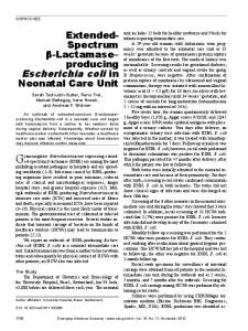
Figure 2 | Example of application. Coexpression of pGEX-4T-1 harboring the gene encoding GST-hAgo2 with pRSF-Duet harboring the gene encoding hHSP90 results in soluble expression of GST-hAgo2 (right, E; red arrow). Analysis by SDS-PAGE shows that expression of pGEX-4T-1-hAgo2 alone yields insoluble GST-Ago2 (left lanes WCL, P; asterisk) and no soluble GSTtagged hAgo2 (left, E). Soluble hHSP90 can be observed in the supernatant (right, S; black arrow). The presence of soluble GST-hAgo2 was verified by mass spectrometry and was shown to be active2. WCL, whole-cell lysate; P, pellet; S, supernatant; E, eluate from the glutathione affinity step. 62 | VOL.3 NO.1 | JANUARY 2006 | NATURE METHODS
© 2006 Nature Publishing Group http://www.nature.com/naturemethods
PROTOCOL To definitively prove that Ago2 itself is the slicer enzyme and to further characterize its activity, we sought to obtain purified human Ago2 from E. coli for biochemical characterization because E. coli does not possess an RNAi pathway. However, expression of GST-tagged human Ago2 (GST-hAgo2) in E. coli yielded insoluble protein (Fig. 2). Ago2-containing complexes purified from human cell lines contained almost stoichiometric amounts of human Ago2 and human heat shock protein 90 (hHSP90; ref. 10). We therefore tested whether coexpression of GST-hAgo2 with hHSP90 in E. coli could overcome the insolubility resulting in functional protein. Indeed, we obtained soluble GST-hAgo2 using this method (Fig. 2)2. With this system, we demonstrated that GST-hAgo2 alone can combine with a small interfering RNA (siRNA) to form a recombinant complex (termed minimal RISC) that can accurately cleave substrate RNAs and that this complex has similar kinetic properties to both fruit fly and human RISC2. A major difference between these complexes, however, is that, unlike fly or human RISC, minimal RISC is not stimulated by ATP, indicating that factors stimulating product release are absent from the recombinant complex. These studies demonstrate that Ago2 catalyzes mRNA cleavage within RISC, and pave the way for complete recombinant reconstitution of the RNAi effector complex.
Table 2 | Strategies for coexpressing different targets One protein plus refolding factor Target protein Factor Vector (MCS) Tag Vector (MCS) Tag Duet (MCS1) His Duet (MCS2) None pGEX
GST
Duet (MCS2)
None
pMAL
MBP
Duet (MCS2)
None
Single-tagged two-protein complex Protein 1 Protein 2 Vector (MCS) Tag Vector (MCS) Duet (MCS1) His Duet (MCS2)
Tag None
pGEX
GST
Duet (MCS2)
None
pMAL
MBP
Duet (MCS2)
None
Notes Both MCSs can be used on a single Duet vector, or two compatible vectors could be used. Any Duet vector can be used, except pET-Duet because of incompatibility with pGEX. Any Duet vector can be used, except pET-Duet because of incompatibility with pMAL.
Notes Both MCSs can be used on a single Duet vector, or two compatible vectors could be used. Any Duet vector can be used, except pET-Duet because of incompatibility with pGEX. Any Duet vector can be used, except pET-Duet because of incompatibility with pMAL.
Double-tagged two-protein complex Protein 1 Protein 2 Vector (MCS) Tag Vector (MCS) pGEX GST Duet (MCS1)
Tag His
Notes Any Duet vector can be used, except pET-Duet because of incompatibility with pGEX.
pMAL
His
Any Duet vector can be used, except pET-Duet because of incompatibility with pMAL.
MBP
Duet (MCS1)
Two-protein complex plus refolding factor Protein 1 Protein 2 Vector (MCS) Tag Vector (MCS) Tag pGEX GST Duet (MCS1) His
Factor Vector (MCS) Tag Duet (MCS2) None
pMAL
Duet (MCS2)
MBP
Duet (MCS1)
© Cold Spring Harbor Laboratory Press
His
None
Notes Any Duet vector can be used, except pET-Duet because of incompatibility with pGEX. Both MCSs can be used on a single Duet vector, or two compatible vectors could be used. Any Duet vector can be used, except pET-Duet because of incompatibility with pMAL. Both MCSs can be used on a single Duet vector, or two compatible vectors could be used. NATURE METHODS | VOL.3 NO.1 | JANUARY 2006 | 63
PROTOCOL ACKNOWLEDGMENTS We thank R.-M. Xu for constructive conversations on coexpression, E. Enemark and D. Chitwood for comments on the manuscript, and members of the Joshua-Tor laboratory for helpful discussions. This work was supported by National Institutes of Health grant GM072689 (to L.J.).
© 2006 Nature Publishing Group http://www.nature.com/naturemethods
SOURCE This protocol is based on new developments to “Expression of cloned genes in E. coli using IPTG-inducible promoters,” in Molecular Cloning: A Laboratory Manual on the Web (eds. Sambrook, J & Russell, D.W.) Chapter 15 Protocol 1 (Cold Spring Harbor Laboratory Press, Cold Spring Harbor, New York, USA (2001); http://www.cshlpress.com/link/molclon3.htm) and “User Protocol TB340” (Novagen). 1.
2.
3.
4.
5.
Li, C., Schwabe, J.W., Banayo, E. & Evans, R.M. Coexpression of nuclear receptor partners increases their solubility and biological activities. Proc. Natl. Acad. Sci. USA 94, 2278– 2283 (1997). Rivas, F.V. et al. Purified Argonaute2 and an siRNA form recombinant human RISC. Nat. Struct. Mol. Biol. 12, 340– 349 (2005). Bross, P. et al. Co-overexpression of bacterial GroESL chaperonins partly overcomes non-productive folding and tetramer assembly of E. coli–expressed human medium-chain acyl-CoA dehydrogenase (MCAD) carrying the prevalent disease-causing K304E mutation. Biochim. Biophys. Acta 1182, 264–274 (1993). Woestenenk, E.A., Hammarstrom, M., van den Berg, S., Hard, T. & Berglund, H. His tag effect on solubility of human proteins produced in Escherichia coli: a comparison between four expression vectors. J. Struct. Funct. Genomics 5, 217– 229 (2004). Kapust, R.B. & Waugh, D.S. Escherichia coli maltose-binding protein is uncommonly effective at promoting the solubility of polypeptides to which it is fused. Protein Sci. 8, 1668– 1674 (1999).
64 | VOL.3 NO.1 | JANUARY 2006 | NATURE METHODS
6.
Selzer, G., Som, T., Itoh, T. & Tomizawa, J. The origin of replication of plasmid p15A and comparative studies on the nucleotide sequences around the origin of related plasmids. Cell 32, 119–129 (1983). 7. Davison, J. Mechanism of control of DNA replication and incompatibility in ColE1-type plasmids—a review. Gene 28, 1–15 (1984). 8. Ho, T.Q., Zhong, Z., Aung, S. & Pogliano, J. Compatible bacterial plasmids are targeted to independent cellular locations in Escherichia coli. EMBO J. 21, 1864–1872 (2002). 9. Song, J.J., Smith, S.K., Hannon, G.J. & Joshua-Tor, L. Crystal structure of Argonaute and its implications for RISC slicer activity. Science 305, 1434–1437 (2004). 10. Liu, J. et al. Argonaute2 is the catalytic engine of mammalian RNAi. Science 305, 1437–1441 (2004). 11. Meister, G. et al. Human Argonaute2 mediates RNA cleavage targeted by miRNAs and siRNAs. Mol. Cell 15, 185–197 (2004). 12. Held, D., Yaeger, K. & Novy, R. New coexpression vectors for expanded compatibilities in E. coli. inNovations 18, 4–6 (2003).