JOURNAL OF MORPHOLOGY 274:108–120 (2013)
Structural Adaptations for Ram Ventilation: Gill Fusions in Scombrids and Billfishes Nicholas C. Wegner,1,2* Chugey A. Sepulveda,3 Scott A. Aalbers,3 and Jeffrey B. Graham1 1
Center for Marine Biotechnology and Biomedicine, Marine Biology Research Division, Scripps Institution of Oceanography, University of California San Diego, La Jolla, California 92093 2 Fisheries Resource Division, Southwest Fisheries Science Center, National Marine Fisheries Service, National Oceanic and Atmospheric Administration, La Jolla, California 92037 3 Pfleger Institute of Environmental Research, Oceanside, California 92054 ABSTRACT For ram-gill ventilators such as tunas and mackerels (family Scombridae) and billfishes (families Istiophoridae, Xiphiidae), fusions binding the gill lamellae and filaments prevent gill deformation by a fast and continuous ventilatory stream. This study examines the gills from 28 scombrid and seven billfish species in order to determine how factors such as body size, swimming speed, and the degree of dependence upon ram ventilation influence the site of occurrence and type of fusions. In the family Scombridae there is a progressive increase in the reliance on ram ventilation that correlates with the elaboration of gill fusions. This ranges from mackerels (tribe Scombrini), which only utilize ram ventilation at fast cruising speeds and lack gill fusions, to tunas (tribe Thunnini) of the genus Thunnus, which are obligate ram ventilators and have two distinct fusion types (one binding the gill lamellae and a second connecting the gill filaments). The billfishes appear to have independently evolved gill fusions that rival those of tunas in terms of structural complexity. Examination of a wide range of body sizes for some scombrids and billfishes shows that gill fusions begin to develop at lengths as small as 2.0 cm fork length. In addition to securing the spatial configuration of the gill sieve, gill fusions also appear to increase branchial resistance to slow the high-speed current produced by ram ventilation to distribute flow evenly and optimally to the respiratory exchange surfaces. J. Morphol. 274:108–120, 2013. Ó 2012 Wiley Periodicals, Inc. KEY WORDS: tuna; mackerel; marlin; swordfish; gill filament; gill lamellae
INTRODUCTION Tunas, bonitos, and mackerels (family Scombridae) and billfishes (families Istiophoridae, Xiphiidae) are continuous swimmers and breathe using ram ventilation, the mechanism through which forward swimming provides the force required to drive water into the mouth and through the branchial chamber (Brown and Muir, 1970; Stevens, 1972; Roberts, 1975, 1978; Stevens and Lightfoot, 1986; Wegner et al., 2012). Ram ventilation transfers the energetic cost of active gill ventilation to the swimming musculature, and because mouth and opercular motions are miniÓ 2012 WILEY PERIODICALS, INC.
mized, both respiratory and swimming efficiency are increased (Freadman, 1979, 1981; Steffensen, 1985). However, at the relatively high swimming speeds attained by scombrids and billfishes, ram ventilation poses two challenges. First, the ramventilatory current must be slowed to create optimal flow conditions for efficient gas exchange at the respiratory lamellae (Brown and Muir, 1970; Stevens and Lightfoot, 1986; Wegner et al., 2012). Second, the gills must be reinforced in order to maintain normal orientation with respect to a high-pressure branchial stream, the force of which increases with swimming speed (Muir and Kendall, 1968; Brown and Muir, 1970). Recent work by Wegner et al. (2010) showed that the morphometrics of scombrid and billfish gills (e.g., shape, size, and number of gill lamellae) increase branchial resistance and help slow ram-ventilatory flow. To increase the overall rigidity of the branchial sieve, some scombrids and billfishes have structural supports in the form of gill fusions (Muir and Kendall, 1968; Muir, 1969; Johnson, 1986; Wegner et al., 2006). In most teleost fishes, the gill filaments are not interconnected and extend independently from the gill arch, with lamellae extending freely from the Contract grant sponsor: National Science Foundation; Contract grant number: IOS-0817774; Contract grant sponsors: The Tuna Industry Endowment Fund at Scripps Institution of Oceanography, the Pfleger Institute of Environmental Research, the George T. Pfleger Foundation, the Moore Family Foundation, the Nadine A. and Edward M. Carson Scholarship awarded by the Achievement Rewards for College Scientists (ARCS), Los Angeles Chapter (N.C.W.), a National Research Council Associateship (N.C.W.), and the Kennel-Haymet Student Lecture Award (N.C.W.). *Correspondence to: Nicholas C. Wegner, National Marine Fisheries Service, Southwest Fisheries Science Center, 8901 La Jolla Shores Dr., La Jolla, CA 92037. E-mail:
[email protected] Received 4 April 2012; Revised 21 July 2012; Accepted 23 August 2012 Published online 29 September 2012 in Wiley Online Library (wileyonlinelibrary.com) DOI: 10.1002/jmor.20082
GILL FUSIONS FOR RAM VENTILATION
109
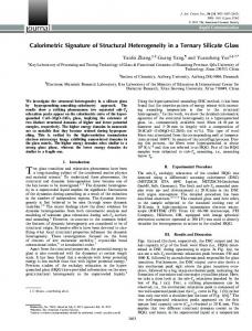
Fig. 1. Basic gill morphology and fusion structure described for tunas and billfishes. (A) First gill arch taken from the left side of the branchial chamber of a scombrid showing the gill filaments emanating from the arch. (B–E) Magnified views of black box in A showing the leading (water-entry) edges of three adjacent gill filaments (each containing two rows of gill lamellae) with different fusion types: (B) No gill fusions, (C) Interlamellar fusions, (D) Complete lamellar fusions, and (E) Filament fusions (with complete lamellar fusions extending underneath). Water flow direction in B-E is into the page. IF, interlamellar fusion; F, filament; FF filament fusions; L, lamellae; LF, complete lamellar fusion. [Color figure can be viewed in the online issue, which is available at wileyonlinelibrary.com.]
gill filaments (Fig. 1A,B). The gill fusions of scombrids and billfishes connect these normally independent structures to form lamellar fusions (Fig. 1C–E) and filament fusions (Fig. 1E). Lamellar fusions, which bind the gill lamellae near their leading (water-entry) edge and thus secure the spatial integrity of the interlamellar channels (5lamellar pores), can be further categorized into two forms. Interlamellar fusions (Fig. 1C) bind adjacent lamellae on the same filament and have been described for wahoo, Acanthocybium solandri (a non-tuna scombrid), and striped marlin, Kajikia audax (Wegner et al., 2006). Complete lamellar fusions (Fig. 1D) connect the lamellae on one filament to the closely positioned and opposing lamellae of the adjacent filament, thus providing support to both the gill lamellae and filaments (Muir and Kendall, 1968; Muir, 1969; Wegner et al., 2006). Complete lamellar fusions have been reported in several tuna species and in the striped marlin, where, in some areas of the gills, the interlamellar fusions of adjacent filaments join together (i.e., the striped marlin has both complete lamellar and interlamellar fusions; Wegner et al., 2006). Filament fusions (Fig. 1E) bridge the interfilament space in a lattice-like pattern, binding adjacent filaments on the same gill hemibranch (Muir and Kendall, 1968; Johnson, 1986; Davie, 1990). These fusions, which form on the trailing (waterexit) and often leading (water-entry) edges of the filaments, have been documented in tunas of the genus Thunnus, the wahoo, and some billfishes
(Istiophorus, Kajikia, and Xiphias; Muir and Kendall, 1968; Johnson, 1986). In the tuna genus, Thunnus, filament fusions are formed by the expansion of the mucosal filament epithelium (Muir and Kendall, 1968). In contrast, wahoo and billfish filament fusions are formed by bony epithelial toothplates, which on the trailing edges of the filaments cover cartilaginous connections of the filament rods (Johnson, 1986). The diversity of gill fusion type and structure can be expected to reflect interspecific differences in reliance upon, or specialization for, ram ventilation. Although all scombrids and billfishes use ram ventilation, basic information about the occurrence of fusions and their structure is not available for many species, and there are few data addressing how fusion structure may change with body size or the range of swimming speeds employed by these fishes. Because the family Scombridae demonstrates a progression in adaptations for fast, continuous swimming (from least derived mackerels to most derived tunas) and an associated increase in reliance on ram ventilation, determination of gill fusion type and pattern along this gradient can provide insight into the structural requirements of ram ventilation. This comparative study thus examines the gills of 28 scombrid and seven billfish species in order to determine how factors such as body size, swimming speed, and the degree of dependence upon ram ventilation correlate with the site of occurrence and elaboration of fusions. Journal of Morphology
110
N.C. WEGNER ET AL. TABLE 1. Scombrids and billfishes examined in relation to gill fusion type
Species name
Common name
Thunnus alalunga Thunnus albacares Thunnus atlanticus Thunnus obesus Thunnus orientalis Thunnus tonggol Katsuwonus pelamis Euthynnus affinis Euthynnus alleteratus Euthynnus lineatus Auxis rochei Auxis thazard Allothunnus fallai Sarda australis Sarda chiliensis Sarda orientalis Sarda sarda Cybiosarda elegans Acanthocybium solandri Scomberomorus commerson
Albacore Yellowfin tuna Blackfin tuna Bigeye tuna Pacific bluefin tuna Longtail tuna Skipjack tuna Kawakawa Little tunny Black skipjack Bullet tuna Frigate tuna Slender tuna Australian bonito Eastern Pacific bonito Striped bonito Atlantic bonito Leaping bonito Wahoo Narrow-barred Spanish mackerel Monterey Spanish mackerel Queensland school mackerel Pacific sierra Shark mackerel Double-lined mackerel Short mackerel Indian mackerel Pacific chub mackerel Sailfish Blue marlin Black marlin Striped marlin Shortbill spearfish Roundscale spearfish Swordfish
Scomberomorus concolor Scomberomorus queenslandicus Scomberomorus sierra Grammatorcynus bicarinatus Grammatorcynus bilineatus Rastrelliger brachysoma Rastrelliger kanagurta Scomber japonicus Istiophorus platypterus Makaira nigricans Istiompax indica Kajikia audax Tetrapturus angustirostris Tetrapturus georgii Xiphias gladius
n total (collected) 4 (2) 25 (19) 11 (5) 6 (5) 5 (5) 9 (2) 14 (8) 14 (4) 2 (0) 22 (10) 3 (0) 25 (2) 2 (0) 1 (0) 25 (25) 2 (0) 2 (0) 7 (7) 9 (9) 6 (5)
Fork length (cm)
Mass (kg)
39.0–82.0 1.0–11.3 29.0–182.0 0.38–94.0 45.5–52.0 1.6–2.4 40.0–136.0 1.5–46.0 86.0–120.0 12.2–32.3 38.5–85.5 0.83–9.6 30.4–77.0 0.48–9.6 19.7–78.0 0.10–6.0 6.5–8.0 2.5–4.6 g 10.9–59.0 16.4 g–2.94 21.8–36.0 0.11–0.76 7.7–44.5 3.4 g–1.48 84.5–85.5 11.3–11.8 33 0.52 27.5–82.0 0.15–6.4 48.0–50.5 1.45–1.56 11.5–48.0 10.0 g–1.58 30.6–35.5 0.44–0.67 75.0–152.0 2.1–24.2 43.5–96.0 0.59–7.0
5 (0)
38.5–55.0
0.35–1.39
6 (5)
22.3–58.5
65.0 g–1.65
5 2 4 2 4 25 2 3 2 9 3 1 16
(5) (2) (0) (0) (2) (25) (2) (0) (0) (8) (0) (0) (9)
47.0–50.0 0.75–0.90 — 3.5–4.5 41.5–51.0 0.59–0.95 10.0–10.3 7.5–9.5 g 12.1–31.5 16.6 g–0.43 9.5–40.0 7.2 g–0.74 200.0–207.0 31.0–34.4 — 53.2–189.5 86.5–90.5 — 95.0–188.5 8.0–70.0 125.0–143.0 — 173.0 — 34.0–223.5 73.1 g–155.3
Lamellar fusion
Filament fusion
Collection location
LF LF LF LF LF LF LF LF LF LF LF LF LF LF LF LF LF LF IF --
ME ME ME ME ME ME ------------ETP --
1 1,2,3 4 3,5 2 6 1,2,3 6 — 2,7 — 2 — — 1,2 — — 6 2,3 6
--
--
—
IF*
--
6
------IF, LF
------ETP ETP ETP ETP ETP ETP ETP
7 6 — — 8 1 7 — — 1,2 — — 1
IF, LF IF, LF --
The total number of specimens examined is given with the number collected (i.e., those not from scientific collections) in parentheses. Collection locations: 1. Southern California, USA; 2. Baja California and Baja California Sur, Mexico; 3. Hawaii, USA; 4. Grand Cayman Island; 5. Central equatorial Pacific; 6. Queensland, Australia; 7. Costa Rica (Pacific coast); 8. Palau. Abbreviations: ETP, epithelial toothplate filament fusion; IF interlamellar fusion; LF, complete lamellar fusion; ME, mucosal epithelium filament fusion. Dashes (--) indicate a lack of fusions; blank spaces indicate that fusion status remains undetermined. Billfish lengths are given as lower jaw fork lengths. *Interlamellar fusions were present in five S. queenlandicus from 46.9–58.5 cm (0.87–1.65 kg) but were not present in a 22.3 cm (65.0 g) specimen. The small yellowfin and sailfish specimens examined to understand the timing of fusion development are not included in this table. Fish masses are given in kg unless otherwise denoted by grams (g).
MATERIALS AND METHODS Gill Tissue Collection and Preservation Fresh gill tissue samples were collected from 19 scombrid and three billfish species from various locations around the world by working with other researchers and fishermen. All collected individuals were sacrificed upon capture by severing the spinal cord at the base of the skull in accordance with protocol S00080 of the University of California, San Diego Institutional Animal Care and Use Committee. Length measurements [fork length for scombrids, lower jaw fork length for billfishes] were taken, and when possible, each specimen was weighed using a digital scale. In cases where body mass could not be directly measured, estimates were made using species-specific length-weight regressions (Chatwin, 1959; Wolfe and Webb, 1975; Faruk Kara, 1979; Muthiah, 1985; Ramos et al., 1986; McPherson, 1992; Hsu et al., 2000; Chiang et al., 2004; Beerkircher, 2005; de la Serna et al., 2005; Moazzam et al., 2005; Vieira et al., 2005). Whenever possible, a large size range of individuals from each species was collected in order to examine changes in fusion structure with body size.
Journal of Morphology
For most specimens, the gills from one or both sides of the branchial chamber were excised immediately and fixed in 10% formalin buffered in sea water. For large fish, a low-pressure seawater hose was used to keep the gills wet during excision, which, depending on the size of the fish, required up to 10 min (prolonged air exposure results in the degradation of fine gill tissue). On an opportunistic basis, specimens from select species were perfused with vascular casting solution in order to determine if the elaboration of gill fusions was associated with changes in gill circulation. Vascular casting procedures were performed on two yellowfin tuna (Thunnus albacares), one skipjack tuna (Katsuwonus pelamis), seven eastern Pacific bonito (Sarda chiliensis), three wahoo, one sailfish (Istiophorus platypterus), three striped marlin, and three swordfish (Xiphias gladius). Each freshly killed fish was placed ventral side up in a V-shaped cradle while a low-pressure seawater hose was used to ventilate the branchial chamber and keep the gills wet. The heart was exposed by midline incision and cannulated to administer heparinized saline (2–5 min) followed by vascular casting solution
GILL FUSIONS FOR RAM VENTILATION
111
Fig. 2. Scombrid and billfish cladograms showing the presence of the different gill fusion types for each genus. The number of species examined as a ratio of the total number in the genus is given in parentheses. Abbreviations: ETP, epithelial toothplate filament fusion; IF, interlamellar fusions; LF, complete lamellar fusions; ME, mucosal epithelium filament fusion. Dashes (--) indicate a lack of fusions; blank spaces indicate that fusion status remains undetermined. Scombrid cladogram based on Collette et al. (2001); billfish cladogram based on Collette et al. (2006). Included in the number of Thunnus and Scomber species examined is the Atlantic bluefin tuna, T. thynnus and the Atlantic mackerel, S. scombrus, for which fusion data were reported by Muir and Kendall (1968). Lamellar fusion status in Makaira and Istiompax remains undetermined due to the poor preservation of lamellar structure in the scientific collection specimens examined.
(Mercox, Mercox II, Ladd Research, Williston, VT). Gills were perfused at 70–95 mmHg, which is consistent with vascular casting preformed in other scombrid and billfish studies (Olson et al., 2003; Wegner et al., 2006; Wegner et al., 2010). Following polymerization, gills from one side of the branchial chamber were fixed to determine fusion status, while the gills from the other side were stored frozen and then macerated in washes of 15–20% KOH to expose the vascular casts. [Note: Both fixed gill tissue and vascular casts from Wegner et al. (2006) and Wegner et al. (2010) are included in the present analysis].
Gill Samples from Scientific Collections Preserved specimens from scientific collections at Scripps Institution of Oceanography, the Smithsonian National Museum of Natural History, the Australian Museum, and the Australian National Fish Collection were also examined. Most of these fish had highly degraded gill tissue (likely caused by prolonged air exposure or freezing prior to fixation), which precluded accurate determination of fusion status. Because degraded gill tissue has led to discrepancies in the description of fusions in the literature (Muir and Kendall, 1968; Muir, 1969), only scientific collection specimens in which fusion status could be clearly determined were included in the present analysis. Although most billfish gill tissue had undergone substantial degradation preventing the determination of lamellar fusion status, billfish filament fusions are composed of tough bony epithelial toothplates, which made it possible to record their presence in some specimens.
Gill Fusion Assessment Fixed gill tissue from each specimen was examined to determine the presence or absence of the different fusion types: filament fusions (either composed of a mucosal epithelium or bony epithelial toothplates), complete lamellar fusions, and interlamellar fusions. For most specimens, filament fusion type and the presence of complete lamellar fusions could be determined by direct observation with the naked eye or aided by a dissection microscope. In specimens for which complete lamellar fusions were not obvious (e.g., species with interlamellar fusions or lacking lamellar fusions), scanning electron microscopy (SEM) was used to assess lamellar fusion status. SEM followed the protocol of Wegner et al. (2006). Small sections of gill tissue (usually 1 cm2 or less) were removed from each specimen, rinsed in deionized water, and either dehydrated in 100% ethanol (20–25% increments over 24 h) and critical-point dried, or dehydrated in 100% tert-butyl alcohol (25% increments over 24 h, rinsed twice at 100%), frozen in the alcohol at 48C, and freeze dried. Dried material was then sputter-coated with gold-palladium and viewed using an FEI Quanta 600 SEM (FEI, Hillsboro, OR) under high-vacuum mode. Vascular casts of the gill circulation were examined using dissection light microscopy and the SEM under low-vacuum mode.
Gill Fusion Development In order to determine the timing and structural changes associated with lamellar fusion development, a size range of small
Journal of Morphology
112
N.C. WEGNER ET AL.
Fig. 3. Scanning electron micrographs of complete lamellar and interlamellar fusions from (A) a 1.45 kg eastern Pacific bonito, (B) a 45.0 kg striped marlin, (C) a 1.07 kg Queensland school mackerel, (D) a 1.07 kg Queensland school mackerel (magnified image of box in C), (E) a 25.0 kg striped marlin, and (F) a 1.45 kg eastern Pacific bonito. B and E are from Wegner et al. (2006). F, filament; IF, interlamellar fusion; L, lamellae; LF, complete lamellar fusion. Water flow direction is into the page.
individuals from one scombrid species (yellowfin tuna, n 5 6, 1.5–5.2 cm, 0.10–2.22 g) and one billfish species (sailfish, n 5 15, 12.4–33.0 cm, 4.5–95.5 g) were examined.
Statistics It is important to note that correlations between fusion type and structure and dependence on ram ventilation are based on general observations only and were not tested statistically. Statistical techniques such as phylogenetic independent contrast can not be employed as the degree of dependence on ram ventilation (as determined through measures of respiratory movements at varying swimming speeds) has not been quantified for most species examined.
RESULTS Table 1 shows the presence and type of gill fusions (along with collection location data) for the 28 scombrid and seven billfish species examined in this study. Fusion status for each genus is mapped onto the scombrid and billfish phylogenies in Figure 2. Journal of Morphology
Lamellar Fusions Scanning electron micrographs depicting the structural details for complete lamellar and interlamellar fusions are shown in Figure 3. Complete lamellar fusions (Fig. 3A) are present in all five tuna genera (i.e., Thunnus, Katsuwonus, Euthynnus, Auxis, and Allothunnus) and the bonito genera Sarda and Cybiosarda (quality specimens of the monotypic Gymnosarda and Orcynopsis were not available for examination). This study further verifies interlamellar fusions (Fig. 3B) in striped marlin and wahoo as described previously (Wegner et al., 2006) and also documents their presence in sailfish, shortbill spearfish, Tetrapturus angustirostris, and one Spanish mackerel species, the Queensland school mackerel, Scomberomorus queenlandicus (Fig. 3C,D). The interlamellar fusions of Queensland school mackerel and wahoo are thinner and less complete (i.e., fusions do not bind all of the lamellae along the length of the filament) than
GILL FUSIONS FOR RAM VENTILATION
Fig. 4. Filament fusions on the leading edge of the anterior hemibranch of the third gill arch near the cerato-epibranchial joint for (A) a 32.3 kg Pacific bluefin tuna, (B) a 72.0 kg yellowfin tuna, (C) a 46.0 kg bigeye tuna, (D) a 24.2 kg wahoo, (E) a 34.4 kg sailfish, (F) a 67.8 kg striped marlin, and (G) a 64.9 kg swordfish. F and G are from Wegner et al. (2010). [Color figure can be viewed in the online issue, which is available at wileyonlinelibrary.com.]
those of striped marlin, shortbill spearfish, and sailfish (cf., Fig. 3C,D with 3B), which are well developed, and, in many cases, fuse together to form complete lamellar fusions (Fig. 3E). Other Spanish mackerels of the genus Scomberomorus and Grammatorcynus, the mackerels (Rastrelliger, Scomber), and the swordfish, Xiphias gladius, lack both forms of lamellar fusions. Although nearly all the lamellae in tunas and bonitos are bound by complete lamellar fusions, remnants of interlamellar fusions from development (see below) are found at the filament tips in some species (Fig. 3F).
Filament Fusions Filament fusions occur in tunas of the genus Thunnus, the wahoo, and all the billfish species sampled (spanning both billfish families and all six billfish genera) (Table 1, Fig. 2). For all species,
113
the lattice-like filament fusions on the trailing (water-exit) edges of the gill filaments occur along nearly their entire length (i.e., from the gill arch to the filament tips). However, on the leading (water-entry) edges, species-specific patterns in filament fusion distribution are prevalent and these are shown for several species in Figure 4A–G (once filament fusions are fully developed their size and location remains relatively conserved within a species). These patterns range from three Thunnus species that lack filament fusions entirely on the leading edge: albacore, Thunnus alalunga, blackfin tuna, Thunnus atlanticus, and longtail tuna, Thunnus tonggol (not shown in Fig. 4), to the swordfish in which the leading filament edges are bound by fusions along nearly their entire length (Fig. 4G). Many striped marlin specimens have a thick fusion of the filament tips (Fig. 4F). All filament fusions examined conform to the structural pattern described by Johnson (1986), in which the filament fusions of Thunnus are extensions of the filament mucosal epithelium, while those of the wahoo and billfishes are formed by epithelial toothplates (Table 1, Fig. 2). Vascular gill casts reveal an additional structural difference in that the more robust toothplate and cartilaginous-based fusions on the trailing edge of wahoo and billfish filaments often contain blood vessels that connect the circulation of adjacent filaments. Three types of vascular junctions occur: 1) connections between afferent filamental arteries on adjacent filaments (Fig. 5A), 2) afferent lamellar arterioles that supply blood from one filament to a limited number of lamellae on the adjacent filament (Fig 5B), and 3) nutrient blood vessels (i.e., non-respiratory vessels), which likely serve to support the extensive fusion latticework (not pictured). Vascular connections between filaments are not found in the mucosal epithelium filament fusions of Thunnus or in lamellar fusions. Fusion Development Figures 6–8 show the progressive development of lamellar fusions in a size series of juvenile yellowfin tuna. At 1.5 cm (103 mg) the gill filaments and lamellae are fully developed but lack fusions (Fig. 6). By 2.0 cm (154 mg), however, some interlamellar fusions have developed near the filament tips (Fig. 7A–E). Interlamellar fusion formation appears to involve the bending of the leading lamellar lateral edge toward the filament tip until it contacts the adjacent lamella (Fig. 7C–E). By 3.0– 3.2 cm (453–915 mg) interlamellar fusions start to grow together to form complete lamellar fusions (Fig. 8). However, interlamellar fusions persist near the filament tip, and lamellae near the base of the filaments remain free of fusions. By 5.5 cm (2.22 g) most of the interlamellar fusions have Journal of Morphology
114
N.C. WEGNER ET AL.
Fig. 5. Scanning electron microscope images of vascular casts from a 31.0 kg sailfish showing the afferent gill filament circulation. (A) Six adjacent afferent filamental arteries (AFA) connected at several locations along their lengths (*). (B) Magnified view of dotted box in A, showing the afferent lamellar arterioles (ALA). The largest ALA (*) bridges the interfilament space providing blood from one filament to the lamellae (L) of the adjacent filament.
grown together to form complete lamellar fusions, which have progressed further towards the base of the filaments, leaving fewer non-fused lamellae. In sailfish the interlamellar fusions also first form near the filament tip and extend toward the filament base as body size increases. Figure 9 shows the distribution of interlamellar fusions on the gill filaments of a 28.5 cm (68.0 g) sailfish. Although interlamellar fusions were observed over the entire size range of small sailfish examined (12.4–33.0 cm lower jaw fork length, 4.5–95.5 g), no complete lamellar fusions were observed. Because of the limited number of specimens available over the required size range, the progression and the exact onset of filament fusion development could not be assessed in the same detail as
lamellar fusions. Among scombrids, filament fusions consistently appear to begin development at relatively small body sizes, first appearing near the gill arch and progressing toward the filament tips with growth. In yellowfin tuna, filament fusions already bind approximately 40% of the length of the trailing filament edges by 29 cm (0.38 kg) and 50–90% of the trailing edges by 32.6 cm (0.60 kg). By 43.5 cm (1.6 kg) filament fusions are completely formed on the trailing edges and partially formed on the leading edges. Likewise, on the smallest available specimens of longtail (38.5 cm, 0.83 kg) and blackfin tuna, (46.0 cm, 1.6 kg), filament fusions are completely formed on the trailing edges, while in the smallest bigeye tuna (40.0 cm, 1.5 kg) and wahoo (75.0 cm, 2.1 kg), fila-
Fig. 6. Scanning electron micrographs of the gill arches, filaments, and lamellae from a 1.5 cm (103 mg) yellowfin tuna.
Journal of Morphology
GILL FUSIONS FOR RAM VENTILATION
115
Fig. 7. Scanning electron micrographs of the gill filaments and lamellae of a 2.0 cm (154 mg) yellowfin tuna. (A) Filaments from the first gill arch. (B) Enlarged image of dotted box in A showing interlamellar fusions (IF) forming near some filament tips. (C) Filament tips with interlamellar fusions. (D) Magnification of dotted box in C (left). (E) Enlarged image of dotted box in C (right) showing the curving of a lamella toward the filament tip to fuse with the adjacent lamella.
ment fusions are also beginning to develop on the leading edges. In billfishes, there appear to be interspecific differences in the timeline of filament fusion development. Although not present in a 33.0 cm (95.5 g) sailfish, filament fusions are already completely formed on the trailing edges of the gill filaments in a 34 cm (73.1 g) swordfish. By 56.5 cm (1.3 kg), swordfish filament fusions also bind approximately 30–50% of the leading edges closest to the gill arch. By 88.5 cm (6.9 kg) swordfish filament fusions are fully formed and cover
both the leading and trailing edges as shown in Figure 4. DISCUSSION This study of 28 scombrid and seven billfish species provides data supporting a direct relationship between branchial fusions and reliance upon ram ventilation. Although many fish groups utilize ram ventilation while swimming at fast velocities (Roberts, 1975; 1978; Wegner and Graham, 2010), gill Journal of Morphology
116
N.C. WEGNER ET AL.
Fig. 8. Scanning electron micrographs of the gill filaments from a 3.2 cm (915 mg) yellowfin tuna. (A) Interlamellar fusions near the filament tips that grow together to form complete lamellar fusions; no fusions are present near the base of the filaments. (B) Magnified image of dashed box in A showing complete lamellar and interlamellar fusions. (C) Gill filaments with interlamellar fusions near the tips, but no complete lamellar fusions.
fusions only appear present in species highly dependent upon this form of respiration. In these fishes, the continuous high-pressure ventilatory stream produced through ram ventilation appears to require fusions to maintain the spatial and structural integrity of the gills and, in some cases, slow and distribute branchial flow evenly to the respiratory surfaces to optimize gas transfer efficiency. Gill Fusions and Ram Ventilation Scombrids. Morphological and physiological comparison of the four major scombrid tribes (mackerels, tribe Scombrini; Spanish mackerels, tribe Scomberomorini; bonitos, tribe Sardini; tunas, tribe Thunnini) demonstrates a correlation between the progressive development of graded adaptations related for high-performance swimming (including a dependence on ram ventilation) and the elaboration of gill fusions (Fig. 2). The mackerels (Scomber and Rastrelliger, tribe Scombrini) are the most basal members of the scombrid subfamily Scombrinae (Fig. 2). Although there have been no studies on the respiratory biomechanics of Rastrelliger, Roberts (1975) showed that the Atlantic mackerel, Scomber scombrus, is not an obligatory ram ventilator. S. scombrus uses active gill ventilation during slow swimming and switches to ram ventilation at speeds of 53–75 cm s21 (2.7–4.7 body lengths s21). While neither Scomber nor Rastrelliger have gill fusions, studies Journal of Morphology
with S. japonicus (Wegner et al., 2010) have identified two lamellar features also present in more derived scombrids that likely facilitate ram ventilation when swimming at fast speeds. First, mackerel gill lamellae have a long rectangular shape that reduces their profile (i.e., height) and provides an extended surface for attachment to the gill filament, thus enhancing lamellar rigidity. Second, mackerel lamellae are closely spaced (i.e., mackerel gills have a high lamellar frequency), which, in addition to augmenting the total respiratory surface area of the gills to power continuous swimming, contributes to gas-transfer efficiency by minimizing physiological dead space and increasing branchial resistance to slow the high-speed branchial current produced by ram ventilation. The Spanish mackerels (Grammatorcynus, Scomberomorus, and Acanthocybium, tribe Scomberomorini) are intermediate between the mackerels and the more derived tunas and bonitos (Collette et al., 2001). Morphological features such as a well-developed lateral keel on the caudal peduncle distinguish this group from the mackerels and suggest an increased capacity for fast, sustainable swimming (Collette and Russo, 1984; Collette et al., 2001). While there are no data on this group’s range of swimming speeds or its dependence upon ram ventilation, the finding of interlamellar fusions in Queensland school mackerel, Scomberomorus queenslandicus, but not in other species of this genus or in Grammatorcynus sug-
GILL FUSIONS FOR RAM VENTILATION
117
Fig. 9. Gill filaments from a 28.5 cm (68.0 g) sailfish. (A) Synoptic view of the entire length of the gill filaments emanating from the gill arch on the left. (B) Enlarged image of dotted box in A (left) showing non-fused lamellae near the base of the filaments. (C) Magnified box in A (right) showing interlamellar fusions near the filament tips.
gests varying levels of dependence on, and specialization for, ram ventilation. The wahoo, considered a specialized offshoot of Scomberomorus (Collette et al., 2001), has interlamellar fusions similar to those of the Queensland school mackerel as well as filament fusions composed of bony epithelial toothplates. The more rigid gill sieve of the wahoo suggests it has a greater dependence on ram ventilation than other members of the tribe. This is supported by the wahoo’s more oceanic distribution (Collette and Nauen, 1983) in comparison to other Scomberomorini, and that its burst swimming speeds are comparable to those of large tunas and billfishes (Walters and Fierstine, 1964), groups that also have filament fusions. Regarded as sister groups, tunas (tribe Thunnini) and bonitos (tribe Sardini) are the most derived scombrids and share several morphological and physiological features related to high-performance swimming (Collette et al., 2001; Graham and Dickson, 2004). Although tunas have a greater degree of physiological and biochemical specialization [e.g., regional endothermy, greater enzymatic activities, higher metabolic rates, and larger gill surface areas (Korsmeyer and Dewar, 2001; Sepulveda et al., 2003; Graham and Dickson, 2004; Wegner et al., 2010)], both tunas and bonitos are
obligate ram ventilators (Brown and Muir, 1970; Sepulveda et al., 2003). All of the tuna and bonito species examined in this study possess complete lamellar fusions, which by connecting lamellae on adjacent filaments (Fig. 3A), bind lamellar pore dimensions. This increases gas-transfer efficiency by limiting non-respiratory shunting associated with anatomical dead space (i.e., the high-pressure ram-ventilatory stream cannot push apart the gill lamellae and filaments to bypass the respiratory exchange surfaces). The addition of filament fusions in the most derived tuna genus, Thunnus (Fig. 4), further enhances the rigidity provided by lamellar fusions and appears to correlate with the generally larger body sizes of species in this genus (and hence their faster cruise speeds) in comparison to other tunas and bonitos. Within Thunnus, the smaller species (i.e., albacore, blackfin tuna, longtail tuna) only have filament fusions on the trailing edges of the gill filaments. However, in the larger body-sized species (e.g., yellowfin, bigeye tuna, Thunnus obesus, and Pacific bluefin tuna, Thunnus orientalis) filament fusions are also present on the leading edges (Fig. 4A–C). Billfishes. All billfishes reach relatively large body sizes and are all thought to be obligate ram ventilators. As a result, a progressive trend in the Journal of Morphology
118
N.C. WEGNER ET AL.
complexity of gill fusions is not evident within this group. All of the seven species examined have toothplate-based filament fusions that are present on both the leading and trailing edges of the filaments. However, some differences occur in lamellar fusion status; sailfish, shortbill spearfish, and striped marlin gills are similar in the presence of complete lamellar and interlamellar fusions, while the swordfish lacks both. Lamellar fusion status remains unresolved in Makaira and Istiompax due to the poor quality of the lamellar structure of the preserved specimens examined. However, based on the billfish phylogeny shown in Figure 2 and the presence of interlamellar and complete lamellar fusions in striped marlin, sailfish, and shortbill spearfish, these two types of lamellar fusion are likely also present in Makaira and Istiompax.
The findings of interlamellar fusions in a 2.0 cm yellowfin tuna (Fig. 7) and complete lamellar fusions in a 3.2 cm specimen (Fig. 8) are consistent with the observation of complete lamellar fusions in a 3 cm skipjack tuna (Muir and Kendall, 1968) and indicate that tunas and bonitos make an early transition to the use of, and possible reliance upon, ram ventilation. Also supporting this contention are the findings of complete lamellar fusions in the smallest specimens (6–12 cm) of other tuna and bonito species examined (Table 1). In contrast, the occurrence of interlamellar rather than complete lamellar fusions in 12.5–33.0 cm sailfish suggests a slower transition to a complete reliance upon ram ventilation in billfishes. The lack of interlamellar fusions in a small (22.7 cm) Queensland school mackerel, but their presence in larger individuals (46.9–58.5 cm), suggests a similar trend (Table 1).
example, lamellar frequency is highest in scombrids lacking filament fusions (e.g., Pacific chub mackerel, eastern Pacific Bonito, and skipjack tuna) and lowest in the swordfish, in which filament fusions cover both the entire leading and trailing edges of the gill filaments (Fig. 4G). Other scombrids and billfishes appear to span the gamut in between: yellowfin tuna and wahoo, which have filament fusions along 30–40% of their leading filament edges (Fig. 4B,D), have lower lamellar frequencies than skipjack, but higher lamellar frequencies than the striped marlin (Wegner et al., 2010), which has filament fusions covering 70–80% of its leading filament edges (Fig. 4F). The role of filament fusions in optimizing ventilatory flow rates to the respiratory lamellae may help to explain their distribution on the leading edges of the gill filaments. With the exception of the swordfish, in which filament fusions bind the entire leading edges (Fig. 4G), fusions in other species are most abundant near the gill arch (Fig. 4A–F). This appears to correlate with the area of highest water inertia, and filament fusions at this location may help to disperse the flow of water evenly along the length of the filaments (i.e., fusions would limit the volume of water entering the interlamellar channels near the gill arch where flow is strongest). The distinctions in filament fusion patterns between species may therefore reflect interspecific differences in the branchial stream. For example, in the smaller bodied Thunnus species (i.e., albacore, blackfin tuna, longtail tuna) water inertia in the branchial chamber may be lower than in the larger members of this genus swimming at faster speeds, and thus filament fusions may not be required on the leading edges to help evenly disperse water flow. In addition, the smaller bodied tunas have shorter filaments over which water must be dispersed.
Gill Fusions and Water Flow
Evolution of Gill Fusions
The complete development of filament fusions in Thunnus, wahoo, and billfishes at relatively small body sizes suggests their function in more than just gill support. Muir and Kendall (1968) proposed that filament fusions may also aid respiration by optimizing both the speed and volume of water entering the lamellar channels. In this way, filament fusions would act in concert with modifications in lamellar shape and frequency to optimize water flow to the gills for gas exchange (i.e., long and closely spaced lamellae resulting in long, narrow interlamellar channels increases branchial resistance and slows flow; Hughes, 1966; Wegner et al., 2010). The finding of a negative correlation between lamellar frequency and the prevalence of filament fusions suggests that the added resistance of filament fusions may relax selection for narrow interlamellar channels (Wegner et al., 2010). For
Figure 2 suggests that filament fusions have independently evolved three times for use in ram ventilation: once in the billfishes, and twice in scombrids (in the wahoo, and again in the genus Thunnus). The similarity of the epithelial toothplate-covered filament fusions of the wahoo and billfishes, along with a number of other shared characters, led Johnson (1986) to propose that the billfishes are sister group to the wahoo and should be included within the family Scombridae. This would indicate the independent evolution of filament fusions occurred only twice. However, recent molecular work (Orrell et al., 2006) suggests separate billfish and scombrid suborders and thus supports the independent evolution of these structures three times. The number of evolutionary appearances of lamellar fusions for ram ventilation remains less
Gill Fusion Development and Correlations with Body Size
Journal of Morphology
GILL FUSIONS FOR RAM VENTILATION
clear. One possibility is that lamellar fusions have independently evolved twice, once in scombrids and once in billfishes. Under this scenario, the interlamellar fusions of Scomberomorus and the wahoo would be the more basal character state that led to the complete lamellar fusions of bonitos and tunas. This hypothesis is supported by the ontogeny of yellowfin lamellar fusion development in that lamellae are first bound by interlamellar fusions which subsequently grow together to form complete lamellar fusions. However, this hypothesis implies the loss of interlamellar fusions in Grammatorcynus and some members of Scomberomorus. While scombrid systematics are fairly well understood (Collette et al., 2001), the additional character states of complete lamellar and interlamellar fusions could be considered in future phylogenetic analyses. Conclusions for Scombrid and Billfish Specializations for Ram Ventilation Gill adaptations for ram ventilation must increase structural rigidity to maintain gill configuration and moderate the high-pressure ventilatory stream to create optimal flow conditions in the interlamellar channels for gas exchange. The results of this study combined with gill morphometric data (Wegner et al., 2010) show that the progressive changes in the gill structure of both scombrids and billfishes for ram ventilation include 1) lamellae that are long and low in profile (height) which creates an extended axis for attachment to the gill filament and increases lamellar stability, 2) a high lamellar frequency which works in conjunction with long lamellae to increase branchial resistance and slow ram-ventilatory flow so as to optimize gas exchange, 3) binding of adjacent lamellae on the same filament (interlamellar fusions) to secure interlamellar spacing, 4) binding of interlamellar fusions to form complete lamellar fusions which further strengthens lamellar pore integrity and increases filament rigidity, and 5) formation of filament fusions that provide additional support to the gills, help to distribute flow evenly along the gill filaments, and increase branchial resistance to slow ram-ventilatory flow, consequently relieving selection for a high lamellar frequency. ACKNOWLEDGMENTS This work would not have been possible without the many scientists and fishermen that helped in the acquisition of gill samples; The authors thank N. Ben-Aderet, D. Bernal, R. Brill, D. Cartamil, P. Davie, K. Dickson, J. Finan, D. Fuller, A. Graham, S. Griffiths, R.K. Kopf, D. McCarthy, M. McGrouther, M. Musyl, J. Pepperell, K. Schaefer, H.J. Walker, L. Williams, J. Young, and the crews of
119
the Polaris Supreme and Oscar Elton Sette. Juvenile yellowfin tuna samples were donated by Jeanne Wexler from the Inter-American Tropical Tuna Commission’s Achotines Laboratory. Pacific bluefin tuna gills were provided compliments of Eric Pedersen. They additionally thank E. York of the SIO Analytical Facility for technical assistance with microscopy work and H. Dewar, P. Hastings, M. McHenry, F. Powell, C. Purcell, R. Rosenblatt, and three anonymous reviewers for their constructive feedback on this manuscript. LITERATURE CITED Beerkircher LR. 2005. Length to weight conversions for wahoo, Acanthocybium solandri, in the Northwest Atlantic. Col Vol Sci Pap ICCAT 58:1616–1619. Brown CE, Muir BS. 1970. Analysis of ram ventilation of fish gills with application to skipjack tuna (Katsuwonus pelamis). J Fish Res Bd Can 27:1637–1652. Chatwin BM. 1959. The relationships between length and weight of yellowfin tuna (Neothunnus macropterus) and skipjack tuna (Katsuwonus pelamis) from the Eastern Tropical Pacific Ocean. Inter-Am Trop Tuna Comm Bull 3:305–352. Chiang W-C, Sun C-L, Yeh S-Z, Su W-C. 2004. Age growth of sailfish (Istiophorus platypterus) in waters off eastern Taiwan. Fish Bull 102:251–263. Collette BB, McDowell JR, Graves JE. 2006. Phylogeny of Recent billfishes (Xiphioidei). Bull Mar Sci 79:455–468. Collette BB, Nauen CE. 1983. FAO species catalogue, Vol 2. Scombrids of the world: An annotated and illustrated catalogue of tunas, mackerels, bonitos, and related species known to date. FAO 125:1–137. Collette BB, Reeb C, Block BA. 2001. Systematics of the tunas and mackerels (Scombridae). In: Block BA, Stevens ED, editors. Tuna: Physiology, Ecology and Evolution. San Diego: Academic Press. pp 5–30. Collette BB, Russo JL. 1984. Morphology, systematics, and biology of the Spanish mackerels (Scomberomorus, Scombridae). Fish Bull 82:545–692. Davie PS. 1990. Pacific Marlins: Anatomy and Physiology. Palmerston North, New Zealand: Simon Print.88 p. de la Serna JM, Ortiz de Urbina JM, Alot E, Garcı´a S, Rioja P. 2005. Biological parameters of bullet tuna (Auxis rochei) observed in the Spanish Mediterranean fisheries. Col Vol Sci Pap ICCAT 58:517–526. Faruk Kara O. 1979. Observations on growth and relationship between length and weight of Sarda sarda (Bloch). Inv Pesq 43:95–105. Freadman MA. 1979. Swimming energetics of striped bass (Morone saxatilis) and bluefish (Pomatomus saltatrix): Gill ventilation and swimming metabolism. J Exp Biol 83:217– 230. Freadman MA. 1981. Swimming energetics of striped bass (Morone saxatilis) and bluefish (Pomatomus saltatrix): Hydrodynamic correlates of locomotion and gill ventilation. J Exp Biol 90:253–265. Graham JB, Dickson KA. 2004. Tuna comparative physiology. J Exp Biol 207:4015–4024. Hsu CC, Liu HC, Wu CL, Huang ST, Liao HK. 2000. New information on age composition and length-weight relationship of bluefin tuna, Thunnus thynnus, in the southwestern North Pacific. Fish Sci 66:485–493. Hughes GM. 1966. The dimensions of fish gills in relation to their function. J Exp Biol 45:177–195. Johnson GD. 1986. Scombroid phylogeny: An alternative hypothesis. Bull Mar Sci 39:1–41. Korsmeyer KE, Dewar H. 2001. Tuna metabolism and energetics. In: Block BA, Stevens ED, editors. Tuna: Physiology, Ecology and Evolution. San Diego: Academic Press. pp 35–78.
Journal of Morphology
120
N.C. WEGNER ET AL.
McPherson GR. 1992. Age and growth of the narrow-barred Spanish mackerel (Scomberomorus commerson Lace´pe`de, 1800) in north-eastern Queensland waters. Aust J Mar Freshwater Res 43:1269–1282. Moazzam M, Osmany HB, Zohra K. 2005. Indian mackerel (Rastrelliger kanagurta) from Pakistan-I. Some aspects of biology and fisheries. Rec Zool Surv Pakistan 16:58–75. Muir BS. 1969. Further observations on gill modifications of oceanic fishes. Copeia 1969:629. Muir BS, Kendall JI. 1968. Structural modifications in the gills of tunas and some other oceanic fishes. Copeia 1968:388–398. Muthiah C. 1985. Fishery and bionomics of tunas at Mangalore. CMFRI Bull 36:51–70. Olson KR, Dewar H, Graham JB, Brill RW. 2003. Vascular anatomy of the gills in a high energy demand teleost, the skipjack tuna (Katsuwonus pelamis). J Exp Zool 297A:17–31. Orrell TM, Collette BB, Johnson GD. 2006. Molecular data support separate scombroid and xiphioid clades. Bull Mar Sci 79:505–519. Ramos A, Alot E, Camin˜as JA. 1986. Relacion talla/peso de la melva, Auxis thazard, para el Atlantico y Mediterraneo. Col Vol Sci Pap ICCAT 25:265–268. Roberts JL. 1975. Active branchial and ram gill ventilation in fishes. Biol Bull 148:85–105. Roberts JL. 1978. Ram gill ventilation in fish. In: Sharp GD, Dizon AE, editors. The Physiological Ecology of Tunas. San Francisco: Academic Press. pp 83–88. Sepulveda CA, Dickson KA, Graham JB. 2003. Swimming performance studies on the eastern Pacific bonito Sarda chiliensis, a close relative of the tunas (family Scombridae) I. Energetics. J Exp Biol 206:2739–2748.
Journal of Morphology
Steffensen JF. 1985. The transition between branchial pumping and ram ventilation in fishes: Energetic consequences and dependence on water oxygen tension. J Exp Biol 114:141–150. Stevens ED. 1972. Some aspects of gas exchange in tuna. J Exp Biol 56:809–823. Stevens ED, Lightfoot EN. 1986. Hydrodynamics of water flow in front of and through the gills of skipjack tuna. Comp Biochem Physiol 83A:255–259. Vieira KR, Oliveira JEL, Barbalho MC, Aldatz JP. 2005. Aspects of the dynamic population of blackfin tuna (Thunnus atlanticus - Lesson, 1831) caught in the northeast Brazil. Col Vol Sci Pap ICCAT 58:1623–1628. Walters V, Fierstine HL. 1964. Measurements of swimming speed of yellowfin tuna and wahoo. Nature 202:208–209. Wegner NC, Graham JB. 2010. George Hughes and the history of fish ventilation: From Du Verney to the present. Comp Biochem Physiol 157A:1–6. Wegner NC, Lai NC, Bull KB, Graham JB. 2012. Oxygen utilization and the branchial pressure gradient during ram ventilation in the shortfin mako, Isurus oxyrinchus: Is lamnid shark-tuna convergence constrained by elasmobranch gill morphology? J Exp Biol 215:22–28. Wegner NC, Sepulveda CA, Bull KB, Graham JB. 2010. Gill morphometrics in relation to gas transfer and ram ventilation in high-energy demand teleosts: Scombrids and billfishes. J Morphol 271:36–49. Wegner NC, Sepulveda CA, Graham JB. 2006. Gill specializations in high-performance pelagic teleosts, with reference to striped marlin (Tetrapturus audax) and wahoo (Acanthocybium solandri). Bull Mar Sci 79:747–759. Wolfe DC, Webb BF. 1975. Slender tuna (Allothunnus fallai Serventy): First record of bulk catches, Tasmania 1974. Aust J Mar Freshwater Res 26:213–221.


![Grommets [ventilation tubes] for hearing loss ... - Wiley Online Library](https://m.moam.info/img/260x300/grommets-ventilation-tubes-for-hearing-loss-wiley-_5b23dab7097c4777668b4599.jpg)