DNA RESEARCH 7, 229-231 (2000)
Short Communication
Structurally and Functionally Distinct Mouse Hsp70 Family Members Mot-1 and Mot-2 Proteins are Encoded by Two Alleles Sunil C. KAUL, 1 Emma DUNCAN,1 Takashi SUGIHARA,2 Roger R. REDDEL, 3 Youji MITSUI, 1 and Renu WADHWA2-* National Institute of Bioscience and Human-Technology, AIST, 1-1 Higashi, Tsukuba, Ibaraki 305-8566, Japan,1 Chugai Research Institute for Molecular Medicine, 153-2 Nagai, Niihari-mum, Ibaraki 30041, Japan,2 and Children's Medical Research Institute, 214 Hawkesbury Road, Westmead-2145, NSW, Australia3
Abstract The mouse mortalin proteins Mot-1 and Mot-2 differ by two amino acids in their carboxy-terminus. These proteins are differentially localized in the cell cytoplasm and have contrasting biological activities. The genetic relationship between Mot-1 and Mot-2 was deciphered by mouse family analyses. Mot-1 and Mot-2 segregated in Fi and F2 progeny, providing direct evidence that the two proteins are encoded by two alleles. Key words: Mot-1; Mot-2; alleles The mouse mortalin genes mot-1 and mot-2 were originally cloned from normal and immortal cells, respectively.1'2 Their protein products are differentially localized in the cell cytoplasm; Mot-1 has a pancytosolic distribution and Mot-2 has a perinuclear distribution.3'4 Moreover, the two proteins have contrasting biological activities.1'2'5 The open reading frames of mot-1 and mot2 cDNA differ by only two nucleotides. These differences translate into two amino acid differences: Mot-1 has valine at residue 618 (V618) and arginine at residue 624 (R624) while Mot-2 has methionine at residue 618 (M618) and glycine at residue 624 (G624).2 Genomic PCR analysis using primers which flank the region harboring the two nucleotide differences demonstrated the existence of both mot-1 and mot-2 sequences in the CD1ICR and Balb/c mouse strains.6 Furthermore, sequence analysis of mot-1 and mot-2 cDNA from different mouse cells and tissues showed that the two-nucleotide differences were always linked. These data had suggested that the two cDNAs are not the product of PCR and/or cloning mutations but rather represent distinct mortalin loci.6 The structure of the mot-2 gene has been reported.7 Based on that information, we amplified six mortalin introns (introns 2, 3, 10, 14, 15, and 16) by PCR from mouse genomic DNA samples containing either mot-1 or *
Communicated by Dai Ayusawa To whom correspondence should be addressed. Tel. +81-29830-6211, Fax. +81-298-30-6270, E-mail:
[email protected]
mot-2. The sequences of these six introns were identical between mot-1 and mot-2 (data not shown). Furthermore, the promoter sequence of mot-1 and mot-2 was also identical. Although this analysis was not conclusive, it indicated that mot-1 and mot-2 have similar genetic structure and could be alleles. To definitively resolve whether mot-1 and mot-2 are different genes or different alleles, we analyzed their segregation in two mouse families. The rationale was that if mot-1 and mot-2 are allelic then they should segregate between generations but if they are distinct genes then they should not. Restriction fragment length polymorphism (RFLP) analysis of PCR-amplified genomic DNA was performed to distinguish between mot1 and mot-2 DNA. This analysis took advantage of a polymorphic Taq I restriction endonuclease recognition site present in mot-1 but not in mot-2 DNA (Fig. 1A). Two pregnant CD1-ICR female mice were obtained from Charles River Japan, Inc. A 466-bp genomic mortalin fragment was amplified by PCR using genomic DNA isolated from tail samples. Two hundred nanograms of genomic DNA was mixed with 2 fiM dNTPs (Boehringer Mannheim, Germany), 1 x Expand11 High Fidelity PCR buffer (with MgCl2) (Boehringer Mannheim), 10 pmol each primer, and 2U Expanda High Fidelity enzyme mix (Boehringer Mannheim) in a total volume of 100 /xl. The sequence of the primers was 5'-ACCAAGATGGAAGAATTTAAGGAC-
Downloaded from http://dnaresearch.oxfordjournals.org/ by guest on May 30, 2013
(Received 10 May 2000; revised 21 May 2000)
Mouse mot-1 and mot-2 are encoded by two alleles
230
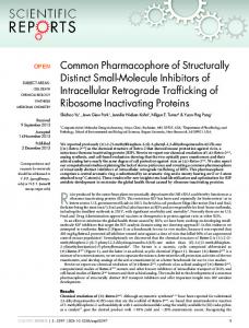
5' Primer
3' Primer
mot-1 - • 298 hp
78 bp
90 bp
mot-2 37« bp
B
/J////tSf/SJ//////•
F2
II Fl
Figure 1. A, Schematic presentation of mot-1 and mot-2 cDNA showing the presence of two Taq I sites in mot-1 and only one in mot-2. B, Taq I restriction analyses for mot-1 and mot-2 genes in mouse families. Presence of mot-1 (lower band) and mot-2 (upper band) genes in different members of two families is seen. C, Pedgree analyses of two mouse families showing segregation of mot-1 and mot-2 genes. Circles (O) indicate females and squares (•) indicate males. The presence of mot-1 and mot-2 is indicated by empty and filled circles or rectangles, respectively.
3' and 5'-ACAGTTTTCTCAATTCTATAAATGGT-3'. Hot-start PCR was performed using the following program: 1 cycle 95°C for 5 min; 40 cycles 95°C 1 min, 52°C for 2 min, 72°C for 3 min; 1 cycle 72°C for 1 min. PCR products were directly purified using the Wizard Prep PCR purification system (Promega, USA) and then digested with Taq I (Takara, Japan) at 65°C. DNA fragments were analyzed on a 3% agarose gel. Both the female mice contained mot-1 and mot-2 DNA (Figs. 2B and 2C) and their progeny were either mot-1 only or mot-
II mot-2. This result strongly suggested that mot-1 and mot-2 are allelic, and that the father was mot-l/mot-1. To further confirm this, mot-1/mot-2 Fi progeny were mated and the F 2 generation was analyzed. Both families showed segregation of mortalin alleles (Figs. IB and 1C) in a Mendelian inheritance pattern. Furthermore, while the intensity of the PCR product was identical between all mice (data not shown) the intensity of the digested bands in mot-l/mot-2 mice was less than that of either mot-1 mice or mot-2 mice (Fig. 2B). This further suggests that the mice that showed only mot-1 or mot-2 are homozygous containing two copies of their mortalin allele. These results together demonstrate that mot-1 and mot-2 are alleles of the same gene. It has been shown that Mot-1 and Mot-2 proteins have different distributions in normal and immortal cells, respectively, and have contrasting biological activities.1 5 Mot-2, but not Mot-1, protein interacts with and inactivates tumor suppressor function8 that may mediate, at least in part, its malignant transformation function. Here we have shown that these structurally and functionally distinct members of mouse hsp70 family of proteins are encoded by allelic versions of the same gene. The presence of both the alleles have also been detected in Balb/c mice6 suggesting that this phenomenon is not limited to the CD1-ICR strain. Our earlier observations that NIH Swiss and C3H mouse had only mot-2 and mot-1, respectively,6 suggest that the fibroblasts or animals from which DNA was obtained for PCR were homologous for one of the two alleles. Mouse Mot-1 and Mot-2 proteins are shown to have different biological functions. Therefore, it would be interesting to determine their effect on mouse longevity and on frequency of spontaneous immortalization in embryonic fibroblasts with different genetic background at the mot locus. References 1. Wadhwa, R., Kaul, S. C , Ikawa, Y., and Sugimoto, Y. 1993, Identification of a novel member of mouse hsp 70 family: its association with cellular mortal phenotype, J. Biol. Chem., 268, 6615-6621. 2. Wadhwa, R., Kaul, S. C , Sugimoto, Y., and Mitsui, Y. 1993, Induction of cellular senescence by transfection of cytosolic mortalin cDNA in NIH 3T3 cells, J. Biol. Chem., 268, 22239-22242. 3. Wadhwa, R., Kaul, S. C , Mitsui, Y., and Sugimoto, Y. 1993, Differential subcellular distribution of mortalin in mortal and immortal mouse, and humanfibroblasts,Exp. Cell Res., 207, 442-448. 4. Wadhwa, R., Pereira-Smith, O. M., Reddel, R., Sugimoto, Y., Mitsui, Y., and Kaul, S. C. 1995, Correlation between complementation group for immortality and the cellular distribution of mortalin, Exp. Cell Res., 216, 101-106. 5. Kaul, S. C , Duncan, E. L., Englezou, A., Reddel, R. R., Mitsui, Y., and Wadhwa, R. 1998, Malignant transforma-
Downloaded from http://dnaresearch.oxfordjournals.org/ by guest on May 30, 2013
Fl
[Vol. 7,
No. 3]
S. C. Kaul et al.
tion of NIH 3T3 cells by overexpression of mot-2 protein, Oncogene, 17, 907-911. 6. Wadhwa, R., Akiyama, S., Sugihara, T., Reddel, R. R., Mitsui, Y., and Kaul, S. C. 1996, Genetic differences between the pancytosolic and perinuclear forms of murine mortalin, Exp. Cell Res. 226, 381-386. 7. Michikawa, Y., Baba, T., Arai, Y., Sakakura, T., and
231
Kusakabe, M. 1993, Structure and organization of the gene encoding a mouse mitochondrial stress-70 protein, FEBS Lett, 20, 27-33. Wadhwa, R., Takano, S., Robert, M., Yoshida, A., Nomura, H., Reddel, R. R., Mitsui, Y., and Kaul, S. C. 1998, Inactivation of tumor suppressor p53 by mot-2, a hsp70 family member, J. Biol. Chem., 273, 29586-29591.
Downloaded from http://dnaresearch.oxfordjournals.org/ by guest on May 30, 2013
Downloaded from http://dnaresearch.oxfordjournals.org/ by guest on May 30, 2013