of avian oncovirus MH2. The MH2E21 genome, measuring only 2.3 kilobases, can be replicated like larger retroviral genomes and hence contains all cis-acting ...
JOURNAL OF VIROLOGY, Aug. 1986, p. 341-353
Vol. 59, No. 2
0022-538X/86/080341-13$02.00/0 Copyright (C 1986, American Society for Microbiology
Structure and Transforming Function of Transduced Mutant Alleles of the Chicken c-myc Gene TILO PATSCHINSKY,' HANS W. JANSEN,1t HELMUT BLOCKER,2 RONALD FRANK,2 AND KLAUS BISTER1* Otto-Warburg-Laboratoriium, Max-Planck-Institut fiir Molekulare Genetik, D-1000 Berlini 33 (Dahlem),' antd DNA-Synthese-Gruppe, Gesellschaft fur Biotechnologische Forschung mnbH, D-3300 Braunschweig,2 Federal Republic of Germany Received 6 January 1986/Accepted 7 April 1986
A small retroviral vector carrying an oncogenic myc allele was isolated as a spontaneous variant (MH2E21) of avian oncovirus MH2. The MH2E21 genome, measuring only 2.3 kilobases, can be replicated like larger retroviral genomes and hence contains all cis-acting sequence elements essential for encapsidation and reverse transcription of retroviral RNA or for integration and transcription of proviral DNA. The MH2E21 genome contains 5' and 3' noncoding retroviral vector elements and a coding region comprising the first six codons of the viral gag gene and 417 v-myc codons. The gag-myc junction corresponds precisely to the presumed splice junction on subgenomic MH2 v-myc mRNA, the possible origin of MH2E21. Among the v-myc codons, the first 5 are derived from the noncoding 5' terminus of the second c-myc exon, and 412 codons correspond to the c-myc coding region. The predicted sequence of the MH2E21 protein product differs from that of the chicken c-myc protein by 11 additional amino-terminal residues and by 25 amino acid substitutions and a deletion of 4 residues within the shared domains. To investigate the functional significance of these structural changes, the MH2E21 genome was modified in vitro. The gag translational initiation codon was inactivated by oligonucleotidedirected mutagenesis. Furthermore, all but two of the missense mutations were reverted, and the deleted sequences were restored by replacing most of the MH2E21 v-myc allele by the corresponding segment of the CMII v-myc allele which is isogenic to c-myc in that region. The remaining two mutations have not been found in the v-myc alleles of avian oncoviruses MC29, CMII, and OK10. Like MH2 and MH2E21, modified MH2E21 (MH2E21m1cI) transforms avian embryo cells. Like c-myc, it encodes a 416-amino-acid protein initiated at the myc translational initiation codon. We conclude that neither major structural changes, such as in-frame fusion with virion genes or internal deletions, nor specific, if any, missense mutations of the c-myc coding region are necessary for activation of the basic oncogenic function of transduced myc alleles.
The transformation-specific oncogenes (v-onc genes) of highly oncogenic retroviruses represent transduced mutant alleles of normal cellular genes (c-onc genes or protooncogenes). Proto-oncogenes are obviously nononcogenic in their normal cellular environment, and most of them presumably fulfil essential physiological functions in normal cell growth and development. The transition from a normal chromsomal allele to a transduced mutant allele with oncogenic function is always accompanied by multiple changes in gene structure and control of gene expression. It is largely unknown which of the qualitative or quantitative changes in gene structure and expression are necessary or even sufficient for oncogenic activation (4, 5, 7, 59; K. Bister and H. W. Jansen, Adv. Cancer Res., in press). The chicken c-myc gene is composed of three exons and two intervening sequences (32, 47, 52, 60, 65). The open reading frame encoding a 416-amino-acid protein extends from a translational initiation codon within the second exon to a termination codon within the third exon (65). The corresponding protein product with an apparent molecular weight of 58,000 was identified in extracts from normal chicken cells (2, 16). Transduced mutant alleles of the chicken c-myc gene have been found in different genetic contexts in the genomes of four highly oncogenic independent retroviral isolates: MC29, CMII, OK10, and MH2 (5, 6; Bister and Jansen, in press). All four v-myc alleles contain
nontruncated and precisely fused coding regions from the second and third exon of the cellular gene, but lack any sequences from the first (noncoding) exon and various 3' complements from the untranslated region of the third exon (1, 17, 22, 26, 45, 56, 57, 62). The MC29, OK10, and MH2 v-myc alleles also contain sequences derived from the first c-mye intron. Within the coding domains, the transduced alleles differ from their common cellular progenitor by various nucleotide substitutions and, in the case of MH2 v-myc, by a small in-frame deletion. The MC29, CMII, and OK10 v-myc alleles are fused in frame with virion genes and are expressed via genome-sized mRNAs as gag-myc (MC29, CMII) or gag-pol-myc (OK10) hybrid proteins with apparent molecular weights of 110,000, 90,000, and 200,000, respectively (8, 9, 11, 44). The OK10 v-mnvc allele is also expressed via subgenomic mRNA as a 60,000-molecular-weight protein which presumably contains 11 amino-terminal amino acid residues specified by the first six gag codons present in the leader sequence and five codons derived from the noncoding 5' terminus of the second c-inve exon in addition to amino acid residues encoded by sequences derived from the c-inye coding region (2, 12, 16, 17, 42). The MH2 v-inyc allele is expressed via subgenomic mRNA as a 59,000/61,000molecular-weight protein whose amino terminus is presumably structured like that of the OK10 60,000-molecular-weight protein (16, 22, 39, 42). MH2 carries another cell-derived oncogene, v-m71il, which is expressed via genome-sized mRNA as a gag-mil hybrid protein (19-21, 23, 25). In summary, the protein products of all transduced myc alleles differ from the c-myc protein by their modified amino
* Corresponding author. t Present address: Hoechst AG, D-6230 Frankfurt 80, Federal Republic of Germany.
341
342
PATSCHINSKY ET AL.
termini and by various substitutions and (for MH2) a small deletion of amino acid residues. Recent analyses of the genetic structures and biological properties of spontaneous or constructed mil deletion mutants of MH2 have revealed that expression of the MH2 v-myc allele is sufficient for the transformation of avian embryo cells (3, 22, 24, 66). This result demonstrated that at least for this allele, in-frame fusion with large complements of virion genes or concomitant expression of a second oncogene is not essential for transforming function. However, the question remained whether the amino-terminal modification and the substitutions and deletions of amino acid residues are relevant to the function of the MH2 v-myc protein product. Based on the molecular and biological properties of new spontaneous and constructed derivatives of MH2, we now present evidence that neither major structural modifications nor specific, if any, missense mutations of the c-myc coding region are necessary for oncogenic activation upon transduction.
MATERIALS AND METHODS Cells and viruses. Quail eggs were purchased from Gutsverwaltung Schomberg, Gemmingen-Schomberg, and chicken eggs were obtained from Lohmann Tierzucht, Cuxhaven. Quail and chicken embryo cells were prepared from 9-day-old embryos as described elsewhere (61). Nonproducer quail cell lines transformed by v-myc-carrying avian retroviruses OK10 or MH2 (line A103) have been described previously (11, 22). Focus and soft-agar colony assays of transformed cells were performed as described previously (8, 61). The nonproducer quail cell line E21 transformed by the MH2 derivative MH2E21 was derived from a focus picked in an assay designed to isolate nonproducer cells transformed by partial mil deletion mutants of MH2 (compare Results section). Analyses of virion production and viral RNA. Viral particle production was analyzed by measuring reverse transcriptase activity essentially as described previously (8), except that the final reaction mixture contained 2 mM each of dATP, dCTP, and dGTP, or by dot blot hybridization analysis of viral RNA. The dot blot analyses were carried out as described elsewhere (10, 58), except that the conditions for the hybridization reactions were as described by Jansen et al. (20, 21). The molar ratio of transforming to helper viral RNAs in pseudotype virus stocks was determined by RNA dot blot analysis with gene-specific (gag and myc) hybridization probes (see below). Molecular cloning and structural analysis of proviral DNA. Preparation of high-molecular-weight DNA from E21 cells, digestion with restriction enzymes, separation of DNA fragments by agarose gel electrophoresis, transfer of DNA to nitrocellulose sheets, and hybridization to DNA probes radioactively labeled by nick translation were performed essentially as described previously (20-23, 62). The following plasmid clones were used as hybridization probes: pMC29-SB, containing a 1.1-kilobase-pair (kbp) SalI-BamHI fragment of MC29 proviral DNA representing the 3' half of v-myc (22); pMH2-B, containing a 1.4-kbp BamHI fragment from the MH2 gag sequences (22); and pRAV-LTR, containing the entire provirus of Rous-associated virus type 1 (RAV-1) (provided by B. Vennstrom). Molecular cloning of integrated proviral DNA from E21 cells was performed essentially by the method described previously (22). Briefly, a partial pBR322 recombinant library of E21 cellular DNA was prepared as follows. A 2-,ug
J. VIROL.
sample of the 2- to 3-kbp fraction of EcoRI-digested E21 DNA was ligated with 0.4 FLg of EcoRI-digested and dephosphorylated pBR322 DNA, and the ligation mixture was used to transform competent bacteria. A total of 2 x 10⁵ independent recombinants was obtained. Colony screening with the 3' myc-specific probe led to the isolation of plasmid clone pMH2E21-E containing the 2.3-kbp EcoRI fragment of MH2E21 proviral DNA. Procedures for the subcloning of proviral DNA fragments have been described previously (62). Plasmid clones pMH2 (formerly termed pMH2-E), containing the entire MH2 proviral genome, and pCMII-E, containing most of the CMII proviral genome, have been isolated and characterized previously (21, 23, 24, 62). Nucleotide sequence analysis by the dideoxy chain termination method (48) with the M13 vectors mpl8 and mpl9 and 35S-labeled dATP-S (650 Ci/mmol; Amersham Buchler GmbH, Braunschweig, Federal Republic of Germany) was carried out as described previously (19, 37, 62). The sequencing strategy employing forced cloning of defined fragments of proviral DNA is outlined in the Results section. Oligonucleotide-directed mutagenesis. The hexadecanucleotide 5'-d(GCTTCCAGGCTTGATC)-3' was synthesized by the segmental solid-support approach and phosphorylated as described previously (14). In vitro mutagenesis was carried out by the gapped-duplex-DNA procedure (29, 37). The 0.6-kbp EcoRI-PstI fragment of MH2E21 proviral DNA containing the gag ATG initiation codon as the target site was cloned into M13mpl8am. This vector was constructed by ligating a 0.7-kbp EcoRI-BglII fragment of M13mpl8 DNA containing the multiple cloning site with the large EcoRI-BglII fragment of M13mp8 DNA carrying the amber mutations. Viral plus-strand M13mpl8am DNA containing the insert DNA was annealed with denatured DNA of the large EcoRI-PstI fragment of wild-type M13mpl8 replicative form DNA. The gapped duplex containing singlestranded insert DNA was annealed with the synthetic oligonucleotide, and remaining gaps on the minus-strand DNA were filled and sealed enzymatically by incubation with DNA polymerase I (large fragment) and T4 DNA ligase, respectively (29). The heteroduplex DNA was used to transform Escherichia coli BMH71-18mutS, which suppresses amber mutations and is deficient in DNA mismatch repair (29). Clones containing the synthetic marker were selected from the phage progeny by plating on strain MK30-3, which does not suppress amber mutations (29). The mutation in the 0.6-kbp EcoRI-PstI fragment was confirmed by nucleotide sequencing, and the fragment was reinserted into MH2E21 proviral DNA. DNA transfection. Transfection of cloned proviral DNAs onto quail embryo cells was performed as described previously (62), except that helper virus was provided by cotransfection with pRAV-LTR DNA rather than by superinfection. A total of 10 ,ug of DNA (including the amount of proviral DNA specified in the Results section and quail embryo cell DNA as carrier) was transfected onto each 6-cm dish of recipient cells. Focus formation of transformed cells was then assayed by two different protocols described in the Results section. Analysis of proteins. Subconfluent cell cultures were labeled for 2 h with 200 ,uCi of [35S]methionine (1,000 Ci/mmol; Amersham Buchler GmbH) per ml of methionine-free minimum essential medium (GIBCO Europe GmbH, Karlsruhe, Federal Republic of Germany) supplemented with 5% dialyzed calf serum. Labeling was done with a total of 200, 400, or 600 ,uCi for culture dishes with diameters of 3.5, 6, or 10 cm, respectively. Heat- and sodium dodecyl sulfate-
VOL. 59, 1986
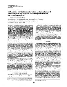
denatured lysates were prepared as described elsewhere (13, 22, 41). Briefly, cells were scraped into 0.1, 0.2, or 0.6 ml (depending on the size of the dish) of 10 mM sodium phosphate (pH 7.2)-0.5% sodium dodecyl sulfate-1% trasylol-1 mM dithiothreitol. Lysates were boiled for 2 min, cooled on ice, and diluted with 4 volumes of a buffer containing 10 mM sodium phosphate (pH 7.2), 150 mM NaCl, 1% sodium deoxycholate, 1% Nonidet P-40, 1% trasylol, and 1 mM dithiothreitol. Proteins were precipitated from portions of clarified lysates with rabbit antisera raised against whole disrupted virions of Rous sarcoma virus (RSV) (8, 21) or against a synthetic peptide specific for the carboxyl terminus of myc-encoded proteins (42). The appropriate amount of serum was applied as a dilution in 200 ,ul of 10 mM sodium phosphate (pH 7.2)-150 mM NaCl-0.1% sodium dodecyl sulfate-1% sodium deoxycholate-1% Nonidet P-40-1% trasylol-2 mg of bovine serum albumin per ml (RIPA/BSA). Immune complexes were collected by the addition of 200 ,ul of a 1% (wt/vol) suspension (in RIPA/BSA) of fixed Staphylococcus aureus cells (Pansorbin; Calbiochem GmbH, Frankfurt, Federal Republic of Germany) which had been pretreated by boiling in 10 mM sodium phosphate (pH 7.2)-150 mM NaCl-3% sodium dodecyl sulfate-10% mercaptoethanol (46). Polyacrylamide-sodium dodecyl sulfate gel electrophoresis and fluorography were performed as described elsewhere (21, 22, 41, 42). RESULTS of MH2E21. We have previstructure and Origin genetic ously described the in vitro construction of a partial mil deletion mutant of MH2 (24). In a focus assay of quail embryo cells infected with a stock of that mutant, a transformed cell clone was isolated and developed into a
nonproducer line (E21). Analysis of virus-specific protein synthesis showed that E21 cells contained a v-myc protein product of 59,000/61,000 molecular weight indistinguishable from that of wild-type MH2, but no proteins precipitable by antiserum against virion structural proteins (Fig. 1). Transforming retrovirus (MH2E21) could be rescued efficiently from E21 nonproducer cells by superinfection with helper virus. MH2E21 pseudotype virus stocks were also obtained by cotransfection of cloned MH2E21 and helper proviral DNAs onto quail embryo cells (see below). Virus was then harvested from completely transformed cultures obtained after repeated passaging of the recipient cells. The titer of focus-forming units (FFU) in these pseudotype virus stocks (approximately 0.5 x 10³ FFU/ml) was about 20- to 30-fold lower than that of wild-type MH2 stocks generated by the same procedure with the same helper virus (RAV-1). The analyses of viral particle production and of viral RNA composition in these particles are largely in agreement with the observed differences in the titers of transforming virus. As determined by both reverse transcriptase assays and RNA dot blot analyses, MH2-transformed quail cells coinfected with helper virus (RAV-1) released about three times more virus particles per milliliter of culture fluid than MH2E21-transformed cells coinfected with the same helper virus. Furthermore, the molar ratio of transforming to helper viral RNAs determined by dot blot analysis with myc- and gag-specific probes was about three times higher in pseudotype particles released by MH2-transformed cells (approximately 0.6) than in those released by MH2E21-transformed cultures (approximately 0.2). Together, these results would account for an approximately 10-fold reduction in the titer of transforming virus in infectious MH2E21 stocks compared with MH2 stocks.
~ p59/61
ONCOGENIC myc ALLELES 1 2 3 4
5 6 7 8
100gag-mitf
-
343
FIG. 1. Proteins encoded by MH2 and MH2E21. Cultures of the nonproducer quail cell lines E21 (lanes 1, 2, 5, and 6) or A103 (lanes 3, 4, 7, and 8) transformed by MH2E21 or MH2, respectively, were labeled with [35S]methionine, and heat- and detergent-denatured proteins in cellular lysates were immunoprecipitated. Each precipitate was prepared from the lysate of 1.8 x 101 E21 cells (containing 107 cpm) or of 0.9 x 10⁵ A103 cells (containing 107 cpm), respectively, with 1 ,ul of either preimmune rabbit serum (lanes 1 and 3) or rabbit serum against whole disrupted RSV (lanes 2 and 4). Precipi-
p.g of the total immunoglobulin G fraction of a rabbit against a carboxy-terminal myc-specific synthetic peptide (lanes 5 and 7) or with the same amount of serum preadsorbed to the peptide (1 ,ug/,ug of immunoglobulin G; lanes 6 and 8) were obtained from lysates of cells labeled in a separate experiment. The precipitates were prepared from the lysate of 2.8 x 10⁵ cells (containing 1.2 x 10⁷ cpm) of the E21 or A103 line, respectively. Equal portions (20%) of all precipitates were analyzed by electrophoresis on 12.5% polyacrylamide-sodium dodecyl sulfate gels. The fluorographs were exposed for 19 h (lanes 1 through 4) or 6 days (lanes 5 through 8). tates with 20 serum
To identify MH2E21 proviral DNA, high-molecularweight DNA from E21 nonproducer cells was digested with EcoRI, and electrophoretically separated digestion products
were transferred to a nitrocellulose filter and hybridized with a myc-specific probe (pMC29-SB). In addition to the endogenous quail c-myc DNA fragment, a single fragment of 2.3
kbp hybridized with the myc probe (Fig. 2A). The same DNA fragment also hybridized with a complete helper proviral probe (pRAV-LTR), but not with an internal gagspecific probe (pMH2-B) (not shown). The 2.3-kbp EcoRI fragment was molecularly cloned, and its restriction enzyme cleavage site map (Fig. 2B) and entire nucleotide sequence (Fig. 3) were determined. The termini of the fragment correspond to EcoRI cleavage sites within the U3 regions of the 5' and 3' long terminal repeat (LTR) sequences of MH2E21 proviral DNA. Between the 5' and 3' noncoding sequences, the proviral DNA contains a coding region composed of the very 5' six codons of the gag gene fused in frame with v-myc coding sequences (417 codons). The gag-myc junction corresponds precisely to the standard splice donor site within the gag gene of avian sarcoma-
344
PATSCHINSKY ET AL.
A kbp
J. VIROL.
B 1
2
U3
RU5
A gag
v-
U3 RU5
myc
-. -
TT
23.1 3 --9.42-656-
ATE 41T
TAG
4.36 5
L232 2 03-
C0.5
10 C:V
1 G
N
}1.5 ctC
1 5
Ct
2.D
cLoup
~~~~~~~~~kbp
0.56-
FIG. 2. Genetic structure of MH2E21. (A) High-molecular-weight DNAs (15 ,ug each) from uninfected quail embryo cells (lane 1) or from cells of the MH2E21-transformed nonproducer quail cell line E21 (lane 2) were digested with EcoRI, and the digests were separated by electrophoresis through a 0.8% agarose gel with fragments of X DNA digested with HindIII as molecular weight markers. DNA was transferred to a nitrocellulose filter and hybridized with nick-translated DNA (2 x 10⁶ cpm of 32P) of a 3' myc-specific probe. The autoradiograph was exposed for 3 days. (B) A plasmid clone, pMH2E21-E, containing the 2.3-kbp DNA fragment hybridizing with the myc probe was isolated from a pBR322 library of the 2- to 3-kbp fraction of EcoRI-digested DNA from the E21 cell line. The genetic structure of MH2E21 proviral DNA was deduced from the restriction map, from hybridization with gene-specific probes (not shown), and from complete nucleotide sequence analysis (compare Fig. 3). The strategy for sequencing fragments of proviral DNA cloned into M13mpl8/19 is shown below the restriction map. The open reading frame on the MH2E21 genome is indicated by the position of the translational initiation codon of the partial .gag gene complement and by the position of the termination codon within v-mnvc (stippled box). The position of the first ATG codon within the transduced invc sequences is also shown (compare Fig. 5).
leukosis viruses (15, 49) and to the splice acceptor site for the second c-myc exon which is conserved in the MH2 v-inyc allele (22, 26, 65). Except for the lack of 5' intronderived sequences, the MH2E21 v-myc allele is nearly isogenic with that of MH2. Of the 27 missense mutations reported for the MH2 v-myc allele in comparison to the chicken c-inyc gene (26), 24 were also found in the MH2E21 v-myc allele, and one codon is different from the homologous counterparts in both c-mvyc and MH2 v-myc. Furthermore, the MH2E21 and MH2 v-mnyc alleles share the same deletion of four consecutive codons in comparison to c-mnyc (Fig. 3). The nucleotide sequence of the 3' noncoding region of MH2E21 proviral DNA between the 3' terminus of v-myc and the 5' terminus of U3 is nearly identical with that reported for the corresponding region of the MH2 provirus (26, 57). This region shows extensive sequence homology to corresponding sequence elements in the genomes of avian sarcoma viruses Y73 and RSV SR-A, but it is less related to the corresponding region of the RSV PR-C genome (49, 57). On the other hand, the R, U5, U3, and 5' leader sequences of the MH2E21 genome are closely related to those of RSV PR-C, but have some distinctive features (Fig. 4). The alignments in Fig. 4 show that sequence duplications and triplications have occurred in the 5' leader sequence and in the U3 region, respectively. The sequence which is triplicated in the U3 region of the MH2E21 genome and present only once in the RSV PR-C U3 region is presumably part of the transcriptional enhancing region of the LTR sequences (30, 33). Essentially the same unusual structural features have recently been reported for the U3 region and 5' leader sequences of ring-necked pheasant virus (RPV) (53), a nondefective weakly oncogenic avian retrovirus often used as a helper virus. Since our wild-type MH2 and spontaneous and constructed mutant derivatives were frequently propagated with RPV as a helper virus (22), it appears very likely
that MH2E21 was derived from a parental MH2 virus that had recombined with RPV. This possibility is also consistent with the observation that the U3 regions of both MH2E21 (Fig. 2 and 3) and RPV proviral DNAs (53) contain EcoRI cleavage sites, whereas the U3 region of our original clone of MH2 proviral DNA contained KpnI cleavage sites (21, 23). The overall genetic design of the MH2E21 genome is strikingly similar to the one proposed for subgenomic v-myc mRNA of MH2 (22, 26, 39) and provides direct evidence that the presumed splicing signals on MH2 genome-sized RNA are actually utilized. The genesis of the transmissible MH2E21 retrovirus presumably involved encapsidation and reverse transcription of subgenomic v-myc mRNA. MH2E21 derivatives designed in vitro. The open reading frame on the MH2E21 genome encodes a 423-amino-acid protein which is distinguished from the cellular c-myc protein by the additional amino acid residues at the amino terminus and by the substitutions and deletions of residues within the shared domains (Fig. 3 and SA). To assess the functional significance of these structural changes, we have constructed derivatives of MH2E21 proviral DNA encoding protein products which are progressively more similar to the c-mnyc protein. The gag translational initiation codon (ATG) was modified to CTG by using the synthetic anticoding hexadecanucleotide 5'-d(GCTTCCAGGCTTGATC)-3' (the substituted nucleotide is underlined) for mutagenesis (Fig. 5; compare Materials and Methods). The mutated MH2E21 derivative, termed MH2E21ml, encodes a 412-amino-acid protein whose synthesis would start at the first translational initiation codon within the myc sequences (Fig. SB). This codon corresponds to the presumed c-myc translational initiation codon near the 5' terminus of the second exon (32, 52, 65). A second derivative, MH2E21c1, was constructed by replacing most of the MH2E21 v-myc allele, i.e., the 1.0-kbp Pstl-Rsal fragment (compare Fig. 2B), by the cor-
VOL. 59, 1986
ONCOGENIC mvc ALLELES
3. ~ ~ ~ ~. ~ ~~~~~~R
: ~ ~
A
A
:A
A
345
:A
CAC66CC66C66C66CTCGGA6GAAGAACAAGAA6AA6ATGA6GAAATCGATGTC6TTAC 1200 ThrAlaIlyGlySlySer6luBluSlu61nGluSluAsp6luSlulleAspValValThr 250
AATTCC6CATT6CA6A6ATATT6TATTTAAGT6CCTA6CTC6ATACAATA A TTT T :U5 R,
6(0
6ACCATTCACcAcATTG6TGGCACCTG66TTGATG6CC66ACCGTT6ATTCCCTGACGA
120
U 5 ""-I CTAC6AGCACAI6CATGAAGCAGAA66CTTCATTTGGT6ACCCC6ACGTGATCGTTAGGG
180
ATTAGCTGAAGC6AACGAGTCTGAATCCA6CACA6AGTCCA6CACA6AAGCATCA6A66A 1260 LeuAlaSluAlIAsn6luSer6luSerSerThr6luSerSerThr6luAlaSer6luSlu 270
AATA6TGGTCGGCCACAS6CGGC6T6GCGATCCTGTCCTCATCCGTCTCGCTTATTCGGG
240
GCACTGTAAGCCCCACCACAGTCCGCJ66TCCTCGA6CGGT6TCAC6TCAACATCCACCA 1320
GAGCG6ACGATGACCCTAGTAGAGGGGGCTGCG6CTTAG6AGGGCAGAAGCTGAGTGACG
300
TCGGA6GGA6CTCCACG6CCGGGGGCCAAGATACCCTACC6AGAACTCAGAGAGTCGTTG
36(1
ACACAACTAC6CTGCTCCTCCCTCCACCAAGT 66AATACCCA6CCGCCAA6A6GCTAAA 1380 HisAsnTyrAlaAlaProProSerThrLysVal6luTyrProAlaAlaLysArgLeuLys 310
GAAGACG6GAA6AAAGCCCGACGACTGAGCGGTCCACCCCAGGCGT6ATTCCGGTTGCTC gg9? myc TGCGTGATTCCGGTCGCCCG6TGAA'CAA6CAT6GAAGCC6TCATAAAGGCA6CAGCCGC
420
6TTGGACA6TGGCA66GTCCTCAAACAGGTCAGCAACAACC6AAAAT6CTCCAGTCCCC6
ThrSerSerAsp
H:sCysLysProHisHisSerProl.euValLeuGluArgCvsH:sVilAsnIleHis6ln 290 Lys
A:
lie
Het6luAlaVa&lleLvsAlaAlaAlaAla
480 10
C6C6ATGCC6CTCA6CGTCAGCCTCCCCAGCAAGAACTACGATTAC6ACTAC6ACTCGGT AlaMetProLeuSerValSerLeuProSerLysAsnTyrAspTyrAspTyrAspSerVal
540 30
C
*
Ala
1440
LeuAspSer6lyArqValLeuLvsSlnValSerAsnAsnArgLvsCysSerSerProArg 330 A
:
CACGTCA6ACTCAGAGGTGAACGACAAGAGGCGAACGCACAACGTCTTGGAGCGCCAGC6
ThrSerAspSerSluValAsnAspLysArgArgThrHKsAsnValLeuGluArg6lnArg
1O50(
350
Slu
T
:
AAGGAATGAGCTGAAGCT6SACTTCTTTGCCCrTC6GGACCAGATACCCGAS6TGGCCAA 1560 ArgAsn6luLeuLysLeuSerPhePheAlILeuArgAsp6lnllePro6luValAlaAsn 370
6CA6CCCTACTTCTACTTCGA66AG6AGGAGGA6AACTTCTACCT6GCGGCGCAGCAGCG
600
GlnProTyrPheTyrPhe6luSluGlu6luGluAsnPheTyrLeuAlaAlaGInG6nArg 50 :C
G
6AGCABC6A6CTGCA6CCTCCA6CCCCGTCC6A66ACATCT6GAAGAAGTTTGA6CTCCT b60 SerSer6luLeu6InProProAlaProSerGluAsplIeTrpLysLysPheGluLeuLeu 70 GlY
C:
A
G
GCCCGCGCC6CCCCTCTCGCCCAGCTGCCGCTCCAACCTGGCCGCCGCCTCCTGCTTCCC 720 ProAlaProProLeuSerProSerCysArgSerAsnLeuAlaAlaAlaSerCysPhePro 90 Thr Ser Arg TTCCACCGCCGACCAGCTGGAGATGGTGACGGAGCTGCTCG66GGGGACATGGTCAACCA 780 SerThrAlaAsoGlnLeu6luMetValThr6luLeuLeu6ly6lyAspMletValAsn6ln 110 A
T
6A6CTCCATCTGCGACCC66AC6AC6AATCCTTCSTCAAATCCATCATCATCCGGGACT6 840 SerSerlleCysAspProAspAspGluSerPheValLysSerllellelleArgAspCys 130 Gin
Phe
CATT 66A6C6GCTTCTCCGCCGCC6CCAAGCT66AGAAGGTSStc6TCAGAASCTCGC MetTrpSerGlyPheSerAlaAlaAlaLysLeu6luLysVaIlValSer6luLysLeuAla 150 C:
ThrTyrLpsAlaSerArgArg6lu6ly6lyProAlaAlaAlaSerArgProGlyProPro G1n
960 170
GCCCTCGGGGCCGCCGCCTCCTCCCSCCGGCCCCGCCGCCTCGGCCGGCCTCTACCTGCA 1020 ProSer6lyProProProProProAla6lyProAlaAlaSerAlalyLeuTyrLeuHlis 190 :
A
AC
Asp
AspPro
:
A
C6ACCTGS6AGCCGCG6CC6CCGGCT6CATC66CTCCTCGTGGTCTTCCCCTSCCCGCT 1090 AspLeuGlyAlaAlaAlaAlaGlyCys5leGlySerSerValValPheProCysProLeu 210 A 6A
AA
Tyr
:
162u
3q(Q
Lys
A
CCAATC66ACGA6CACA6ACT6ATC6CA6A6AAA6A6CAGTr6A66C66A66A6A6AACA 1680 61 nSerAspGl uHi sArgLeull eAl aG 1uLysGl uGl nLeuArqArgArqArg6luG1n 410 GTTGAAACACAAACTTGAGCAGCTAAGGAACTCTCGTGCATA66AACTCTTGGACATCAC I1740 423 LeuLysHisLysLeuGlu61nLeuArgAsnSerArgAIa*l ' myc q-1
TTAGAATACCCCAAACTASACTCCSTSGTATA6CT66TTGGATC6TTAATC66ACSGCTG
GCACACG6AAT6TA66A66TCGCTGAGTAAGTACGAACAAAATTTAC6TlT6AATAA66T GAGGCTT6ACCIACAATTGTICAAATAATGCTTCT6TAGAAATGTTTA6CATTASSCATC 1920
900
CACCTACAAAGCCTCCCGCC66SAG6GGGCCCCGCC6CCGCCTCCCGACCCGGCCCGCC
A
CAAC6A6AAG6C6CCCAAG6TT6TCATCCTGAAAA6A6CCAC66A6TAC6TTCTG6CTAT Asn6luLysAlaProLysValVallleLeuLysArgAlaThr6luTyrValLeuSerlle
TTGCGCTGCTCCGCGAT6TACGSGTCAGGTATAATSTGCA6TTTGACTGAG666ACCATG 1980
ATATGTATAGGC6AAA66CS666CTTCr66TT6TAC6C66TTA66A6CCCCTCA66ATAT 2040 ,o0-U3 , A6TASTTTCGCTTTT6CATA66SAS6SSSAAAT6TAGTCTTATGCAATACTCITTTASTC 2100
TTGCAACATGCTTATGTAACGATGA6TTASCAACATGCCTTATAA66AAA6AAAAA6CAC '160 CGTGCAT6CCGATT6GT66AA6TAA6GT6GTAT6AtCGTGGTAIGATC6GTGSAT6ATCG 22220 TGCCTTATTA66AAGGCAACA6ACGSGICTAACAC66ATT66AC6AACCACTG
2273
: A
CGGCAGGC6CGGCtCGCCCGGC&CCGGCCCCGCGGCTCTGCTGGGGGTCGACGCGCCGCC 114(1 ProProSlyAlaGIyProAlaAlaLeuLeuGlyValAspAlaProPro 230 51vArqArg6ql SerGlu Ali Thr Asn 'Pr oArqA! aAl a
FIG. 3. Nucleotide sequence of MH2E21 proviral DNA. The sequence of 2,273 nucleotides (numbered in the margin) between the EcoRI cleavage sites within the U3 regions of the LTRs (compare Fig. 2) is shown. The predicted sequence of amino acid residues (numbered in the margin) of the protein product of MH2E21 is shown below the nucleotide sequence. Within the mnvc domain, all changes in nucleotide sequence and predicted amino acid sequence compared with those of the chicken c-myc gene (65) are indicated above and below the MH2E21 sequences, respectively. The gag-myc junction on the MH2E21 genome corresponds precisely to the standard splice donor site within the gag gene of avian retroviruses from the leukosis-sarcoma group (15, 49) and to the splice acceptor site of the second exon of the chicken c-tnvc gene (32, 52, 65).
346
PATSCHINSKY ET AL.
A
1T dCfti1
r-oR I-,t') fr
rb ,,;1
RH1,'U5 ,r T1 5(-m G,1T1'3 tI H;i f ,I C C
..
6HC.,-) T-r T ih CIC',T T
f&,
TI'TC IT G; 1CLC.CJi)C";rI CCl-l'T r(Ai I C c
C,-r J G-Gyf31 r G -1- 3C IT C-0 G TT T ,GT -i1 C2iT, I-I CI
61
C T'" A hCG TC *;P-*iC5A-i GCAC-.i';C: T'( Ci ii- iCIff'ii G'("Ir ~T l- UC.(-5 T P,t i ACt'"
61
1C T( vIC.U
r;,C, (-; CA C(.C,T1 G';"I; T13(iAit:;i.G.i17(iGiH( U:'T:
1TTtI,Si ' - f-iTi
121
-C G HT I-`)G TI;I iGt(-IA ,CrUAi
r
f 'r "I
( IT 1iC, frIu Iv
GGc- T G f ;C,C.,C: C.{; C' (", T GX,I T C,
1 A33 1T 1T T(741 T r1
3111C'.C.C.C G;il C; f:i
lU I CTI
i !C1 f.( IT ) 121 )(,I rI
TJ 0
1 1c I
-, T r- Tl-,'G(,hG - r~i-:`lCJ r-.( , :l T F-C,-rt- C-1*- C.i .iT T F-- C -1- rf Ci C s i: i0:l-,C C l; r- TrrfC -. 1 11 J 11117132 HTTIC Ui~ L-u 6 _4 qit HL iI( I 41(A HiI 11(1 111i211112111 c T T fr A,(-A ;-` C(- (:Cl* i-, xC:;--n:-s- .0 t1 C I(: TC.t`(_ ('S'.C:o 16;-, C. C-'i (.( F- C.,r,-:: C.' ,3: Ct
121 1 Li 1
T,~
r, ,"-- ,i A C,
c'
,A,v
2I 1( :. 4 t
C.C I.. 'I I.j
..
C)'iT i-, ,r r:C' t. It",, Jitt1 liC. T C- i- i Cr.T(, Ci..j li J_ I-i If Tt7,; Ci,C i-t 1C :I ii"IC'i si iCj,;_-,i(;l- .7 t UiFF11213~6i~ IUUi. ~iIi(II Hi IC2 li Ii
1~11iIi'2I I(tI
24 1
;3ij! 1 31
1T
J. VIROL.
T
I ,-
1
C I ,'
T ,i,l
If
IT,rI tI IIT ffIll1X.*=( 11 3'lii 1
fr
B
.,
,_
C
F
,)
.1
T
i(. IIR ,iiii,-ls I.--tI;: ii 1 14 12
I
,) It
:,:-2121 (:::i',(! (:
i 1;il
C Cl,-t.tXJ:t*,V> (, -fj ,i:(::;-r(:tl, fl;S J
r,; cl - T ,r tlJT
r7mU3
T1 T C,1 , f t(. 1 r r,I
;C r2sc.c1 A
C
U
1
. ( ti 'T (- .t I t r -f G; T fi- T f ,C .f G) 1, 1( ,1 T1, t' G(
; r ; ST
'(I (i"
A;11(;T1i '11 1C 1`i C t I 1i( CC Ci;( I -J f
i;i
C
C
C. .
(above)
rresponding
C I T Td Mf-, 2T eqr 2; 31 1'i t';idets '1
e
'r i .lS D;in
Ia1
(i A C i:*:C ; C
t
f-) A ,:9C,
T
(,('C3
I
T
r("Gf1(
G' rfr (-( C'SG'§;: j-,,T
r 7*`1
I'e
T
i cl
C
1i(-'I................... O t-I IrV 1C C C Cr r(T C"
C "I :(.'
C (::C :T ('.' A.' 1- T(:.(,l
C;i' t (-r-r(:( 6f 1-,(:I - (" (;i TkrU,;3C.-R ; "f T -f' C')t
'th n-
F"
si
C rrlC;-1(t
.-i-
I
l T tg', ' C
C e( T -s lG TcCo
af1 t'h
TT G tI' T r(1; I.*,( 1 I
I(,-1' i-f 1'-.
1-,t T 'I T
OAG(' 1' (41f;( T6i
1: 1'1r[-F- ,IC R
FIG. 4. Alignment of proviral nucleotide sequences of MH2E21 (above) and RSV (below) corresponding to the S' (A) and 3' (B) termini of the viral RNA genomes. (A) Nucleotides 54 through 469 of the MH2E21 sequence shown in Fig. 3 are aligned with nucleotides 1 through 397 of the RSV PR-C sequence (49). SD indicates the position of the splice donor site in the RSV genome and of the splice junction site on the MH2E21 genome. (B) Nucleotides 2071 through 2273 and 1 through 74 of the MH2E21 sequence (Fig. 3) are aligned with nucleotides 9058 through 9312 of the RSV PR-C sequence (49). Duplications in the MH2E21 sequence are indicated by brackets.
responding region of the CMII v-myc allele. For the construction, the 5' 0.6-kbp EcoRI-PstI fragment and the 3' 0.7-kbp RsaI-EcoRI fragment (obtained by partial digestion) of MH2E21 proviral DNA (compare Fig. 2B) were ligated with the 1.0-kbp PstI-RsaI fragment from the v-myc region of cloned CMII proviral DNA (62). Within that fragment, the nucleotide sequence of the CMII v-myc allele is identical with that of the chicken c-myc gene (62; N. Walther, H. W. Jansen, C. Trachmann, and K. Bister, Virology, in press). The PstI site on MH2E21 proviral DNA maps at nucleotide position 611 on the sequence shown in Fig. 3, and the RsaI site is located at position 1606. Hence, the MH2E21c1 provirus has retained only two missense mutations (5' of the PstI site) within the v-myc allele which encodes a 427-amino-
acid protein (Fig. 5A). By the same strategy as was used for the construction of MH2E21c1 from MH2E21, a third derivative, MH2E21mlcl, was constructed by replacing most of the MH2E21ml v-myc allele by the corresponding segment of the CMII v-myc allele. MH2E21mlcl proviral DNA encodes a 416-amino-acid protein (Fig. SB) which differs from the c-myc protein product by only 2 amino acid
substitutions, an alanine-valine and a glycine-serine exchange (the mutations map at nucleotide positions 498 and 602, respectively, on the sequence shown in Fig. 3). Notably, neither of these two mutations has been found in the
oncogenic v-myc alleles of MC29, CMII, or OK10 (1, 17, 45, 62; Walther et al., in press). To obtain biologically active proviral DNA of MH2E21
VOL. 59, 1986
ONCOGENIC myc ALLELES
A
Lfs
v-myc
T
A
G
Agag
347
C
XZRm
T
C= _
6
1
421.
[OGH -ftoodD
4:42 3427
4---
aa -%mom
am
--_
me t
-ATC[AG
A'L Va L e L y sAaaA IcaA;, :A .Me t P- - W [Z-rr 6TCZA'`QCT G A CZ CA
A,
AT
Li
_
J my
B
G A Agag LI
lI
T
C
v-myc
[=Z1E
I
NH27;-,41 2 41 6 aa 5
-
-
AT W AA G C CTEA GOAM -
CGTEAT !A'AG5 CA G ,,sg
M e t P rc -- -
G ATG c
5'
3'
myc
FIG. 5. Mutagenesis of MH2E21. (A) Protein-coding domain on the proviral genomes of MH2E21 and MH2E21cl. MH2E21 specifies a 423-amino-acid protein with 6 residues encoded by gag and 417 residues encoded by v-myc sequences. MH2E21cl encodes a 427-amino-acid protein with 6 residues encoded by gag and 421 residues encoded by v-myc sequences (numbers in parentheses). MH2E21cl was derived from MH2E21 by in vitro recombination restoring the four myc codons deleted from the MH2E21 v-myc allele (see Fig. 3 and text). The section of the sequencing gel (shown here for MH2E21cl DNA; the sequence of MH2E21 DNA in this region is identical) shows the sequence of the anticoding strand complementary to the 5' 28 nucleotides of the coding-strand sequence shown in the diagram. (B) Protein-coding domain on the proviral genomes of MH2E21m1 and MH2E21mlcl. MH2E21m1 specifies a 412-amino-acid protein, and MH2E21m1c1 encodes a 416-amino-acid protein (number in parentheses). Synthesis of both proteins is initiated at the myc ATG codon. MH2E21m1 was derived from MH2E21 by oligonucleotide-directed mutagenesis of the gag translational initiation codon, and MH2E21mlcl was derived from MH2E21m1 by in vitro recombination restoring the four myc codons deleted from the MH2E21m1 v-myc allele (see text). The section of the sequencing gel (shown here for MH2E21mlcl DNA; the sequence of MH2E21m1 DNA in this region is identical) shows the sequence of the anticoding strand complementary to the 5' 28 nucleotides of the coding-strand sequence shown in the diagram. The single nucleotide substitution is indicated by the arrow.
(and, by an analogous strategy, of its derivatives), the partial LTR sequences at the 5' and 3' termini of the 2.3-kbp EcoRI fragment (compare Fig. 2B) were completed in vitro. The 3' 0.7-kbp BalI-EcoRI fragment of proviral DNA (Fig. 2B) was cloned into the PvuII and EcoRI sites of pBR328. The 5' 0.3-kbp EcoRI-SacI fragment of proviral DNA (Fig. 2B) was cloned into the corresponding sites of pUC12. DNAs from the pBR328 and pUC12 clones were then cleaved with EcoRI and HindIII. The large fragment of the pBR328 digest and the small fragment of the pUC12 digest were ligated, yielding a plasmid clone containing a complete MH2E21 LTR flanked by partial proviral sequences. The 2.3-kbp EcoRI fragment of pMH2E21-E (Fig. 2B) was now inserted in the appropriate orientation into the EcoRI site within the U3 sequences of the LTR-containing plasmid clone. The final product, pMH2E21 (Fig. 6A), contained a complete and biologically active (see below) provirus of MH2E21. By the same procedure, pMH2E21m1, pMH2E21cl, and pMH2E21mlcl were constructed, containing biologically active proviral DNAs of the MH2E21 derivatives. The structural integrity of all constructs was verified by restriction enzyme digestion analysis of DNA from the complete proviral plasmid clones (Fig. 6B). Within the MH2E21 v-myc sequences, the 5' PvuII site (nucleotide position 682 in Fig. 3), the 3' Sacl site (position 781), and the XhoI site (position 1292) are specific for this allele, whereas all other restriction
enzyme sites shown in Fig. 2B and 6A are shared with the chicken c-myc and the CMII v-myc alleles (47, 60, 62, 65). Hence, the particular digestions shown in Fig. 6B directly confirm the substitution of CMII v-myc sequences for MH2E21 v-myc sequences in MH2E21c1 and MH2E21m1c1 proviral DNAs. The expected restoration in those DNAs of the deleted sequences is also confirmed by the slightly larger size of their 0.5-kbp PstI-SalI fragment (Fig. 6B). Further confirmation of the expected structures of the constructs was obtained by partial nucleotide sequence analysis. In particular, it was confirmed that the MH2E21m1c1 v-myc allele, like that of CMII (62; Walther et al., in press) and like the chicken c-myc gene (65), contains an ACG codon at a position where the v-myc alleles of MH2 (26), MH2E21 (Fig. 3, nucleotide position 665), MC29 (1, 45), and OK10 (17) all have a missense mutation. Protein products and transforming properties of MH2E21 and its derivatives. DNAs from the plasmid clones containing complete proviral genomes of MH2E21 or its derivatives (Fig. 6) were transfected onto quail embryo cells in the presence of cloned helper proviral DNA. In all cases, transformed cell clones were obtained (see below). Analysis of v-myc-specific protein products in these cells (Fig. 7A) revealed that the apparent molecular weights deduced from their relative electrophoretic mobilities agree precisely with the predictions about their relative sizes (Table 1) based on
348
PATSCHINSKY ET AL.
B
A
*-1~ ~ 2.30
J. VIROL.
k bp
23 !
~ 2313 ~~~~~~~~ 9 .4
6.56 -~~~~~~~--. 4. 36 _~~~~~~
_
-
_ Z
.5 'b
Un
a-
FIG. 6. Construction of biologically active proviral DNAs of MH2E21 and its derivatives. (A) The 5'-terminal EcoRI-SacI fragment and the 3'-terminal BaiI-EcoRI fragment of the 2.3-kbp EcoRI insert fragment of pMH2E21-E (compare Fig. 2 and 3) were subcloned into pUC12 and pBR328, respectively, and then joined to the 3' and 5' termini, respectively, of the 2.3-kbp EcoRI fragment (see text). The final product of that construction, pMH2E21, contains a complete and biologically active provirus. All restriction enzyme cleavage sites utilized for the construction or for the digestions shown in panel B are indicated, except the Sall and PstI sites within the remnant of the pUC12 multiple cloning site. By the same strategy, pMH2E21ml, pMH2E21c1, and pMH2E21mlcl, containing biologically active proviruses of the MH2E21 derivatives, were constructed. (B) DNAs (1 ,ug for each digestion) of pMH2E21 (lanes 1, 5, 9, and 13), pMH2E21c1 (lanes 2, 6, 10, and 14), pMH2E21ml (lanes 3, 7, 11, and 15), and pMH2E21mlcl (lanes 4, 8, 12, and 16) were digested with EcoRI and XhoI (lanes 1 through 4), EcoRI and PvuII (lanes 5 through 8), EcoRI and Sacl (lanes 9 through 12), or PstI and Sall (lanes 13 through 16). The digestion products were separated by electrophoresis through a 1% agarose gel. Molecular weight markers are as explained in the legend to Fig. 2. Note that within the v-myc sequences, the 5' PvuII, 3' Sacl, and XhoI sites are specific for the MH2E21 and MH2E21ml v-myc alleles (see text).
the sequence analysis (Fig. 3) and the strategies of the in vitro constructions (Fig. 5). While all these proteins show the substantial difference between calculated and apparent molecular weight (Table 1) typical for all myc protein products (1, 2, 8, 16, 42, 45, 65), their differences in size (electrophoretic mobility) relative to each other agree with the predicted differences in numbers of amino acid residues (Table 1). MH2E21 encodes a 59,000/61,000-molecularweight protein (p59/61v-1Yc) which is indistinguishable from the MH2 v-myc protein product (Fig. 1 and 7A). MH2E21ml, MH2E21cl, and MH2E21mlcl specify v-myc protein products with apparent molecular weights of 57,000/59,000 (p57/59V-MYC), 60,000 (p6O`vnY% and 58,000 (p58V-mYc), respectively (Fig. 7A). Interestingly, the appearance as a characteristic doubiet in gel electrophoresis is typical for those v-myc proteins which are distinguished by the MH2- and MH2E21-specific amino acid substitutions and deletions, but is not dependent on the utilization of the gag translational initiation codon (Fig. 1 and 7A). As expected (Table 1), the MH2E21c1 p60V-MYc protein comigrated with the protein product of OK10 subgenomic v-myc mRNA (Fig. 7A). To test the prediction (Table 1 and Fig. SB) that the MH2E21mlcl p58v-myc protein would be nearly identical with the chicken c-myc protein, its electrophoretic mobility was compared with that of the chicken c-myc protein product and also that of the quail. The analysis showed that the apparent molecular weights of the protein products from the transduced and the chromosomal myc alleles are identical (Fig. 7B). Based on the amount of cellular lysate used for the immunoprecipitation and the relative intensities of the radioactive signals of these protein bands (Fig. 7B), it was estimated that there is at least 30 to 50 times more p58v-myc in MH2E21mlcl-transformed cells than there is p58c-mYc in normal avian cells.
The transforming function of MH2E21 and its derivatives was measured by transfection of their proviral DNAs onto quail embryo cells. Most importantly, all proviral DNAs were capable of inducing focus formation of transformed cells. A quantification of the transforming activities of MH2E21 and its derivatives relative to that of MH2 is shown in Table 2. Under assay conditions which largely prohibit the spread of infectious virus, MH2E21 and MH2E21ml were about as efficient as MH2 in the induction of foci, whereas the substitution of sequences isogenic to c-myc for most of the MH2E21 v-myc sequences in MH2E21cl and MH2E21mlcl caused a significant and reproducible reduction in the titer of FFU per picomole of transfected proviral TABLE 1. Sizes of proteins encoded by transduced or chromosomal myc alleles Mol wt No. of Origin of amino acid protein product Apparentb Calculateda residues
v-myc, v-myc, v-myc, v-myc, v-myc, v-myc, c-myc,
MH2 MH2E21 MH2E21ml
MH2E21c1 MH2E21mlcl OKlOd chicken
423c 423c 412 427c 416 427C 416
46,109 46,137 45,111 47,073 46,047 47,015 45,989
59,000/61,000 59,000/61,000 57,000/59,000 60,000 58,000 60,000 58,000
Calculated molecular weights of myc protein products are based on amino acid sequences deduced from nucleotide sequence analyses of v-myc or c-myc alleles: MH2 (26); MH2E21 (Fig. 3); MHZE21 derivatives (Fig. 5; see text); OK10 (17); chicken (65). b Apparent molecular weights of myc protein products were determined by electrophoresis on polyacrylamide-sodium dodecyl sulfate gels (Fig. 1 and 7). c Including 6 amino-terminal amino acids encoded by 5' gag sequences. d Only the v-myc protein product translated from subgenomic mRNA is considered here. a
ONCOGENIC myc ALLELES
VOL. 59, 1986
DNA (Table 2). Under assay conditions which allow virus spread, the relative increase in titer was larger in transfections with MH2 proviral DNA than with the other DNAs. Similarly, the titer of FFU was higher in infectious virus stocks of MH2 than in stocks of MH2E21 (see above) or its derivatives, even when the same helper virus was used for pseudotype formation. Interestingly, the comparison between the transforming activities of MH2E21 and MH2E21m1 or between those of MH2E21cl and MH2E21m1c1 revealed that the presence of additional amino-terminal amino acid sequences not encoded by authentic c-myc coding sequences is not essential for the transforming function of v-myc protein products (Table 2). Based on average size and morphology of induced foci and on the cellular morphology of transformed cell lines, the transforming properties of MH2E21 and its derivatives were indistin-
A
B 2 3 4 5 6 7 8 9 10
2 3 4 5
349
TABLE 2. Focus formation of quail embryo cells trdnsformed by cloned proviral DNAs of MH2, MH2E21, or MH2E21 derivatives DNAa
pRAV-LTR pRAV-LTR pRAV-LTR pRAV-LTR pRAV-LTR pRAV-LTR
+ + + + +
pMH2 pMH2E21 pMH2E21ml
pMH2E21c1 pMH2E21mlcl
FFUb
0 39 36 38 10 4
(326) (95) (141) (28) (42)
Transforming activity % pMH2
FFU/pmolc
81 77 75 21 9
(677) (203) (278) (59) (95)
100 (100) 95 (30) 93 (41) 26 (9) 11 (14)
a Plasmid DNAs containing proviral genomes of helper virus RAV-1, MH2, MH2E21, or its derivatives were transfected onto quail embryo cells as described in Materials and Methods. Cells on two 6-cm dishes received a total of 2 p.g of pRAV-LTR DNA or mixtures of 2 ,ug of pRAV-LTR DNA with approximately 4 or 2 p.g of plasmid DNAs containing the MH2 provirus or proviruses of MH2E21 and its derivatives, respectively. b The total number of FFU in transfected DNAs was determined in focus assays. The recipient cells were either overlaid with agar 1 day after DNA transfection or transferred on two 10-cm dishes 3 days after the transfection and then overlaid on the next day. The first number is the total count of foci on two 6-cm dishes in the direct assay, and the number in parentheses is the total count of foci on two 10-cm dishes in the secondary assay. c FFU per picomole of transfected proviral DNA.
guishable from each other, but distinct from those of MH2 (Fig. 8). On average, MH2-induced foci are substantially larger than those induced by MH2E21 or its derivatives, and MH2-transformed cell cultures show a more disordered arrangement of cells, which also frequently pile up in clumps
(Fig. 8). iO
61 00
."DtOC Df
00,'.;
Mo_
-
58,000
--5o
FIG. 7. Protein products of transduced and chromosomal myc alleles. (A) Quail embryo cells transformed by MH2E21 (lanes 1 and 6), MH2E21m1 (lanes 2 and 7), MH2E21cl (lanes 3 and 8), MH2E21mlcl (lanes 4 and 9), or OK10 (lanes 5 and 10) were labeled with [35S]methionine, and proteins were immunoprecipitated from cellular lysates. Each precipitate was prepared from the lysate of 5 x 10⁶ cells (containing between 5 x 10⁷ and 7 x 10⁷ cpm) with the rabbit anti-myc-peptide serum either without (lanes 1 through 5) or with (lanes 6 through 10) preadsorption to the peptide. Equal portions (20%) of all precipitates were analyzed by gel electrophoresis as for Fig. 1. The fluorograph was exposed for 3 days. The apparent molecular weights of the following v-myc protein products are indicated in the margin: MH2E21 p59/61v-mYc, MH2E21ml p57/59V nYC, MH2E21cl p60'VYC, MH2E21mlcl p58v' Yc, OK10 p64V-myC, and OK10 p2gag-Pol-myc. (B) Proteins from uninfected chicken embryo cells (lanes 1 and 2) and uninfected quail embryo cells (lanes 4 and 5) were labeled and analyzed as described for panel A. Each immunoprecipitate was prepared from the lysate of 4.5 x 10⁶ chicken embryo cells (containing 3.3 x 10⁸ cpm) or of 5.6 x 10⁶ quail embryo cells (containing 3.9 x 101 cpm), with the anti-mycpeptide serum either without (lanes 2 and 4) or with (lanes 1 and 5) preadsorption to the peptide. Equal portions (30%) of all precipitates were analyzed on the gel. For comparison, a portions (5%) of the precipitate from 5 x 10⁶ MH2E21mlcl-transformed cells (panel A, lane 4) was coelectrophoresed in lane 3. Chicken p58c-myc, quail p58c-myc, and MH2E21m1c1 p8v-myc have identical apparent molecular weights of 58,000.
DISCUSSION The structure of MH2E21 proviral DNA described in this study apparently comes close to that of the smallest possible vector that contains an oncogenic myc mutant allele and that can be replicated like a retroviral genome. The nucleotide sequence of the MH2E21 genome indicates that it is almost certainly derived from subgenomic v-myc mRNA of MH2. In fact, the sequence directly proves that the presumed splicing signals on the MH2 genome are utilized for the expression of its v-myc allele. Apparently, all cis-acting sequence elements necessary for encapsidation and reverse transcription of viral RNA and for integration and transcription of proviral DNA are contained within the 5' leader sequence and the 3' noncoding region of the MH2E21 genome. This finding is consistent with the genetic analyses of packaging mutants of RSV which were shown to contain deletions mapping between the primer binding site and the start of the gag gene, hence at locations 5' of the splice donor site utilized for the generation of subgenomic mRNAs (38, 50), or within the 3' noncoding region (54). The titer of transforming virus in infectious virus stocks obtained by superinfection of transformed nonproducer cells with helper virus is lower for MH2E21 than for MH2. Similarly, upon transfection of proviral DNAs onto avian embryo cells under assay conditions allowing virus spread, MH2 is more efficient than MH2E21 in focus induction. This difference could reflect less-efficient packaging or stabilization of the MH2E21 genome owing either to its small size or to the lack of sequences which may enhance efficiency of encapsidation or stabilization of viral RNA, like the proposed RNA dimer linkage site which maps 3' of the splice donor site of RSV RNA (49) or the proposed additional packaging locus that also maps downstream of the splice donor site (43). For murine leukemia virus and for avian reticuloendotheliosis and spleen necrosis viruses (which are unrelated to the avian
350
PATSCHINSKY ET AL.
J. VIROL.
AFLQ¢#A,
WK lu
' _1
G
H
f
*.3.5.:
FIG. 8. Cell transformation by MH2, MH2E21, and MH2E21 derivatives. Morphology of quail embryo cells transformed by MH2 (line A103) (A), MH2E21 (line E21) (B), MH2E21ml (C), MH2E21cl (D), or MH2E21mlcl (E) (phase contrast micrographs, x79). The cell cultures transformed by the MH2E21 derivatives were obtained by transfection of the respective proviral DNAs in the presence of helper (RAV-1) proviral DNA. Focus formation of quail embryo cells transformed by MH2 (F), MH2E21 (G), MH2E21ml (H), MH2E21cl (I), or MH2E21mlcl (K) (bright field micrographs, x31). Proviral DNAs were transfected onto quail embryo cells in the presence of helper (RAV-1) proviral DNA, the recipient cells were transferred after 3 days and overlaid with agar on the next day, and foci were photographed after 13 more days (compare Table 2).
leukosis-sarcoma viruses), cis-acting encapsidation sequences have been mapped downstream of the splice donor site utilized for the generation of subgenomic mRNAs, providing a straightforward explanation for the preferential incorporation of unspliced genome-sized RNAs into viral particles (34, 63, 64). For the avian leukosis-sarcoma viruses, however, there is precedence for the encapsidation and presumably also reverse transcription of env and possibly also src mRNAs, although in most cases subsequent transmission of the detected subgenomic proviruses through a normal retroviral life cycle was not directly demonstrated (18, 27, 28, 35, 55). Nevertheless, the efficiency of incorporation of subgenomic mRNAs of avian leukosis-sarcoma viruses is generally low. Hence, the relatively high efficiency with which the MH2E21 virus can be propagated may be partially due to unusual structural features of its genome, like the observed peculiar changes due to the apparent recombination with RPV or like an undefined fortuitous packaging sequence provided by the v-myc allele. After submission of this manuscript, Martin et al. (36) reported the isolation of an MH2 variant very similar and, in its genetic design, possibly identical to MH2E21. The independent isolation of such variants from different stocks of MH2 is another direct indication for the relatively high frequency of the events leading to their formation. The oncogenic transduced mutant alleles of the chicken c-myc gene found in the retroviral isolates MC29, CMII, OK10, and MH2 all encode protein products that are structurally different from the normal c-myc protein product. The v-myc alleles are fused in frame either with large complements of virion genes on genome-sized mRNAs, or, on subgenomic mRNAs, with a small gag complement on the leader sequence. All v-myc alleles contain nucleotide substi-
tutions or substitutions and deletions (MH2) within the c-myc-derived coding region, and sequences derived from the 5' noncoding terminus of the second c-myc exon are always included in the v-myc coding region. Furthermore, OK10 encodes two v-myc protein products, one specified by genome-sized mRNA and the other specified by subgenomic mRNA, and MH2 encodes a gag-mil hybrid protein in addition to the v-myc protein product. In summary, all natural oncogenic retroviral isolates containing transduced myc alleles carry and express genetic information other than authentic c-myc coding sequences. Hence, in the absence of a classical genetic definition, a rigorous biochemical definition of sequences necessary or sufficient, or both, for cell transformation by v-myc oncogenes could not be directly deduced from the genetic structure of these viruses. However, it has recently been demonstrated for the v-myc alleles of MH2 and MC29 that neither a fusion with a large complement of virion genes nor, in the case of MH2, the concomitant expression of a second oncogene is necessary for transformation of avian embryo cells (3, 22, 24, 51, 66). The analyses of the molecular and biological properties of MH2E21 and its defined derivatives now confirm and extend these observations. The direct comparison between the protein products and the transforming properties of MH2E21 and MH2E21ml shows that additional amino-terminal amino acid residues not encoded by the authentic myc coding region are not essential for cell transformation and do not even have an enhancing effect on the transforming function of the v-myc protein product. Comparisons between MH2E21 and MH2E21cl or between MH2E21ml and MH2E21m1c1 directly show that most of the structural alterations within the MH2E21 (or MH2E21m1 or MH2) v-myc coding region are not essential for transformation of
VOL. 59, 1986
ONCOGENIC myc ALLELES
avian embryo cells. MH2E21mlcl encodes a protein product that differs from the normal c-myc protein by only two amino acid substitutions. Since apparently neither the deletion of 4 amino acids nor the substitutions of 23 amino acid residues are essential for the basic transforming function of the MH2E21 v-myc protein product, it appears unlikely, but cannot be ruled out, that just the two remaining mutations in the MH2E21mlcl construct are of functional significance. Also, these two mutations have not been found in the oncogenic v-myc alleles of MC29, CMII, and OK10. It has recently been postulated that mutation of a specific c-myc codon (61 of the c-myc coding region, corresponding to codon 72 of the coding region of the MH2E21 sequence shown in Fig. 3) may be essential for oncogenic activation of transduced myc alleles, based on the observation that this codon is changed in the v-myc sequences of MC29, OK10, and MH2 (40). However, the v-myc alleles of CMII (Walther et al., in press) and of the MH2E21cl and MH2E21m1c1 constructs do not have a mutation of that codon. Hence, the analyses presented here strongly suggest that neither major structural changes nor specific, if any, missense mutations of the c-myc coding region are necessary for activation of the basic oncogenic function of transduced myc alleles. It is possible, however, and indeed suggested by the observation of reduced transformation efficiency of the MH2E21cl and MH2E21mlcl constructs, that mutations may enhance transforming function and possibly play an important role for the evolution of fully tumorigenic v-myc oncogenes. Aside from the two missense mutations in their shared coding domains, the major structural difference between the MH2E21mlcl v-myc allele and the normal chicken c-myc allele is the complete substitution of retroviral for cellular transcriptional control elements in the transduced allele. Concordantly, the abundance of the protein product of the transduced allele in transformed cells is substantially higher than that of the c-myc gene product in normal cells. It has recently also been reported that augmented expression of a c-myc gene with unaltered coding sequence is sufficient for cotransformation of rat embryo cells with a mutant ras gene (31). Hence, quantitative changes in gene expression rather than qualitative changes in the structure of the gene product may be essential for the activation of the basic oncogenic function of the myc oncogene. ACKNOWLEDGMENTS We thank Birgit Schroeer and Christiane Trachmann for excellent technical assistance. This work was supported by the Max-Planck-Gesellschaft, the Deutsche Forschungsgemeinschaft, and the Fonds der Chemischen Industrie. LITERATURE CITED 1. Alitalo, K., J. M. Bishop, D. H. Smith, E. Y. Chen, W. W. Colby, and A. D. Levinson. 1983. Nucleotide sequence of the v-myc oncogene of avian retrovirus MC29. Proc. Natl. Acad. Sci. USA 80:100-104. 2. Alitalo, K., G. Ramsay, J. M. Bishop, S. Ohlsson-Pfeiffer, W. W. Colby, and A. D. Levinson. 1983. Identification of nuclear proteins encoded by viral and cellular myc oncogenes. Nature
(London) 306:274-277. 3. Bechade, C., G. Calothy, B. Pessac, P. Martin, J. Coll, F. Denhez, S. Saule, J. Ghysdael, and D. Stehelin. 1985. Induction of proliferation or transformation of neuroretina cells by the mil and myc viral oncogenes. Nature (London) 316:559-562.
351
4. Bishop, J. M. 1981. Enemies within: the genesis of retrovirus oncogenes. Cell 23:5-6. 5. Bister, K. 1984. Molecular biology of avian acute leukemia viruses, p. 38-63. In J. M. Goldman and 0. Jarrett (ed.), Leukemia and lymphoma research, vol. 1. Mechanisms of viral leukaemogenesis. Churchill Livingstone, Ltd., Edinburgh. 6. Bister, K., and P. H. Duesberg. 1980. Genetic structure of avian acute leukemia viruses. Cold Spring Harbor Symp. Quant. Biol. 44:801-822. 7. Bister, K., and P. H. Duesberg. 1982. Genetic structure and transforming genes of avian retroviruses, p. 3-42. In G. Klein (ed.), Advances in viral oncology, vol. 1. Raven Press, New York. 8. Bister, K., M. J. Hayman, and P. K. Vogt. 1977. Defectiveness of avian myelocytomatosis virus MC29: isolation of long-term nonproducer cultures and analysis of virus-specific protein synthesis. Virology 82:431-448. 9. Bister, K., H.-C. Loliger, and P. H. Duesberg. 1979. Oligoribonucleotide map and protein of CMII: detection of conserved and nonconserved genetic elements in avian acute leukemia viruses CMII, MC29, and MH2. J. Virol. 32:208219. 10. Bister, K., M. Nunn, C. Moscovici, B. Perbal, M. A. Baluda, and P. H. Duesberg. 1982. Acute leukemia viruses E26 and avian myeloblastosis virus have related transformation-specific RNA sequences but different genetic structures, gene products, and oncogenic properties. Proc. Natl. Acad. Sci. USA 79:36773681. 11. Bister, K., G. Ramsay, M. J. Hayman, and P. H. Duesberg. 1980. OK10, an avian acute leukemia virus of the MC29 subgroup with a unique genetic structure. Proc. Natl. Acad. Sci. USA 77:7142-7146. 12. Chiswell, D. J., G. Ramsay, and M. J. Hayman. 1981. Two virus-specific RNA species are present in cells transformed by defective leukemia virus OK10. J. Virol. 40:301-304. 13. Cooper, J. A., and T. Hunter. 1983. Identification and characterization of cellular targets for tyrosine protein kinases. J. Biol. Chem. 258:1108-1115. 14. Frank, R., W. Heikens, G. Heisterberg-Moutsis, and H. Blocker. 1983. A new general approach for the simultaneous chemical synthesis of large numbers of oligonucleotides: segmental solid supports. Nucleic Acids Res. 11:4365-4377. 15. Hackett, P. B., R. Swanstrom, H. E. Varmus, and J. M. Bishop. 1982. The leader sequence of the subgenomic mRNA's of Rous sarcoma virus is appoximately 390 nucleotides. J. Virol. 41:527-534. 16. Hann, S. R., H. D. Abrams, L. R. Rohrschneider, and R. N. Eisenman. 1983. Proteins encoded by v-myc and c-myc oncogenes: identification and localization in acute leukemia virus transformants and bursal lymphoma cell lines. Cell 34:789798. 17. Hayflick, J., P. H. Seeburg, R. Ohlsson, S. Pfeiffer-Ohlsson, D. Watson, T. Papas, and P. H. Duesberg. 1985. Nucleotide sequence of two overlapping myc-related genes in avian carcinoma virus OK10 and their relation to the myc genes of other viruses and the cell. Proc. Natl. Acad. Sci. USA 82:27182722. 18. Hughes, S. H., P. R. Shank, D. H. Spector, H.-J. Kung, J. M. Bishop, H. E. Varmus, P. K. Vogt, and M. L. Breitman. 1978. Proviruses of avian sarcoma virus are terminally redundant, co-extensive with unintegrated linear DNA and integrated at many sites. Cell 15:1397-1410. 19. Jansen, H. W., and K. Bister. 1985. Nucleotide sequence analysis of the chicken gene c-mil, the progenitor of the retroviral oncogene v-mil. Virology 143:359-367. 20. Jansen, H. W., R. Lurz, K. Bister, T. I. Bonner, G. E. Mark, and U. R. Rapp. 1984. Homologous cell-derived oncogenes in avian carcinoma virus MH2 and murine sarcoma virus 3611. Nature (London) 307:281-284. 21. Jansen, H. W., T. Patschinsky, and K. Bister. 1983. Avian oncovirus MH2: molecular cloning of proviral DNA and structural analysis of viral RNA and protein. J. Virol. 48:61-73. 22. Jansen, H. W., T. Patschinsky, N. Walther, R. Lurz, and K.
352
23.
24.
25.
26.
27.
28.
29.
30. 31.
32. 33.
34. 35.
36.
37. 38. 39.
40. 41. 42.
PATSCHINSKY ET AL. Bister. 1985. Molecular and biological properties of MH2D12, a spontaneous mil deletion mutant of avian oncovirus MH2. Virology 142:248-262. Jansen, H. W., B. Ruckert, R. Lurz, and K. Bister. 1983. Two unrelated cell-derived sequences in the genome of avian leukemia and carcinoma inducing retrovirus MH2. EMBO J. 2:1969-1975. Jansen, H. W., C. Trachmann, T. Patschinsky, and K. Bister. 1985. The millraf and myc oncogenes: molecular cloning and in vitro mutagenesis, p. 280-283. In R. Neth, R. C. Gallo, M. Greaves, and G. Janka (ed.), Modern trends in human leukemia VI. Springer-Verlag KG, Berlin. Kan, N. C., C. S. Flordellis, C. F. Garon, P. H. Duesberg, and T. S. Papas. 1983. Avian carcinoma virus MH2 contains a transformation-specific sequence, mht, and shares the myc sequence with MC29, CMII, and OK10 viruses. Proc. Natl. Acad. Sci. USA 80:6566-6570. Kan, N. C., C. S. Flordellis, G. E. Mark, P. H. Duesberg, and T. S. Papas. 1984. Nucleotide sequence of avian carcinoma virus MH2: two potential onc genes, one related to avian virus MC29 and the other related to murine sarcoma virus 3611. Proc. Natl. Acad. Sci. USA 81:3000-3004. Kawai, S., and T. Koyama. 1984. Characterization of a Rous sarcoma virus mutant defective in packaging its own genomic RNA: biological properties of mutant TK15 and mutant-induced transformants. J. Virol. 51:147-153. Koyama, T., F. Harada, and S. Kawai. 1984. Characterization of a Rous sarcoma virus mutant defective in packaging its own genomic RNA: biochemical properties of mutant TK15 and mutant-induced transformants. J. Virol. 51:154-162. Kramer, W., V. Drutsa, H. W. Jansen, B. Kramer, M. Pflugfelder, and H.-J. Fritz. 1984. The gapped duplex DNA approach to oligonucleotide-directed mutation construction. Nucleic Acids Res. 12:9441-9456. Laimins, L. A., P. Tsichlis, and G. Khoury. 1984. Multiple enhancer domains in the 3' terminus of the Prague strain of Rous sarcoma virus. Nucleic Acids Res. 12:6427-6442. Lee, W. M. F., M. Schwab, D. Westaway, and H. E. Varmus. 1985. Augmented expression of normal c-myc is sufficient for cotransformation of rat embryo cells with a mutant ras gene. Mol. Cell. Biol. 5:3345-3356. Linial, M., and M. Groudine. 1985. Transcription of three c-myc exons is enhanced in chicken bursal lymphoma cell lines. Proc. Natl. Acad. Sci. USA 82:53-57. Luciw, P. A., J. M. Bishop, H. E. Varmus, and M. R. Capecchi. 1983. Location and function of retroviral and SV40 sequences that enhance biochemical transformation after microinjection of DNA. Cell 33:705-716. Mann, R., R. C. Mulligan, and D. Baltimore. 1983. Construction of a retrovirus packaging mutant and its use to produce helperfree defective retrovirus. Cell 33:153-159. Martin, G. S., K. Radke, S. Hughes, N. Quintrell, J. M. Bishop, and H. E. Varmus. 1979. Mutants of Rous sarcoma virus with extensive deletions of the viral genome. Virology 96:530546. Martin, P., C. Henry, F. Ferre, C. Bechade, A. Begue, C. Calothy, B. Debuire, D. Stehelin, and S. Saule. 1986. Characterization of a myc-containing retrovirus generated by propagation of an MH2 viral subgenomic RNA. J. Virol. 57:1191-1194. Messing, J. 1983. New M13 vectors for cloning. Methods Enzymol. 101:20-78. Nishizawa, M., T. Koyama, and S. Kawai. 1985. Unusual features of the leader sequence of Rous sarcoma virus packaging mutant TK15. J. Virol. 55:881-885. Pachl, C., B. Biegalke, and M. Linial. 1983. RNA and protein encoded by MH2 virus: evidence for subgenomic expression of v-myc. J. Virol. 45:133-139. Papas, T. S., and J. A. Lautenberger. 1985. Sequence curiosity in v-myc oncogene. Nature (London) 318:237. Patschinsky, T., B. Schroeer, and K. Bister. 1986. Protein product of proto-oncogene c-mil. Mol. Cell. Biol. 6:739-744. Patschinsky, T., G. Walter, and K. Bister. 1984. Immunological analysis of v-myc gene products using antibodies against a
J. VIROL.
myc-specific synthetic peptide. Virology 136:348-358. 43. Pugatsch, T., and D. W. Stacey. 1983. Identification of a sequence likely to be required for avian retroviral packaging. Virology 128:505-511. 44. Ramsay, G., and M. J. Hayman. 1980. Analysis of cells transformed by defective leukemia virus OK10: production of noninfectious particles and synthesis of Pr769"9 and an additional 200,000-dalton protein. Virology 106:71-81. 45. Reddy, E. P., R. K. Reynolds, D. K. Watson, R. A. Schultz, J. Lautenberger, and T. S. Papas. 1983. Nucleotide sequence analysis of the proviral genome of avian myelocytomatosis virus (MC29). Proc. Natl. Acad. Sci. USA 80:2500-2504. 46. Richert, N. D., P. J. A. Davies, G. Jay, and I. H. Pastan. 1979. Characterization of an immune complex kinase in immunoprecipitates of avian sarcoma virus-transformed fibroblasts. J. Virol. 31:695-706. 47. Robins, T., K. Bister, C. Garon, T. Papas, and P. Duesberg. 1982. Structural relationship between a normal chicken DNA locus and the transforming gene of the avian acute leukemia virus MC29. J. Virol. 41:635-642. 48. Sanger, F., S. Nicklen, and A. R. Coulson. 1977. DNA sequencing with chain-terminating inhibitors. Proc. Natl. Acad. Sci. USA 74:5463-5467. 49. Schwartz, D. E., R. Tizard, and W. Gilbert. 1983. Nucleotide sequence of Rous sarcoma virus. Cell 32:853-869. 50. Shank, P. R., and M. Linial. 1980. Avian oncovirus mutant (SE21Q1b) deficient in genomic RNA: characterization of a deletion in the provirus. J. Virol. 36:450-456. 51. Shaw, J., M. J. Hayman, and P. J. Enrietto. 1985. Analysis of a deleted MC29 provirus: gag sequences are not required for fibroblast transformation. J. Virol. 56:943-950. 52. Shih, C.-K., M. Linial, M. M. Goodenow, and W. S. Hayward. 1984. Nucleotide sequence 5' of the chicken c-myc coding region: localization of a noncoding exon that is absent from myc transcripts in most avian leukosis virus-induced lymphomas. Proc. Natl. Acad. Sci. USA 81:4697-4701. 53. Smith, D. R., B. Vennstrom, M. J. Hayman, and P. J. Enrietto. 1985. Nucleotide sequence of HBI, a novel recombinant MC29 derivative with altered pathogenic properties. J. Virol. 56: 969-977. 54. Sorge, J., W. Ricci, and S. H. Hughes. 1983. cis-acting RNA packaging locus in the 115-nucleotide direct repeat of Rous sarcoma virus. J. Virol. 48:667-675. 55. Stacey, D. W. 1980. Expression of a subgenomic retroviral messenger RNA. Cell 21:811-820. 56. Sutrave, P., T. I. Bonner, U. R. Rapp, H. W. Jansen, T. Patschinsky, and K. Bister. 1984. Nucleotide sequence of avian retroviral oncogene v-mil: homologue of murine retroviral oncogene v-raf. Nature (London) 309:85-88. 57. Sutrave, P., H. W. Jansen, K. Bister, and U. R. Rapp. 1984. 3'-Terminal region of avian carcinoma virus MH2 shares sequence elements with avian sarcoma viruses Y73 and SR-A. J. Virol. 52:703-705. 58. Thomas, P. S. 1980. Hybridization of denatured RNA and small DNA fragments transferred to nitrocellulose. Proc. Natl. Acad. Sci. USA 77:5201-5205. 59. Varmus, H. E. 1984. The molecular genetics of cellular oncogenes. Annu. Rev. Genet. 18:553-612. 60. Vennstrom, B., D. Sheiness, J. Zabielski, and J. M. Bishop. 1982. Isolation and characterization of c-myc, a cellular homolog of the oncogene (v-myc) of avian myelocytomatosis virus strain 29. J. Virol. 42:773-779. 61. Vogt, P. K. 1969. Focus assay of Rous sarcoma virus, p. 198-211. In K. Habel and N. P. Salzman (ed.), Fundamental techniques in virology. Academic Press, Inc., New York. 62. Walther, N., R. Lurz, T. Patschinsky, H. W. Jansen, and K. Bister. 1985. Molecular cloning of proviral DNA and structural analysis of the transduced myc oncogene of avian oncovirus CMII. J. Virol. 54:576-585. 63. Watanabe, S., and H. M. Temin. 1982. Encapsidation sequences for spleen necrosis virus, an avian retrovirus, are between the 5' long terminal repeat and the start of the gag gene. Proc. Natl. Acad. Sci. USA 79:5986-5990.
VOL. 59, 1986 64. Watanabe, S., and H. M. Temin. 1983. Construction of a helper cell line for avian reticuloendotheliosis virus cloning vectors. Mol. Cell. Biol. 3:2241-2249. 65. Watson, D. K., E. P. Reddy, P. H. Duesberg, and T. S. Papas. 1983. Nucleotide sequence analysis of the chicken c-myc gene reveals homologous and unique coding regions by comparison
ONCOGENIC
myc
ALLELES
353
with the transforming gene of avian myelocytomatosis virus MC29, Agag-myc. Proc. Natl. Acad. Sci. USA 80:2146-2150. 66. Zhou, R.-P., N. Kan, T. Papas, and P. Duesberg. 1985. Mutagenesis of avian carcinoma virus MH2: only one of two potential transforming genes (bgag-myc) transforms fibroblasts. Proc. Natl. Acad. Sci. USA 82:6389-6393.