J. Med. Chem. 2002, 45, 1607-1623
1607
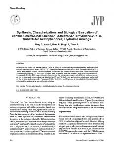
Structure-Based Design, Synthesis, and Biological Evaluation of Irreversible Human Rhinovirus 3C Protease Inhibitors. 6. Structure-Activity Studies of Orally Bioavailable, 2-Pyridone-Containing Peptidomimetics Peter S. Dragovich,* Thomas J. Prins, Ru Zhou, Edward L. Brown, Fausto C. Maldonado, Shella A. Fuhrman, Leora S. Zalman, Tove Tuntland, Caroline A. Lee, Amy K. Patick, David A. Matthews, Thomas F. Hendrickson,† Maha B. Kosa, Bo Liu, Minerva R. Batugo, Jean-Paul R. Gleeson, Sylvie K. Sakata, Lijian Chen, Mark C. Guzman, James W. Meador, III, Rose Ann Ferre, and Stephen T. Worland‡ Pfizer Global Research and Development-La Jolla/Agouron Pharmaceuticals, Inc., 10777 Science Center Drive, San Diego, California 92121-1111 Received October 11, 2001
The structure-based design, chemical synthesis, and biological evaluation of various 2-pyridonecontaining human rhinovirus (HRV) 3C protease (3CP) inhibitors are described. These compounds are comprised of a peptidomimetic binding determinant and a Michael acceptor moiety, which forms an irreversible covalent adduct with the active site cysteine residue of the 3C enzyme. The 2-pyridone-containing inhibitors typically display improved 3CP inhibition properties relative to related peptide-derived molecules along with more favorable antiviral properties. The cocrystal structure of one pyridone-derived 3CP inhibitor complexed with HRV-2 3CP is also described along with certain ab initio conformation analyses. Optimization of the 2-pyridone-containing compounds is shown to provide several highly active 3CP inhibitors (kobs/ [I] > 500 000 M-1 s-1) that function as potent antirhinoviral agents (EC50 ) 100) makes the development of vaccines seem unlikely.4 Previous attempts at antirhinoviral therapeutic identification include the use of interferon,2a,c,e the disruption of virusreceptor (ICAM-1) interactions,5 the examination of capsid-binding antipicornaviral compounds,6 and the use of other miscellaneous agents.7,8 In contrast, the antirhinoviral development efforts at Agouron have focused on disruption of an early, essential step in the HRV replication cycle that involves the proteolytic processing of the large polypeptide produced by cellular translation of the positive strand viral RNA genome. The majority of this processing is effected by the HRV 3C protease (3CP),9,10 a cysteine protease possessing minimal homology with known mammalian enzymes but which structurally resembles members of the trypsin protein family.10,11 Because of its importance in the viral replication cycle and the expected high conservation of its active site residues among all known HRV serotypes,12 it is believed that potent and selective 3CP * To whom correspondence should be addressed. Tel.: (858)6227918. Fax: (858)622-7998. E-mail:
[email protected]. † Present address: Schro ¨ dinger, Inc., 3655 Nobel Drive, Suite 550, San Diego, CA 92122. ‡ Present address: Anadys Pharmaceuticals, 9050 Camino Santa Fe, San Diego, CA 92121.
inhibitors might function as broad-spectrum antirhinoviral therapeutic agents. Indeed, 3CP has been the target of several research programs that sought to identify such antirhinoviral compounds,13 although only one 3CP inhibitor has currently advanced to the stage of human clinical trials (see below). Previously, we described the design and development of substrate-derived9,14 peptidyl and peptidomimetic 3CP inhibitors that incorporate C-terminal Michael acceptor moieties (e.g., compound 1, Figure 1).15-17 These compounds irreversibly inhibit 3CP by forming a covalent adduct with the active site cysteine residue of the enzyme and exhibit broad-spectrum antirhinoviral activity in cell culture assays. Our earlier studies culminated with the selection of one particularly active peptidomimetic compound, ruprintrivir (USAN, AG7088), for clinical development as a nasally delivered antirhinoviral agent.18-20 This molecule recently demonstrated encouraging activity in human clinical trials in which healthy volunteers were deliberately exposed to several HRVs and is currently the subject of large-scale phase II studies to examine its effectiveness against naturally acquired colds.21 Concurrent with these advances, we also wanted to examine orally bioavailable 3CP inhibitors for potential treatment of the common cold and other picornaviral infections.22 Unfortunately, potent 3CP inhibitors with sufficient oral bioavailability to warrant further development were not encountered in the pool of peptide and peptidomimetic compounds that led to ruprintrivir (AG7088).23 Therefore, we continued to seek additional potent, 3CP inhibitors with nonpeptidic chemical structures that were distinct from the molecules that comprised our earlier studies. It was
10.1021/jm010469k CCC: $22.00 © 2002 American Chemical Society Published on Web 03/08/2002
1608
Journal of Medicinal Chemistry, 2002, Vol. 45, No. 8
Figure 1. Design of 2-pyridone-containing HRV 3CP inhibitors.
envisioned that such compounds would aid in the identification of orally bioavailable 3CP inhibitors by further diversifying the physical and biological properties (e.g., mw, mp, water solubility, metabolic stability, etc.) of our existing anti-3CP agents. The discovery and development of one such series of potent, orally bioavailable, 3CP inhibitors are described below.
Inhibitor Design and Initial Structure-Activity Studies Analysis14 of the HRV-2 3CP-1 X-ray crystal structure15 suggested that the P3 amino acid residue contained in the inhibitor 1 could be replaced with a 3-amino-2-pyridone moiety without significantly affecting key protein-ligand interactions (e.g., compound 3, Figure 1). Such replacement seemed plausible since (i) the P3 side chain of 1 did not appreciably contact the 3CP protein, (ii) the P2-P3 amide NH of 1 did not contribute significantly to 3CP recognition and could be
Dragovich et al.
substituted with a ketomethylene24 or an N-methyl moiety1 (e.g., compound 2, Figure 1), (iii) the β-sheet hydrogen-bonding interactions observed between the P3 residue of 1 and the 3CP would be maintained, and (iv) the previous utilization of related 2-pyridones afforded potent inhibitors of other trypsinlike proteases that are structurally similar to 3CP.25,26 The above crystal structure analysis also suggested that in contrast to most previous applications of 2-pyridone-containing enzyme inhibitors, access to the 3CP S2 binding pocket would best be achieved from a position adjacent to the P2 amide carbonyl moiety and not from the pyridone ring itself. In the event, incorporation of an appropriate P3 3-amino-2-pyridone moiety into the inhibitor design afforded a compound (3), which displayed greatly improved anti-3CP and antiviral activity relative to the related peptidyl molecule 1 (Table 1).27 Although the precise nature of these improvements could not be identified with certainty,28 they confirmed the above inhibitor design hypothesis and prompted an extensive structure-activity study of 2-pyridone-containing 3CP inhibitors. Initial 2-pyridone modifications involved variation of the P2 benzyl moiety of 3 through the introduction of functional groups that were guided by previous studies of tripeptidyl 3CP inhibitors.29 Thus, a compound containing a 4-fluorobenzyl P2 substituent (4) displayed anti-3CP and antiviral activities nearly equal to that of 3, while a molecule that incorporated a 3,4-difluorobenzyl moiety (5) exhibited significantly improved inhibition of both the 3C enzyme and the rhinovirus virulence in cell culture (Table 1). The antiviral activity trends of compounds 4 and 5 were also observed with two HRV serotypes other than type 14, suggesting that they might apply to a large number of rhinoviruses (Table 1). Somewhat surprisingly, and in contrast to our earlier studies of tripeptidyl molecules, saturation of the P2 benzyl moiety resulted in a drastic loss of both 3CP inhibition activity and antirhinoviral properties (compound 6, Table 1). The origins of the activity reductions observed with compound 6 have not been rigorously determined but may result from an unfavorable solution conformation of the unbound ligand. Importantly, a 2-pyridone-containing compound that lacked the P2 substituent entirely (7) was an extremely poor 3CP inhibitor and antiviral agent (Table 1). These results suggested that 2-pyridone 3CP inhibitors required some type of P2 moiety for effective 3CP recognition but did not establish a minimum size for such a substituent. Having completed the brief exploration of P2 structure-activity relationships in the 2-pyridone series of 3CP inhibitors described above, we next examined modification of the N-terminal (P4) substituent. Substitution of the Cbz moiety present in 3 with a simple acetyl group resulted in drastic loss of both 3CP inhibitory properties and antirhinoviral activity (compare 8 with 3, Table 1). Both of these activities were regained somewhat by incorporation of a P4 amide derived from cyclopentanecarboxylic acid into the inhibitor design (compound 9, Table 1). Interestingly, introduction of a 1,3-dithiolane-2-carboxylic acid-derived N-terminal amide afforded high levels of 3CP inhibition and antiviral activity when the resulting compound (10) was tested against HRV serotype 14. Unfortunately, the molecule
Human Rhinovirus 3C Protease Inhibitors
Journal of Medicinal Chemistry, 2002, Vol. 45, No. 8 1609
Table 1. 2-Pyridone-Containing 3CP Inhibitors
a Method of preparation: see Schemes 1 and 2. b Elemental analyses (C, H, N) of all compounds agreed to within (0.4% of theoretical values. c Inhibition activity against HRV 3CP; see ref 15 for assay method and error. d Antirhinoviral activity; see ref 15 for assay method and error. e Cytotoxicity; see ref 15 for assay method and error. f 1:1 mixture of diastereomers. ND ) not determined. NA ) not applicable.
1610 Journal of Medicinal Chemistry, 2002, Vol. 45, No. 8
was significantly less potent when examined against two other HRV serotypes in cell culture, suggesting that the improvements observed with type 14 HRV would not be realized against a majority of HRV strains. A variety of other aliphatic N-terminal amides could be incorporated into the 2-pyridone-containing inhibitor series to provide active compounds, although most were not as potent as the Cbz-containing molecule originally studied (compare compounds 11 and 12 with 3, Table 1). However, as was observed in our earlier studies of peptidyl and ketomethylene-containing peptidomimetic 3CP inhibitors, a compound containing an N-terminal amide derived from 5-methylisoxazole-3-carboxylic acid (13) displayed improved anti-3CP and in vitro antirhinoviral properties relative to the corresponding benzyl carbamate 3 (Table 1).30 Interestingly, the magnitude of these improvements was not nearly as great as that noted in our previous work. Again, the source of the activity differences between the current series of 2-pyridone 3CP inhibitors and the related peptide and peptidomimetic molecules is not known with certainty. Utilization of a P4 moiety derived from 5-chloroisoxazole-3-carboxylic acid also afforded a relatively potent 3CP inhibitor and antirhinoviral agent (compound 14, Table 1) suggesting that the 5-chloro and 5-methyl isoxazole substituents could be accommodated nearly equally in the 3CP active site. In addition to examining modification of the P2 and P4 substituents contained within the lead compound 3, we also studied variation of the P1 moiety. Our earlier studies identified an (S)-γ-lactam fragment that imparted high levels of 3CP inhibition activity to peptidyl and peptidomimetic molecules in which it was incorporated.18a Accordingly, this fragment was introduced into the 3-amino-2-pyridone-containing 3CP inhibitor series of the present work. As expected, the resulting compound displayed improved anti-3CP and antiviral properties relative to the glutamine-containing molecule originally studied (compare 15 with 3, Table 1). However, as noted above for N-terminal isoxazole-containing 2-pyridone 3CP inhibitors, the activity improvements realized by P1-lactam incorporation were not as great in the 2-pyridone inhibitor series when compared to those observed earlier during our studies of peptide and peptidomimetic compounds. As anticipated from those earlier studies, introduction of the corresponding P1 (R)γ-lactam into the 2-pyridone-containing inhibitor design resulted in drastic loss of 3CP inhibition activity and antirhinoviral properties when compared with the molecule that contained the (S)-lactam isomer (compound 16, Table 1, compare to 15). X-ray Structure Analysis The iterative analysis of protein-ligand interactions by X-ray crystallography is a critical component of any structure-based inhibitor design program.31 Accordingly, several crystal structures of 3CP-inhibitor complexes were obtained during the development of the 2-pyridone-containing compounds described above. These structures confirmed the binding geometries that the inhibitors adopted when complexed with 3CP and suggested possibilities for compound modification. As in our earlier work, the X-ray data were utilized somewhat cautiously for inhibitor design since the
Dragovich et al.
Figure 2. Crystal structure of 3 complexed with HRV-2 3CP (2.3 Å resolution).
Figure 3. Schematic diagram of 3 bound in the HRV-2 3CP active site. Hydrogen bonds are represented as dashed lines, and the residues that make up the enzyme binding subsites are depicted.
structures depicted the covalent protein-inhibitor products and not the presumably more relevant transition states for adduct formation (see below). The specific details of one particular protein-inhibitor complex are discussed below. The 2.3 Å X-ray crystal structure of the covalent adduct formed between compound 3 and HRV-2 3CP32 is shown in Figure 2, and key protein-inhibitor interactions are illustrated in Figure 3. In general, the inhibitor bound to the enzyme in a manner similar to that observed previously for related tripeptidyl molecules15,29 and filled a series of shallow grooves on the protein surface N-terminal to the scissile amide bond of the substrate (S1-S4).14 As expected, a covalent bond was observed between the 3CP active site cysteine residue (Cys-147) and the β-carbon of the Michael acceptor of 3. As with related peptidyl 3CP inhibitors, a hydrogen bond between the Michael acceptor ester moiety and the 3CP Cys-147 amide NH was present in the above complex, but it is uncertain whether this interaction facilitates addition of the cysteine to 3 or merely arises after the covalent adduct has been formed. Several other important protein-ligand interactions were apparent in addition to those noted in the vicinity of the active site cysteine, which closely paralleled those observed previously with related tripeptidyl 3CP inhibi-
Human Rhinovirus 3C Protease Inhibitors
Journal of Medicinal Chemistry, 2002, Vol. 45, No. 8 1611
tors. For example, the glutamine amide of 3 formed hydrogen bonds with the His-161 side chain and both the Thr-142 side chain and the amide carbonyl in the bottom and on the edge of the 3CP S1 binding pocket, respectively. In addition, the inhibitor phenylalanine residue bound to 3CP in a canyon formed by the side chains of Leu-127, Ser-128, and Asn-130 on one side and His-40 on the other. As expected, the greatest differences between the 3CP binding of the 2-pyridonecontaining inhibitor 3 and the related tripeptidyl molecules were noted in the vicinity of the pyridone ring itself. The conformationally mobile serine residue that forms a hydrogen bond with the P2-P3 amide NH of most peptidyl 3CP inhibitors (Ser-128) was positioned within van der Waals contact distance adjacent to the pyridone ring (Figure 3). As was noted previously during studies of peptidyl 3CP inhibitors, the N-terminal Cbz moiety of 3 was situated in a hydrophobic pocket on the surface of the protein formed by the side chains of Tyr122, Ile-125, and Phe-170. Also as expected, an antiparallel β-sheet hydrogen-bonding interaction was noted between the pyridone moiety and the Gly-164 (Figure 3), which mimicked that crystallographically observed between the 3CP and the P3 amino acid residue of related tripeptidyl 3CP inhibitors. The other backbone amide NH present in 3 formed a hydrogen bond with 3CP Val-162, while an additional hydrogen bond between the inhibitor Cbz carbonyl moiety and the 3CP Ser-128 amide NH was also evident (Figure 3). Thus, a total of eight hydrogen bonds involving both the inhibitor amino acid residues and the 2-pyridone moiety were observed in the complex between compound 3 and HRV-2 3CP. Analysis of the above complex suggested several possibilities for beneficial modification of 3CP inhibitors that contained the 2-pyridone fragment. In particular, the crystal structure indicated that the pyridone-P4 carbamate moiety was significantly twisted out of the plane defined by the pyridone ring. However, molecular mechanics calculations (MM2) conducted with uncomplexed 3-carbamoyl-2-pyridones predicted a coplanar relationship between the carbamate and the heterocyclic ring. Together, these observations suggested that alkyl substitution of the pyridone ring adjacent to the carbamate moiety might minimize the conformational differences between the 3CP-complexed and the uncomplexed structures. This suggestion was supported by ab initio studies conducted at the HF 6-31G* level of theory. The crystal structure analysis also indicated that introduction of such an alkyl moiety should not sterically interfere with the binding of such compounds to the 3CP enzyme. In the event, however, a 3-carbamoyl4-methyl-2-pyridone-containing compound displayed significantly reduced 3CP inhibition activity when compared to a related nonmethylated molecule (compare 17 with 3, Table 2). An additional comparison was made between two amide-containing molecules (compounds 13 and 18, Table 2). Again, the methylated pyridone 18 displayed reduced anti-3CP activity when compared to its nonmethylated analogue 13, although the difference in activity between the two amide-containing compounds was not as dramatic as that noted above for the benzyl carbamates. The failure of the 4-methyl pyridones to improve 3CP inhibition properties is currently
Table 2. 4-Methyl-2-Pyridone-Containing 3CP Inhibitors
a Method of preparation: see Scheme 1. b Elemental analyses (C, H, N) of all compounds agreed to within (0.4% of theoretical values. c Inhibition activity against HRV-14 3CP; see ref 15 for assay method and error.
not well understood but may involve uncertainties associated with modeling efforts conducted with irreversible Michael reaction products rather than the transition states of such transformations. Inhibitor Optimization On the basis of the experimental results described above, additional inhibitor optimization efforts were conducted utilizing 3-amino-2-pyridones that lacked substituents at the pyridone 4-position. Accordingly, various beneficial functional groups identified during the initial phase of structure-activity relationship studies described above (Table 1) were combined to form new 3CP inhibitors. Thus, a 2-pyridone containing a P1 (S)-γ-lactam moiety, a P2 benzyl substituent, and an N-terminal (P4) isoxazole fragment displayed very good levels of 3CP inhibition activity as well as extremely potent antirhinoviral properties in cell culture (compound 19, Table 3). This molecule also exhibited potent antiviral activity against two HRV strains other than type 14. However, the compound was extensively metabolized when incubated with human liver microsomes (Table 3), and additional efforts were therefore made to improve upon the biological stability of the molecule. Analysis of the incubation mixtures of 19 with human liver microsomes identified several metabolites including compounds derived from oxidation of the 4-fluorophenyl aromatic ring and hydrolysis of the carboxylic acid ethyl ester.33 In an attempt to reduce the formation of metabolites resulting from the former process, an additional fluorine atom was introduced at the 3-position of the P2 benzyl substituent present in 19. The resulting difluorinated compound (20) displayed improved anti-3CP and antirhinoviral properties relative to the monofluorinated molecule (Table 3, compare 19 with 20), and this result was consistent with similar improvements noted previously for other “unoptimized” 2-pyridone 3CP inhibitors (see Table 1, compounds 4
1612
Journal of Medicinal Chemistry, 2002, Vol. 45, No. 8
Dragovich et al.
Table 3. Optimized 2-Pyridone-Containing 3CP Inhibitors
compd no.
R
X
prepa
formulab
19
CH2CH3
H
B
C30H32FN5O7‚0.50H2O
20
CH2CH3
F
B
C30H31F2N5O7
21
CH(CH3)2
F
C
C31H33F2N5O7‚0.75H2O
serotype
kobs/[I] (M-1 s-1)c
EC50 (µM)d
CC50 (µM)e
HLM (% met)f
plasma T1/2 (h)g
14 1A 10 14 1A 10 14 1A 10 2 3 9 16 25 39 13 78 11 19 23 Hanks
1 250 000 ND ND 1 800 000 ND ND 548 000 ND ND ND ND ND ND ND ND ND ND ND ND ND ND
0.002 0.015 0.004 0.001 0.006 0.005 0.003 0.132 0.085 0.048 0.033 0.109 0.036 0.100 0.044 0.044 0.012 0.003 0.009 0.010 0.003
>10 >10 >10 >10 >10 >10 >10 >10 >10 >10 >10 >10 >10 >10 >10 >10 >10 >10 >10 >10 >10
95
ND
80
1.4
55
9.7
a Method of preparation: see Schemes 2 and 3. b Elemental analyses (C, H, N) of all compounds agreed to within (0.4% of theoretical values. c Inhibition activity against HRV 3CP; see ref 15 for assay method and error. d Antirhinoviral activity; see ref 15 for assay method and error. e Cytotoxicity; see ref 15 for assay method and error. f Loss of compound (% metabolized) after 30 min exposure to human liver microsomes; see Experimental Section for additional details. g Half-life of compound upon exposure to human plasma; see Experimental Section for additional details.
and 5). In addition, the difluorinated compound 20 displayed improved stability toward human liver microsomes as compared with 19, although the metabolism of 20 was still somewhat facile in the in vitro assay (Table 3). Unfortunately, and somewhat surprisingly based on our earlier studies of peptidyl and peptidomimetic 3CP inhibitors,18a compound 20 exhibited a short half-life when exposed to human plasma (Table 3), and efforts were accordingly focused on improving this important biological stability parameter. We anticipated that the instability of compound 20 toward human plasma was primarily due to enzymemediated hydrolysis of the ethyl ester contained in the molecule. Accordingly, the ethyl ester was replaced with a sterically more demanding isopropyl ester (compound 21). This alteration afforded improved performance in the human liver microsomal stability assay and dramatically improved the half-life of the compound in the presence of human plasma (Table 3). These benefits, however, were realized with a simultaneous worsening of anti-3CP and antiviral potency of 21 relative to the ethyl ester-containing molecule 20. Despite these reductions, compound 21 displayed good absolute levels of antirhinoviral potency when tested against 15 HRV serotypes in cell culture (Table 3). Because this molecule combined good levels of antiviral potency with relatively good biological stability, it was therefore chosen for subsequent oral bioavailability studies.
Oral Bioavailability Studies The R,β-unsaturated esters contained within tripeptidyl molecules related to the 2-pyridones of the present study were previously shown to be quite unstable toward a variety of rodent plasmas (e.g., compound 1, t1/2 ) 10 min in rat plasma).15 This rodent plasma instability precluded the use of rats and/or mice for oral bioavailability testing of the compounds of the present study.34 Accordingly, the dog was selected as an appropriate species in which to conduct the desired analysis. In the event, inhibitor 21 displayed good bioavailability and pharmacokinetic properties after oral administration in the dog (Figure 4, Table 4). Encouragingly, plasma levels of the molecule were observed after initial oral dosing, which substantially exceeded the compound’s previously determined average in vitro EC50 antirhinoviral potency (0.045 µM, 0.028 µg/mL, 15 HRV serotypes), and these levels remained above this average potency for more than 8 h. Because no animal model predictive of rhinovirus-associated human symptomatology has been identified to date, the ultimate significance of the above pharmacokinetic profile cannot be determined with certainty. However, the PK results do demonstrate that 2-pyridone-containing, peptidomimetic 3CP inhibitors can display good oral bioavailability properties in the dog and suggest that additional study of such compounds for use as orally bioavailable HRV 3CP inhibitors is warranted.
Human Rhinovirus 3C Protease Inhibitors
Journal of Medicinal Chemistry, 2002, Vol. 45, No. 8 1613
done ring that was encountered when more direct synthetic methods (e.g., P4-P3-P2 + P1 coupling) were explored.
Figure 4. Pharmacokinetic profile of 21 in dogs after IV and oral administration (n ) 3). Vehicle ) 80:20 propylene glycol: H2O. The average antirhinoviral potency of 21 as determined against 15 HRV serotypes in cell culture is indicated by the arrow. An additional sample was collected 24 h after both IV and PO dosing, but the concentration of 21 was below the limit of detection (5 ng/mL) in each.
Synthesis The 2-pyridone-containing 3CP inhibitors described in this study were prepared by three related synthetic methods (A, B, and C). The particular method employed to synthesize a given compound is indicated in Tables 1-3, and representative examples of each are given below. These syntheses differ primarily in the nature of the P1 fragment utilized to begin each sequence and employ several synthetic transformations and intermediates from previously reported preparations of related tripeptidyl 3CP inhibitors.15,18a The first method (method A) is illustrated in Scheme 1 with the preparation of compound 13 and involves alkylation of a Boc-protected pyridone entity (P3 fragment) with an activated P2-P1 moiety. Thus, commercially available N-R-Cbz-γ-tritylL-glutamine was reduced to primary alcohol 22 by a two step process, and this entity was subsequently converted to the corresponding tert-butyldimethylsilyl ether 23 in good yield. Deprotection of the Cbz moiety present in 23 followed by coupling of the resulting amine (not shown) with commercially available (2R)-2-hydroxy-3phenylpropionic acid afforded intermediate 24 in good yield. The secondary alcohol moiety present in 24 was then converted to the corresponding mesylate (25), and the crude material thus obtained was condensed with the sodium salt of (2-hydroxypyridin-3-yl)carbamic acid tert-butyl ester (26, see Experimental Section) at elevated temperature to afford product 27 as a single diastereomer in moderate yield. Attempts to form the triflate corresponding to 25 utilizing trifluoromethanesulfonic anhydride resulted in the unwanted removal of both the primary silyl ether and the trityl protecting group present in 24. These side reactions were presumably caused by the formation of trifluoromethanesulfonic acid in the reaction medium and occurred even in the presence of a large excess of various amine bases (e.g., 2,6-lutidine).35 The synthesis of intermediate 27 predominantly provided the N-alkylated pyridone product as evidenced by thin-layer chromatography (TLC) analysis of the reaction mixture and 1H nuclear magnetic resonance (NMR) analysis of the pyridone product.36,37 The described preparation of intermediate 27 minimized racemization of the stereocenter adjacent to the pyri-
The silyl protecting group present in 27 was removed, and the resulting alcohol (28) was subjected to an oxidation/olefination sequence to provide intermediate 29 in good overall yield. As was encountered during previous syntheses of peptidyl and peptidomimetic 3CP inhibitors,15,18a the above olefination process afforded the desired trans isomer with 2σ(F)]. The rootmean-square deviations from ideal bond lengths and angles were 0.019 Å and 3.2 deg, respectively. The final model consisted of all atoms for residues 1-180 (excluding side chains of residues 12, 45, 55, and 65) plus 141 water molecules.
Acknowledgment. We are grateful for many helpful discussions throughout the course of this work with Prof. Larry Overman and Dr. Steven Bender. We also thank Dr. Xinjun Hou for assistance with the preparation of Figure 2. References (1) For part 5 in this series, see Dragovich, P. S.; Webber, S. E.; Prins, T. J.; Zhou, R.; Marakovits, J. T.; Tikhe, J. G.; Fuhrman, S. A.; Patick, A. K.; Matthews, D. A.; Ford, C. E.; Brown, E. L.; Binford, S. L.; Meador, J. W., III; Ferre, R. A.; Worland, S. T. Structure-Based Design of Irreversible, Tripeptidyl Human Rhinovirus 3C Protease Inhibitors Containing N-Methyl Amino Acids. Bioorg. Med. Chem. Lett. 1999, 9, 2189-2194. (2) (a) Couch, R. B. Rhinoviruses. In Fields Virology, 3rd ed.; Fields, B. N., Knipe, D. M., Howley, P. M., et al., Eds.; Lippincott-Raven Publishers: Philadelphia, 1996; Vol. 1, Chapter 23, pp 713-734. (b) McKinlay, M. A.; Pevear, D. C.; Rossmann, M. G. Treatment of the Picornavirus Common Cold by Inhibitors of Viral Uncoating and Attachment. Annu. Rev. Microbiol. 1992, 46, 635-654. (c) Phillpotts, R. J.; Tyrrell, D. A. J. Rhinovirus Colds. Br. Med. Bull. 1985, 41, 386-390. (d) Gwaltney, J. M. The Common Cold. In Principles and Practices of Infectious Diseases; Mandell, G. L., Douglas, R. G., Bennett, J. E., Eds.; John Wiley & Sons: New York, 1985; Chapter 38, pp 351-355. (e) Gwaltney, J. M. Rhinoviruses. In Viral Infections of Humans; Evans, A. S., Ed.; Plenum Publishing Corp.: New York, 1982; Chapter 20, pp 491517. (3) (a) Rueckert, R. R. Picornaviridae: The Viruses and Their Replication. In Fields Virology, 3rd ed.; Fields, B. N., Knipe, D. M., Howley, P. M., et al., Eds.; Lippincott-Raven Publishers: Philadelphia, 1996; Vol. 1, Chapter 21, pp 609-654. (b) Kra¨usslich, H.-G.; Wimmer, E. Viral Proteinases. Annu. Rev. Biochem. 1988, 57, 701-754. (4) Hamparian, V. V.; Colonno, R. J.; Cooney, M. K.; Dick, E. C.; Gwaltney, J. M., Jr.; Hughes, J. H.; Jordan, W. S., Jr.; Kapikian, A. Z.; Mogabgab, W. J.; Monto, A.; Phillips, C. A.; Rueckert, R. R.; Schieble, J. H.; Stott, E. J.; Tyrrell, D. A. J. A Collaborative Report: Rhinoviruses-Extension of the Numbering System From 89 to 100. Virology 1987, 159, 191-192. (5) Turner, R. B.; Wecker, M. T.; Pohl, G.; Witek, T. J.; McNally, E.; St. George, R.; Winther, B.; Hayden, F. G. Efficacy of Tremacamra, a Soluble Intercellular Adhesion Molecule 1, for
1622
(6)
(7)
(8) (9)
(10) (11)
(12)
(13)
Journal of Medicinal Chemistry, 2002, Vol. 45, No. 8 Experimental Rhinovirus Infection. JAMA 1999, 281, 17971804 and references therein. See also Bella, J.; Rossmann, M. G. Rhinoviruses and Their ICAM Receptors. J. Struct. Biol. 1999, 128, 69-74. (a) Oren, D. A.; Zhang, A.; Nesvadba, H.; Rosenwirth, B.; Arnold, E. Synthesis and Activity of Piperazine-containing Antirhinoviral Agents and Crystal Structure of SDZ 880-061 Bound to Human Rhinovirus 14. J. Mol. Biol. 1996, 259, 120-134 and references therein. (b) Diana, G. D.; Rudewicz, P.; Pevear, D. C.; Nitz, T. J.; Aldous, S. C.; Aldous, D. J.; Robinson, D. T.; Draper, T.; Dutko, F. J.; Aldi, C.; Gendron, G.; Oglesby, R. C.; Volkots, D. L.; Reuman, M.; Bailey, T. R.; Czerniak, R.; Block, T.; Roland, R.; Oppermann, J. Picornavirus Inhibitors: Trifluoromethyl Substitution Provides a Global Protective Effect Against Hepatic Metabolism. J. Med. Chem. 1995, 38, 1355-1371 and references therein. (c) Rogers, J. M.; Diana, G. D.; McKinlay, M. A. Pleconaril, a Broad Spectrum Antipicornaviral Agent. Antiviral Chem. Chemother. 1999, 5, 69-76. Hamdouchi, C.; Ezquerra, J.; Vega, J. A.; Vaquero, J. J.; AlvarezBuilla, J.; Heinz, B. A. Short Synthesis and Anti-Rhinoviral Activity of Imidazo[1,2-a]Pyridines: The Effect of Acyl Groups at 3-Position. Bioorg. Med. Chem. Lett. 1999, 9, 1391-1394 and references therein. Carrasco, L. Picornavirus Inhibitors. Pharmacol. Ther. 1994, 64, 215-290. (a) Cordingley, M. G.; Callahan, P. L.; Sardana, V. V.; Garsky, V. M.; Colonno, R. J. Substrate Requirements of Human Rhinovirus 3C Protease for Peptide Cleavage in Vitro. J. Biol. Chem. 1990, 265, 9062-9065. (b) Orr, D. C.; Long, A. C.; Kay, J.; Dunn, B. M.; Cameron, J. M. Hydrolysis of a Series of Synthetic Peptide Substrates by the Human Rhinovirus 14 3C Proteinase, Cloned and Expressed in Escherichia coli. J. Gen. Virol. 1989, 70, 29312942. (c) Cordingley, M. G.; Register, R. B.; Callahan, P. L.; Garsky, V. M.; Colonno, R. J. Cleavage of Small Peptides In Vitro by Human Rhinovirus 14 3C Protease Expressed in Escherichia coli. J. Virol. 1989, 63, 5037-5045. Bergmann, E. M.; James, M. N. G. Proteolytic Enzymes of the Viruses of the Family Picornaviridae. Proteases Infect. Agents 1999, 139-163. (a) Matthews, D. A.; Smith, W. W.; Ferre, R. A.; Condon, B.; Budahazi, G.; Sisson, W.; Villafranca, J. E.; Janson, C. A.; McElroy, H. E.; Gribskov, C. L.; Worland, S. Structure of Human Rhinovirus 3C Protease Reveals a Trypsin-like Polypeptide Fold, RNA-Binding Site, and Means for Cleaving Precursor Polyprotein. Cell 1994, 77, 761-771. (b) Allaire, M.; Chernaia, M. M.; Malcolm, B. A.; James, M. N. G. Picornaviral 3C Cysteine Proteinases Have a Fold Similar to Chymotrypsin-like Serine Proteinases. Nature 1994, 369, 72-76. (c) Bazan, J. F.; Fletterick, R. J. Viral Cysteine Proteases are Homologous to the Trypsin-like Family of Serine Proteases: Structural and Functional Implications. Proc. Natl. Acad. Sci. U.S.A. 1988, 85, 78727876. (d) Gorbalenya, A. E.; Blinov, V. M.; Donchenko, A. P. Poliovirus-encoded Proteinase 3C: A Possible Evolutionary Link Between Cellular Serine and Cysteine Proteinase Families. FEBS Lett. 1986, 194, 253-257. (a) Lee, W.-M.; Wang, W.; Rueckert, R. R. Complete Sequence of the RNA Genome of Human Rhinovirus 16, a Clinically Useful Common Cold Virus Belonging to the ICAM-1 Receptor Group. Virus Genes 1994, 9, 177-181. (b) Aschauer, B.; Werner, G.; McCray, J.; Rosenwirth, B.; Bachmayer, H. Biologically, Active Protease 3C of Human Rhinovirus 1A is Expressed from a Cloned cDNA Segmant in Escherichia coli. Virology 1991, 184, 587-594. (c) Hughes, P. J.; North, C.; Jellis, C. H.; Minor, P. D.; Stanway, G. The Nucleotide Sequence of Human Rhinovirus 1B: Molecular Relationships within the Rhinovirus Genus. J. Gen. Virol. 1988, 69, 49-58. (d) Duechler, M.; Skern, T.; Sommergruber, W.; Neubauer, C.; Gruendler, P.; Fogy, I.; Blaas, D.; Kuechler, E. Evolutionary Relationships Within the Human Rhinovirus Genus: Comparison of Serotypes 89, 2, and 14. Proc. Natl. Acad. Sci. U.S.A. 1987, 84, 2605-2609. (e) Werner, G.; Rosenwirth, B.; Bauer, E.; Siefert, J.-M.; Werner, F.-J.; Besemer, J. Molecular Cloning and Sequence Determination of the Genomic Regions Encoding Protease and Genome-Linked Protein of Three Picornaviruses. J. Virol. 1986, 57, 1084-1093. (f) Skern, T.; Sommergruber, W.; Blaas, D.; Gruendler, P.; Fraundorfer, F.; Pieler, C.; Fogy, I.; Kuechler, E. Human Rhinovirus 2: Complete Nucleotide Sequence and Proteolytic Processing Signals in the Capsid Protein Region. Nucleic Acids Res. 1985, 13, 2111-2126. (g) Callahan, P. L.; Mizutani, S.; Colonno, R. J. Molecular Cloning and Complete Sequence Determination of RNA Genome of Human Rhinovirus Type 14. Proc. Natl. Acad. Sci. U.S.A. 1985, 82, 732-736. (h) Stanway, G.; Hughes, P. J.; Mountford, R. C.; Minor, P. D.; Almond, J. W. The Complete Nucleotide Sequence of a Common Cold Virus: Human Rhinovirus 14. Nucleic Acids Res. 1984, 12, 7859-7875. (a) Dragovich, P. S. Recent Advances in the Development of Human Rhinovirus 3C Protease Inhibitors. Exp. Opin. Ther. Pat. 2001, 11, 177-184. (b) Wang, Q. M. Protease Inhibitors as
Dragovich et al.
(14)
(15)
(16)
(17)
(18)
(19)
(20)
(21)
(22)
Potential Antiviral Agents for the Treatment of Picornaviral Infections. Prog. Drug Res. 1999, 52, 197-219. (c) Wang, Q. M. Human Rhinovirus 3C Protease Inhibitors: Recent Developments. Exp. Opin. Ther. Pat. 1998, 8, 1151-1156. The nomenclature used for describing the individual amino acid residues of a peptide substrate (P2, P1, P1′, P2′, etc.) and the corresponding enzyme subsites (S2, S1, S1′, S2′, etc.) is described in Schechter, I.; Berger, A. On the Size of the Active Site in Proteases. I. Papain. Biochem. Biophys. Res. Commun. 1967, 27, 157-162. Dragovich, P. S.; Webber, S. E.; Babine, R. E.; Fuhrman, S. A.; Patick, A. K.; Matthews, D. A.; Lee, C. A.; Reich, S. R.; Prins, T. J.; Marakovits, J. T.; Littlefield, E. S.; Zhou, R.; Tikhe, J.; Ford, C. E.; Wallace, M. B.; Meador, J. W., III.; Ferre, R. A.; Brown, E. L.; Binford, S. L.; Harr, J. E. V.; DeLisle, D. M.; Worland, S. T. Structure-Based Design, Synthesis, and Biological Evaluation of Irreversible Human Rhinovirus 3C Protease Inhibitors. 1. Michael Acceptor Structure-Activity Studies. J. Med. Chem. 1998, 41, 2806-2818. A similar series of HRV 3CP inhibitors was also reported. See Kong, J.-s.; Venkatraman, S.; Furness, K.; Nimkar, S.; Shepherd, T. A.; Wang, Q. M.; Aube´, J.; Hanzlik, R. P. Synthesis and Evaluation of Peptidyl Michael Acceptors That Inactivate Human Rhinovirus 3C Protease and Inhibit Virus Replication. J. Med. Chem. 1998, 41, 2579-2587. For other examples of Michael acceptor-containing cysteine protease inhibitors, see (a) Roush, W. R.; Gwaltney, S. L., II; Cheng, J.; Scheidt, K. A.; McKerrow, J. H.; Hansell, E. Vinyl Sulfonate Esters and Vinyl Sulfonamides: Potent, Irreversible Inhibitors of Cysteine Proteases. J. Am. Chem. Soc. 1998, 120, 10994-10995. (b) McGrath, M. E.; Klaus, J. L.; Barnes, M. G.; Bro¨mme, D. Crystal Structure of Human Cathepsin K Complexed with a Potent Inhibitor. Nat. Struct. Biol. 1997, 4, 105109. (c) Bro¨mme, D.; Klaus, J. L.; Okamoto, K.; Rasnick, D.; Palmer, J. T. Peptidyl Vinyl Sulphones: A New Class of Potent and Selective Cysteine Protease Inhibitors. Biochem. J. 1996, 315, 85-89. (d) Palmer, J. T.; Rasnick, D.; Klaus, J. L.; Bro¨mme, D. Vinyl Sulfones as Mechanism-Based Cysteine Protease Inhibitors. J. Med. Chem. 1995, 38, 3193-3196. (e) Liu, S.; Hanzlik, R. P. Structure-Activity Relationships for Inhibition of Papain by Peptide Michael Acceptors. J. Med. Chem. 1992, 35, 1067-1075. (f) Hanzlik, R. P.; Thompson, S. A. Vinylogous Amino Acid Esters: A New Class of Inactivators for Thiol Proteases. J. Med. Chem. 1984, 27, 711-712. (a) Dragovich, P. S.; Prins, T. J.; Zhou, R.; Webber, S. E.; Marakovits, J. T.; Fuhrman, S. A.; Patick, A. K.; Matthews, D. A.; Lee, C. A.; Ford, C. E.; Burke, B. J.; Rejto, P. A.; Hendrickson, T. F.; Tuntland, T.; Brown, E. L.; Meador, J. W., III; Ferre, R. A.; Harr, J. E. V.; Kosa, M. B.; Worland, S. T. Structure-Based Design, Synthesis, and Biological Evaluation of Irreversible Human Rhinovirus 3C Protease Inhibitors. 4. Incorporation of P1 Lactam Moieties as L-Glutamine Replacements. J. Med. Chem. 1999, 42, 1213-1224. (b) Matthews, D. A.; Dragovich, P. S.; Webber, S. E.; Fuhrman, S. A.; Patick, A. K.; Zalman, L. S.; Hendrickson, T. F.; Love, R. A.; Prins, T. J.; Marakovits, J. T.; Zhou, R.; Tikhe, J.; Ford, C. E.; Meador, J. W.; Ferre, R. A.; Brown, E. L.; Binford, S. L.; Brothers, M. A.; DeLisle, D. M.; Worland, S. T. Structure-assisted Design of Mechanism-based Irreversible Inhibitors of Human Rhinovirus 3C Protease With Potent Antiviral Activity Against Multiple Rhinovirus Serotypes. Proc. Natl. Acad. Sci. U.S.A. 1999, 96, 11000-11007. (a) Patick, A. K.; Binford, S. L.; Brothers, M. A.; Jackson, R. L.; Ford, C. E.; Diem, M. D.; Maldonado, F.; Dragovich, P. S.; Zhou, R.; Prins, T. J.; Fuhrman, S. A.; Meador, J. W.; Zalman, L. S.; Matthews, D. A.; Worland, S. T. In Vitro Antiviral Activity of AG7088, a Potent Inhibitor of Human Rhinovirus 3C Protease. Antimicrob. Agents Chemother. 1999, 43, 2444-2450. (b) Kaiser, L.; Crump, C. E.; Hayden, F. G. In vitro Activity of Pleconaril and AG7088 Against Selected Serotypes and Clinical Isolates of Human Rhinoviruses. Antiviral Res. 2000, 47, 215-220. Note that the anti-3CP and antirhinoviral activities of ruprintrivir (AG7088) listed in ref 19a differ slightly from those originally disclosed in ref 18a. These differences result from additional biological assays performed with the compound after the publication of ref 18a. The values listed in ref 19a (and in the present work) are considered to be more accurate due to the larger number of experimental determinations that comprise them. Schmidt, A. C.; Couch, R. B.; Galasso, G. J.; Hayden, F. G.; Mills, J.; Murphy, B. R.; Chanock, R. M. Current Research on Respiratory Viral Infections: Third International Symposium. Antiviral Res. 2001, 50, 157-196. Ruprintrivir (AG7088) has exhibited antiviral activity against several nonrhinoviral picornaviruses.19a Treatment of many such picornaviral-mediated diseases would presumably be best ef-
Human Rhinovirus 3C Protease Inhibitors
(23)
(24)
(25)
(26)
(27) (28)
(29)
(30)
(31)
fected with an orally bioavailable therapeutic agent. For additional details, see Rotbart, H. A. Antiviral Therapy for Enteroviruses and Rhinoviruses. Antiviral Chem. Chemother. 2000, 11, 261-271. A detailed account of these studies will be disclosed in the future. The oral bioavailability of ruprintrivir (AG7088) in the dog was determined to be 8% (Tuntland, T.; Lee, C. A. Unpublished results). Dragovich, P. S.; Prins, T. J.; Zhou, R.; Fuhrman, S. A.; Patick, A. K.; Matthews, D. A.; Ford, C. E.; Meador, J. W., III; Ferre, R. A.; Worland, S. T. Structure-Based Design, Synthesis, and Biological Evaluation of Irreversible Human Rhinovirus 3C Protease Inhibitors. 3. Structure-Activity Studies of Ketomethylene-Containing Peptidomimetics. J. Med. Chem. 1999, 42, 1203-1212. (a) Sanderson, P. E. J.; Dyer, D.; Naylor-Olsen, A. M.; Vacca, J. P.; Gardell, S. J.; Lewis, S. D.; Lucas, B. J., Jr.; Lyle, E. A.; Lynch, J. J., Jr.; Mulichak, A. M. L-373,890, An Achiral, Noncovalent, Subnanomolar Thrombin Inhibitor. Bioorg. Med. Chem. Lett. 1997, 7, 1497-1500. (b) Tamura, S. Y.; Semple, J. E.; Reiner, J. E.; Goldman, E. A.; Brunck, T. K.; Lim-Wilby, M. S.; Carpenter, S. H.; Rote, W. E.; Oldeshulte, G. L.; Richard, B. M.; Nutt, R. F.; Ripka, W. C. Design and Synthesis of a Novel Class of Thrombin Inhibitors Incorporating Heterocyclic Dipeptide Surrogates. Bioorg. Med. Chem. Lett. 1997, 7, 1543-1548. (c) Warner, P.; Green, R. C.; Gomes, B.; Strimpler, A. M. NonPeptidic Inhibitors of Human Leukocyte Elastase. 1. The Design and Synthesis of Pyridone-Containing Inhibitors. J. Med. Chem. 1994, 37, 3090-3099. For other examples of 2-pyridone-containing enzyme inhibitors, see (a) Golec, J. M. C.; Mullican, M. D.; Murcko, M, A.; Wilson, K. P.; Kay, D. P.; Jones, S. D.; Murdoch, R.; Bemis, G. W.; Raybuck, S. A.; Luong, Y.-P.; Livingston, D. J. Structure-Based Design of Non-Peptidic Pyridone Aldehydes as Inhibitors of Interleukin-1β Converting Enzyme. Bioorg. Med. Chem. Lett. 1997, 7, 2181-2186. (b) Semple, G.; Ashworth, D. M.; Baker, G.; Batt, A. R.; Baxter, A. J.; Benzies, D. W. M.; Elliot, L. H.; Evans, D. M.; Franklin, R. J.; Hudson, P.; Jenkins, P. D.; Pitt, G. R.; Rooker, D. P.; Sheppard, A.; Szelke, M.; Yamamoto, S.; Isomura, Y. Pyridone-Based Peptidomimetic Inhibitors of Interleukin-1β-Converting Enzyme (ICE). Bioorg. Med. Chem. Lett. 1997, 7, 1337-1342. In the subsequent discussion of structure-activity relationships, the notation “3CP” indicates 3C protease derived from HRV-14 unless otherwise specified. The improved 3CP inhibition activity exhibited by 3 relative to inhibitor 1 may result from reduced entropy costs encountered upon binding of the former to the 3CP active site. The improved antiviral properties of 3 relative to 1 presumably are derived from both the former’s improved 3CP inhibition activity and its altered cell membrane permeability properties (3 contains one less amide NH than 1). For a discussion of the relationship between membrane permeability and the number of amide NHs contained within peptidyl molecules, see (a) Conradi, R. A.; Hilgers, A. R.; Ho, N. F. H.; Burton, P. S. The Influence of Peptide Structure on Transport Across Caco-2 Cells. Pharmacol. Res. 1991, 8, 1453-1460. (b) Conradi, R. A.; Hilgers, A. R.; Ho, N. F. H.; Burton, P. S. The Influence of Peptide Structure on Transport Across Caco-2 Cells II. Peptide Bond Modification Which Results in Improved Permeability. Pharmacol. Res. 1992, 9, 435-439. Dragovich, P. S.; Webber, S. E.; Babine, R. E.; Fuhrman, S. A.; Patick, A. K.; Matthews, D. A.; Reich, S. H.; Marakovits, J. T.; Prins, T. J.; Zhou, R.; Tikhe, J.; Littlefield, E. S.; Bleckman, T. M.; Wallace, M. B.; Little, T. L.; Ford, C. E.; Meador, J. W., III; Ferre, R. A.; Brown, E. L.; Binford, S. L.; DeLisle, D. M.; Worland, S. T. Structure-Based Design, Synthesis, and Biological Evaluation of Irreversible Human Rhinovirus 3C Protease Inhibitors. 2. Peptide Structure-Activity Studies. J. Med. Chem. 1998, 41, 2819-2834. Dragovich, P. S.; Zhou, R.; Skalitzky, D. J.; Fuhrman, S. A.; Patick, A. K.; Ford, C. E.; Meador, J. W., III; Worland, S. T. SolidPhase Synthesis of Irreversible Human Rhinovirus 3C Protease Inhibitors. 1. Optimization of Tripeptides Incorporating NTerminal Amides. Bioorg. Med. Chem. 1999, 7, 589-598. (a) Murcko, M. A.; Caron, P. R.; Charifson, P. S. Structure Based Drug Design. Annu. Rep. Med. Chem. 1999, 34, 297-306. (b) Babine, R. E.; Bender, S. L. Molecular Recognition of ProteinLigand Complexes: Applications to Drug Design. Chem. Rev. 1997, 97, 1359-1472. (c) Bohacek, R. S.; McMartin, C.; Guida, W. C. The Art and Practice of Structure-Based Drug Design: A Molecular Modeling Perspective. Med. Res. Rev. 1996, 16, 3-50. (d) Greer, J.; Erickson, J. W.; Baldwin, J. J.; Varney, M. D. Application of the Three-Dimensional Structures of Protein Target Molecules in Structure-Based Drug Design. J. Med. Chem. 1994, 37, 1035-1054.
Journal of Medicinal Chemistry, 2002, Vol. 45, No. 8 1623 (32) In the discussion of the X-ray crystal structure, the notation “3CP” indicates 3C protease derived from HRV-2. This protein provided diffraction quality crystals more routinely than the serotype 14-derived protease. The inhibitor-binding regions of the two enzymes do not differ significantly. (33) A full account of these metabolism studies will be presented elsewhere (Lee, C. A. Manuscript in preparation). (34) Several representative 2-pyridone-contating 3CP inhibitors that incorporated various R,β-unsaturated esters were also shown to be unstable toward rodent plasma. Full details of these studies will be reported elsewhere (Tuntland, T. Manuscript in preparation). (35) Alternate methods for triflate formation using reagents other than trifluoromethanesulfonic anhydride were not examined. (36) No rigorous effort was made to detect and/or quantitate the possible O-alkylation reaction product using more stringent analytical methods. (37) For general references on the alkylation of 2-hydroxypyridines, see (a) Scriven, E. F. V. In Comprehensive Heterocyclic Chemistry; Katritzky, A. R., Rees, C. W., Eds.; Pergamon Press: Oxford, 1984; Vol. 2, pp 165-314. (b) Tieckelmann, H. In Pyridine and its Derivatives; Abramovitch, R. A., Ed.; WileyInterscience: New York, 1974; Vol. 14, Suppl. 3, Chapter 12, pp 597-1180. (38) Warner, P.; Green, R. C.; Gomes, B.; Strimpler, A. M. Nonpeptidic Inhibitors of Human Leukocyte Elastase. 1. The Design and Synthesis of Pyridone-Containing Inhibitors. J. Med. Chem. 1994, 37, 3090-3099. (39) The chloroisoxazole required for the synthesis of compound 14 was prepared by a method analogous to that described in Stevens, R. V.; Albizati, K. F. Synthesis and Nucleophilic Substitutions of 3-Alkyl-5-chloroisoxazoles. Tetrahedron Lett. 1984, 25, 4587-4590. (40) Cahiez, G.; Metais, E. Enantioselective Preparation of R-Acyloxy Ketones from R-Hydroxy and R-Amino Acids. Tetrahedron Lett. 1995, 36, 6449-6452 and references therein. (41) Mori, S.; Iwakura, H.; Takechi, S. A New Amidoalkynylation Using Alkynlyzinc Reagent. Tetrahedron Lett. 1988, 29, 53915394 and references therein. (42) Overman, L. E.; Osawa, T. A Convenient Synthesis of 4-Unsubstituted β-Lactams. J. Am. Chem. Soc. 1985, 107, 1698-1701 and references therein. (43) (a) Tian, Q.; Nayyar, N. K.; Babu, S.; Chen, L.; Tao, J.; Lee, S.; Tibbetts, A.; Moran, T.; Liou, J.; Guo, M.; Kennedy, T. An Efficient Synthesis of a Key Intermediate for the Preparation of the Rhinovirus Protease Inhibitor AG7088 via Asymmetric Dianionic Cyanomethylation of N-Boc-L-(+)-Glutamic Acid Dimethyl Ester. Tetrahedron Lett. 2001, 42, 6807-6809. (b) Tian, Q.; Nayyar, N. K.; Babu, S.; Tao, J.; Moran, T. J.; Dagnino, R., Jr.; Remarchuk, T. P.; Melnick, M. J.; Mitchell, L. J., Jr.; Bender, S. L. 2001 WO 01/14329. (44) Okuma, K.; Tachibana, Y.; Sakata, J.-i.; Komiya, T.; Kaneko, I.; Komiya, Y.; Yamasaki, Y.; Yamamoto, S.-i.; Ohta, H. Reactions of Wittig Reagents With Episulfides or Elemental Sulfur. Bull. Chem. Soc. Jpn. 1988, 61, 4323-4327 and references therein. (45) Dess, D. B.; Martin, J. C. A Useful 12-I-5 Triacetoxyperiodinane (the Dess-Martin Periodinane) for the Selective Oxidation of Primary or Secondary Alcohols and a Variety of Related 12-I-5 Species. J. Am. Chem. Soc. 1991, 113, 7277-7287. (46) Otwinowski, Z. DENZO in Data Collection and Processing. In Proceedings of the CCP4 Study Weekend; Sawyer, L., Isaacs, N., Baily, S., Eds.; SERC Daresbury Laboratory: Warrington, U.K., 1993; pp 55-62. (47) Bru¨nger, A. T. XPLOR, version 3.1. XPLOR v3.1 Manual; Yale University Press: New Haven, CT, 1992. (48) Webber, S. E.; Okano, K.; Little, T. L.; Reich, S. H.; Xin, Y.; Fuhrman, S. A.; Matthews, D. A.; Love, R. A.; Hendrickson, T. F.; Patick, A. K.; Meador, J. W., III; Ferre, R. A.; Brown, E. L.; Ford, C. E.; Binford, S. L.; Worland, S. T. Tripeptide Aldehyde Inhibitors of Human Rhinovirus 3C Protease: Design, Synthesis, Biological Evaluation, and Cocrystal Structure Solution of P1 Glutamine Isosteric Replacements. J. Med. Chem. 1998, 41, 2786-2805. (49) Andries, K.; Dewindt, B.; Snoeks, J.; Willebrords, R.; Van Eemeren, K.; Stokbroekx, R.; Janssen, P. A. J. In Vitro Activity of Pirodavir (R 77975), a Substituted Phenoxy-Pyridazinamine with Broad-Spectrum Antipicornaviral Activity. Antimicrob. Agents Chemother. 1992, 36, 100-107.
JM010469K