from a homologous domain of Drosophila a actinin did not bind ... with calmodulin binding to vertebrate spectrin (4, 17) which suggests the ... Alpha Spectrin. 10.
Vol. 266, No. 11, Issue of April 15, pp. 7189-7193,1991 Printed in U.S.A.
THEJOURNAL OF BIOLOGICAL CHEMISTRY 0 1991 by The American Society for Biochemistry and Molecular Biology, Inc.
Structure, Calmodulin-binding, and Calcium-binding Properties of Recombinant CY Spectrin Polypeptides* (Received for publication, August 9, 1990)
Ronald R.DubreuilS, Eric Brandin, Jenny H. Sun Reisberg, Lawrence S. B. Goldstein, and Daniel Branton From the Departmentof Cellular and Developmental Biology,Haruard University, Cambridge, Massachusetts 02138
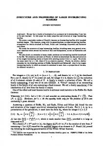
W e examined the structure and the distribution of binding activities within bacterially produced fragments of Drosophila a spectrin. By electron microscopy, purified spectrin fragments resembled the corresponding regions of native spectrin. The contour lengths of recombinant spectrin molecules were proportional to the length of their coding sequences, which is consistent with current models of spectrin structure in which individual segments of the polypeptide contribute independently to the structure of the native molecule. We localized two sitesat which calcium may regulate spectrin function. First, a site responsible for calmodulinbinding to Drosophila a spectrin was identified near the junction of repetitive segments 14 and 15. Second, a domain of Drosophila CY spectrin that includes two EF hand calcium-binding sequences bound 46Cain blot overlay assays. EF hand sequences from a homologous domain of Drosophila a actinin did not bind calcium under the same conditions.
nonmuscle a actinin inhibitsthe interaction of a actinin with actin (10, 11). Calcium also inhibits the interaction of some isoforms of spectrin with actin (12, 13). EF hand sequences are found in the carboxyl-terminal segment of a spectrins, but calcium binding to this region of spectrin has not been examined. A second effect of calcium on the properties of vertebrate brain spectrinoccurs through the calcium-dependent binding of calmodulin to the a chain (14-16). Drosophila a spectrin bindscalmodulin, but lacks the sequence associated with calmodulin binding to vertebrate spectrin (4, 17) which suggests the presence of an alternate calmodulin binding site that may be unique to Drosophila. Here, we use selected regions of spectrin produced from Drosophila spectrin cDNA in bacteria to investigate these aspects of the structure and function of the a subunit. Our results show that various regions of the molecule exhibit structural autonomy thus enabling fragments of the molecule to fold properly when expressed in bacteria. Our results also localize calmodulin binding and calcium binding sites within specific segments of Drosophila a spectrin.
Predictions based on cDNA sequence analysis, as well as biochemical studies using purified proteins, have attributed several properties of the spectrin tetramer, such as its elongated shape, its calmodulin-binding activity, and calcium regulation of the spectrin-actin interaction,to specific regions of the a chain (1-3). Sequence analysis shows that thea chain is composed of 20 repetitive segments and two nonrepetitive segments that are conserved between vertebrates and invertebrates (4-7). The repetitive sequence unit is thought to form a repetitive structural unit, largely a-helical, that independently contributes to the length of the polypeptide and may thus account for the elongated shape of the molecule (8). Indeed, the related protein a actinin is also an elongated molecule, but it consists of a smaller number of these repetitive segments and is substantially shorter than spectrin (9). However, an independent contribution of individual segments to contour length has not been demonstrated experimentally. Sequence comparisons between a spectrin and a actinin suggest a mechanism bywhich calcium may regulate the interaction of spectrin with actin. Calcium binding to theEF hand sequences located in the carboxyl-terminal segment of
MATERIALS AND METHODS’
* This work was supported by Grants GM39686 and HL17411 and an American Cancer Society postdoctoral fellowship (to R. R. D.). The costs of publication of this article were defrayed in part by the payment of page charges. This articlemust therefore be hereby marked “advertisement” in accordance with 18 U.S.C. Section 1734 solely to indicate this fact. $ To whom correspondence should be addressed Dept. of Cellular and Developmental Biology, Harvard University, 16 Divinity Ave., Cambridge, MA 02138. Tel.: 617-495-2646; Fax: 617-495-9300.
RESULTS
Purified recombinant spectrin fragments (shown schematically in Fig. 4) were examined by electron microscopy to assess the contribution of selected domains to the structure of the native molecule. By comparing the contour length of proteins 9AB, 9A, and 10 to the length of purified a spectrin (Table I), we found that the fractional length of these molecules correlated with the fraction of full length coding sequence used to generate them. Purified protein 9AB exhibited calcium-dependent binding to calmodulin-Sepharose (not shown) and derivatives of protein 9AB also bound calmodulin in nitrocellulose blot assays (Fig. 5). Differential binding was observed with a series of nested deletions that had been progressively shortened from their carboxyl terminus (see Fig. 4). Protein 9A, deletion A34, and deletion A44 bound calmodulin, whereas deletion of the sequence between A44 and A48 resulted in a loss of calmodulin-binding activity and clones A48, A52, A50, and A55 did not bind calmodulin (not shown). DNA sequence analysis of these constructs showed that clone A48 ends at amino acid residue 1392 of the a spectrin sequence and A44 ends at residue 1513. Thus, the sequence of amino acids between Portions of this paper (including “Materials and Methods,” and Figs. 1-3) are presented in miniprint atthe end of this paper. Miniprint is easily read with the aid of a standard magnifying glass. Full size photocopies are included in the microfilm edition of the Journal thatis available from Waverly Press.
7189
7190
Recombinant Alpha Spectrin
10
9AB
c I LC
55
(Y
Spectrin Polypeptides u.
8 1AA 205110 97.4
Modllcdlonr d cDNA PA
--
68-
45-
w c. ., 29-
FIG. 4. Top, the arrangement of repetitive (ellipses) and nonrepetitive segments (rectangles) within Drosophila a spectrin. Segment numbers are indicated at the top of the diagram. Bottom, the boundaries of cDNAclones and modified constructs; restriction sites include ClaI (C),StuI (S), and EcoRI ( R ) . cDNA clone 10 begins within segment 2 of a spectrin (amino acid residue 199) and terminates within segment 15 (residue 1512). cDNA clone 9AB begins within segment 10 (residue 1010) and continues through the authentic stop codon. cDNA clone 9A is identical to 9AB except that it terminates at the internal EcoRI site. Clones A22 through A55 represent nested exonuclease digestions of clone 9A. Clone A22(-) differs from clone A22by excision of a 384-bp StuI fragment, without affecting the reading frame of the downstream sequence. Cla6-1F is a 687-bp ClaI fragment from A22 that was subcloned into the ClaI site of pBS (see “Materials and Methods” for details). Clone 9B represents the DNA sequence downstream of the EcoRI site (referred to as “SM” in Ref. 4). Clones 10 and 9B are inserts in the pGEX2 vector, all others were cloned in pBS.
TABLE I Length measurements of recombinant spectrin molecules visualized by electron microscopy Samples of the electron microscopic observations are shown in Fig. 2 of the accomDanvinc!minimint section.
1 2 3 14 2 3 14 2 3 14 2 3 4
A B C D FIG. 5. Identification of a calmodulin binding domain in protein 9AB. Lysates of IPTG-induced bacteria harboring spectrin constructs (as shown in Fig. 4) were solubilized and separated on an SDS gel. A, proteins A22, A22(-), and Cla6-1F were the major products detected in bacterial lysates stained with Coomassie Blue. Identical gels were transferred to nitrocellulose and reacted with a spectrin antibody (B) or with biotinylated calmodulin in the presence of 1 mM CaCI2 (C). The two probes were detected with alkaline phosphatase-conjugated anti-rabbit antibody or alkaline phosphatase-conjugated streptavidin, respectively. In the presence of 5 mM EGTA ( D ) , none of the spectrin fusion proteins bound calmodulin, and the staining pattern at the dye front in C was unchanged if calmodulin was left out of the reaction (not shown). The Cla6-IF coding sequence was cloned as a tandem dimer and the protein product (55 kDa) is approximately twice the size predicted from the single cDNA sequence (26 kDa). Mobility standards are shown in kDa.
failed to bind calmodulin (lane 3). Attempts to express this 124-residue peptidewere unsuccessful. However, an overlapProtein Length % lengthb % length‘ ping 229-residue peptide was expressed and bound calmodulin (expected) (measured) (C6-1F, lane 1 ) . Thus a polypeptide includingthe above 124nrn residue sequence was both necessary for and sufficient for 9A 58 f 4 (n = 44) 63 1280 53 calmodulin-binding activity. 9AB 58 65 6 0 f 6 ( n = 86) 1406 54 10 60-e 11 ( n = 65) 65 1313 Each of the EF hand sequences in segment 22 ofDrosophila 100 100 ad 91 2 2 2 ( n = 6 5 ) 2415 a spectrin has 14 of the consensus residues that are charac’The number of residues in each protein is based on the sequence teristic of calcium-binding proteins (4, 19). To test whether predicted from its corresponding cDNA. or not these EF hand sequences actually bind calcium, a % length (expected) = (number of amino acids in expressed spectrin fragments were reacted with 45CaC12.The termini of protein/number of amino acids in a spectrin coding sequence) X 100. the recombinant fragments relative to the EF hand sequences % length (measured) = (length of recombinant proteins measured are shown in Fig. Protein 3. 9AB bound calcium in blot overlay by electron microscopy/length of native a spectrin measured by electron microscopy) X 100. Each of the length measurements were experiments but protein 9A, which terminates near the end corrected by a factor of -2 nm to compensate for the overestimation of EF hand 1, did not (not shown). For unknown reasons, of length due to platinum shadowing (27). neither purified solubleprotein bound detectable amounts of From Ref. 17. calcium in equilibrium dialysis experiments. The carboxylterminal EcoRI fragment of clone 9AB (127 amino acid resiresidues 1392 and 1513appears to be necessary for calmodulindues; clone 9B in Fig. 4) was subcloned in the pGEX vector binding. Protein 10 also binds calmodulin in blot overlays and expressed as a fusion withglutathione transferase. While (18)and because it extends through amino acid residue 1512, glutathione transferase alone did not bind calcium in blot it further implicates the sequence between residues 1392 and overlays (Fig.6, lane I ) the fusion protein, which includes EF hand 2 of (Y spectrin, did bind calcium(lane 2). 1512 in calmodulin binding. The calcium insensitivity of the muscle a actinin interacWhile clone A22 showed calmodulin-bindingactivity (Fig. 6, lune 2) clonc h22( ), which lackcd rcsiduce 1416 1630, t.inn wit.h F-mt.in is assumed to he due to a lack of calcium binding to EF hand sequences (20). The formal possibility that muscle a actinins bind calcium but fail to respond to ‘The abbreviations usedare: pGEX, Glutagene plasmid; SDS, sodium dodecyl sulfate; PAGE, polyacrylamide gel electrophoresis; calcium binding hadnot been tested. We expressed an EcoRI IPTG, isopropyl-1-thio-@-D-galactopyranoside;EGTA, [ethylene- fragment of Drosophila muscle a actinin (21) as a fusion with glutathione transferase and, although the resulting construct bis(oxyethylenenitrilo)]tetraaceticacid; pBS, Bluescript plasmid.
Recombinant Polypeptides a Spectrin included both of the a actinin EF hand sequences, it failed to bind calcium (Fig. 6,lane 3). DISCUSSION
The amino acid sequence of both the a and p subunits of spectrin are largely composed of 106-residue repetitive segments (ellipses, Fig. 7) that are thought to incrementally contribute to the elongated shape of the molecule (8). If the segmented sequence of spectrin is indicativeof a truly modular structure, then recombinant spectrin fusion proteins encoded by parts of the spectrin sequence should express the corresponding parts of its structure. Our results confirm this prediction and also indicate that local interactions within spectrin fragments are sufficient to direct proper folding of the polypeptide. Our results disagree with an alternate model (22) in which a spectrin is predicted to be a continuous a-helix that folds back upon itself twice. According to the latter model, the partial sequences expressed in the present study should be approximatelythe same length as native a spectrin. The recombinant spectrin fragments (proteins 9AB and 10)
--=i "
SPEC '
ACT' GST"
s
1
2
C. Blue
3
1
2
3
%a
FIG. 6. Detection of a calcium binding site within (Ispectrin. Left, Coomassie Blue-stained gel of IPTG-induced bacterial lysates expressing pGEX with no cDNA insert (GST,lane 1 ), pCEX with a spectrin 9B insert that includes EF hand 2 (SPEC, lane 2), pCEX with Drosophila muscle a actinin EF hand sequences (ACT, lane 3). Right, identical gel lanes were transferred to nitrocellulose, reacted with '"a, and exposed to x-ray film for 6 h. Solid arrows mark the positions of the recombinant protein products in their respective lanes. An open arrow marks the position of bacterial calcium binding protein that varies in intensityaccording to theamount of background bacterial protein. Mobility standards are shown to the left (lane S ) in kDa.
7191
span -13 segments of the a spectrin sequence. Because each of these fragments has a length of -60 nm, it appears that the repetitive segments of spectrin contribute independently to the length of the polypeptide (4-5 nm/segment). An important implication is that spectrin constructs with alterations in segment number or composition are likely to fold properly as long as the minimal folding units are intact. In future studies it maybe possible to address the structural contribution of individual segments, repetitive or nonrepetitive, by assembling and expressing multimers of the coding sequence froma single segment of interest. Drosophila a spectrin binds calmodulin at a site near the junction of repetitive segments 14 and 15. While there is no diagnostic consensus sequenceof calmodulin-binding protein domains (23), most of them, including the vertebrate brain spectrin peptide (15, 16), appear to be amphipathic a helices with a high proportion of basic residues.A 22-residue sequence within the calmodulin binding peptide of Drosophila spectrin appears to meet these criteria (Fig. 7). It is predictedto be ahelical (4), 10 of the 22 residues are basic, and a helical projection of the sequence reveals segregation of charged and apolar residues alongthe helix (not shown). The segments that make upthe ends of the spectrin tetramer (Fig.7, shaped segments) are homologousto thesegments at the ends of the a-actininhomodimer (24). Calcium binding to the EFhands of nonmuscle a-actinin diminishes its actinbinding activity (10, ll),and calcium binding diminishes the actin-binding activity of some spectrins (12, 13), but not others (14). The demonstration that Drosophila spectrin EF hands bind calcium supports the interesting possibility that calcium may regulate the spectrin-actin interaction in the same way that itregulates the a-actinininteraction. Are the repetitive segments of spectrin simplyuniform spacers that separate the functional sites within the molecule, or do they have more specialized roles?The identification of . a unique calmodulin bindingsite within one of the repetitive segments and the pattern of sequence conservation among repetitive segments are both consistent with specialized roles. When a spectrins from various sources are compared it is found that thedifferences betweenrepetitive segments (nonconsensusresidues) are conserved as often as their similarities (consensusresidues, (4)). Future genetic studies in Drosophila shouldprovevaluable in assessing the contributions (and interchangeability) of individual repetitive segments to the
Spectrin Tetramer Actirkbinding C
I"uiin
vertebates
KRKQVLERWRHLKEGLIEKRSRin
binding
1473 FIG.7. A , schematic diagramof the spectrin tetramer. The a and /3 subunits of spectrin consist of repetitive segments (ellipses) and nonrepetitive segments (rectangles, see text for details). The carboxyl-terminal segment of a spectrin includes EF hand sequences that areresDonsible for calcil~mhinrline. Tho nppncnd am;nn-torm;nal domain of the subunit is thought to contain the actin binding site (25,26) anda similar juxtaposition of domains is thought to form the actin binding site in the related protein a-actinin (24). The calmodulin binding site of vertebrate nonerythroid a spectrin resides within a nonrepetitive sequence insert between segments 11 and 12 (arrowhead)that is absentfrom Drosophila a spectrin. The calmodulin binding site of Drosophila a spectrin occurs near the boundary of repetitive segments 14 and 15. B, a candidate 22-residue sequence for the calmodulin binding site begins at residue 1473 within segment 15 (4).
Recombinant
7192
a!
Spectrin Polypeptides
function of the spectrin molecule. Acknowledgment-We actinin genomic DNA.
thank Dr. Eric Fyrberg for providing aREFERENCES
1. Bennett, V. (1985) Annu. Reu. Biochern. 54, 273-304 2. Coleman, T. R., Fishkind, D. J., Mooseker, M. S., and Morrow, J. S. (1989) Cell Motil. Cytoskeleton 12, 225-247 3. Morrow, J. S. (1989) Curr. Opinion Cell Biol. 1 , 23-29 4. Dubreuil, R. R., Byers, T. J., Sillman, A. L., Bar-Zvi, D., Goldstein, L. S. B., and Branton,D. (1989) J. Cell Biol. 1 0 9 , 21972205 5. Moon, R. T., and McMahon, A. P. (1990) J. Biol. Chem. 265, 4427-4433 6. Sahr, K. E., Laurila, P., Kotula, L., Scarpa, A. L., Coupal, E., Leto, T. L., Linnenbach, A. J., Winkelman, J. C., Speicher, D. W., Marchesi, V. T., Curtis, P. J., and Forget, B. G. (1990) J. Bid. Chem. 2 6 5 , 4434-4443 7. Wasenius, V.-M., Saraste, M., SalvBn, P., Eramaa, M., Holm, L., and Lehto, V.-P. (1989) J. Cell Biol. 108, 79-93 8. Speicher, D. W., and Marchesi, V. T. (1984) Nature 3 1 1 , 177180 9. Blanchard, A., Ohanian, V., and Critchley, D. (1989) J. Muscle Res. Cell Motil. 1 0 , 280-289 10. Bennett, J. P., Zaner, K. S., and Stossel, T. P. (1984) Biochemistry 23,5081-5086 11. Noegel, A., Witke, W., and Schleicher, M. (1987) FEBS Lett. 221,391-396 12. Fowler, V., and Taylor, D. L. (1980) J. Cell Biol. 85,361-376 13. Fishkind, D. J., Bonder, E. M., and Begg, D. A. (1987) Cell Motil. Cytoskeleton 7,304-314 14. Harris, A. S., and Morrow, J. S. (1990) Proc. Natl. Aead. Sci. U. S. A. 87,3009-3013 15. Harris, A. S., Croall, D. E., and Morrow, J. S. (1988) J . Bid. Chem. 263,15754-15761
16. Leto, T. L., Pleasic, S., Forget, B.G., Benz, E. J., Jr., and Marchesi. V. T. (1989) J. Bid. Chem. 264. 5826-5830 17. Dubreuil, R . , Byers, T. J., Branton, D., Goldstein, L. S. B., and Kiehart, D. P. (1987) J. Cell Biol. 105,2095-2102 18. Byers, T. J., Dubreuil, R. R., Branton, D., Kiehart, D. P., and -Goldstein, L. S. B. (1987) J. Cell Biol. 1 0 5 , 2103-2110 19. Kretsinper, R. H. (1980) Ann. N. Y. Acad. Sci. 356, 14-19 20. Baron, M..D., Davison, M. D., Jones, P., and Critchley, D. R. (1987) J. Biol. Chem. 262,17623-17629 21. Fyrberg, E., Kelly, M., Ball, E., Fyrberg, C., and Reedy, M. C. (1990) J. Cell Biol. 1 1 0 , 1999-2011 22. Widada, J. S., Asselin, J., Colote, S., Ferraz, C., Trave, G., Afshar, M., Haiech, J., and Liautard, J. P. (1990) Biochirnie (Paris) 72, 19-24 23. O'Neil, K. T., and DeGrado, W. F. (1990) Trends Biochern. Sci. 15,59-64 24. Dubreuil, R. R. (1991) BioEssays, in press 25. Karinch, A. M., Zimmer, W. E., and Goodman, S. R. (1990) J. Biol. Chem. 265,11833-11840 26. Winkelmann, J. C., Chang, J.-G., Tse,Scarpa, A. L.,W. T., Marchesi, V. T., and Forget, B. G. (1990) J. Biol. Chem. 2 6 5 , 11827-11832 27. Shotton, D. M., Burke, B. E., and Branton, D. (1979) J. Mol. Biol. 131, 303-329 28. Sambrook, J., Fritsch, E. F.,and Maniatis, T. (1989) Molecular Cloning:A Laboratory Manual, Cold Spring HarborLaboratory, Cold Spring Harbor, NY 29. Smith, D. B., and Johnson, K. S. (1988) Gene (Amst.) 67,31-40 30. Henikoff, S. (1984) Gene (Amst.) 28,351-359 31. Yanisch-Perron, C., Vieira, J., and Messing, J. (1985) Gene (Amst.) 33, 103-119 32. Laemmli, U. K. (1970) Nature 227,680-685 33. Maruvama. K.. Mikawa., T.., and Ebashi.. S. (1984) . , J. Biochem. 95,"511-519' 34. Tyler, J. M., and Branton, D. (1980) J. Ultrastruct. Res. 7 1 , 95102 35. Babu, Y. S., Sack, J. S., Greenhough, T. J., Bugg, C. E., Means, A. R., and Cook, W. J. (1985) Nature 3 1 5 , 37-40
Supplementary material to:
Struclure, cslmodulin.binding. and esleiumbinding propertiesof r-mbinant alpha
sprelrin pdypeplides
Ronald R. Dubreuil. Enc Brandin,Jenny H. Sun Rcirkrg. Lawrence S.B. Goldstea. Daniel Branwn
MATERIALS & METHODS
bmmochloromdolylphosphate.
and c x p s e d
IO
film a1 -7CPC.
Recombinant CY Spectrin Polypeptides 1
A
2
3
4
5
6
205-, 11697-
c
66-
45-
29-
B
1
116
97 66
IS
2
3
7
7193