THE JOURNAL OF BIOLOGICAL CHEMISTRY © 2000 by The American Society for Biochemistry and Molecular Biology, Inc.
Vol. 275, No. 14, Issue of April 7, pp. 10484 –10491, 2000 Printed in U.S.A.
Structure, Expression, and Function of the Xenopus laevis Caspase Family* (Received for publication, October 18, 1999, and in revised form, January 11, 2000)
Keisuke Nakajima‡, Atsushi Takahashi§, and Yoshio Yaoita‡¶ From the ‡Department of Molecular Neurobiology, Tokyo Metropolitan Institute for Neuroscience, 2-6 Musashidai, Fuchu, Tokyo 183-8526 and the §Department of Hematology and Oncology, Clinical Sciences for Pathological Organs, Graduate School of Medicine, Kyoto University, Kyoto 606-8397, Japan
Caspases, a family of cysteine proteases, have been recognized as the central executors of programmed cell death. Nonetheless, the information on the caspase family has been limited to mammals, Drosophila, and nematodes. To examine the structure and characterization of the Xenopus caspase family, we have cloned the cDNAs encoding caspase-2 and -6 –10 in addition to caspase-1 and -3, which we characterized previously (Yaoita, Y., and Nakajima, K. (1997) J. Biol. Chem. 272, 5122–5127). First, the existence of these caspases in frog suggests that the caspase cascades clarified in mammals are conserved at least from Amphibia. Interestingly, Xenopus caspase-1, -8, and -10 (especially caspase-8) showed a lower degree of identity to human equivalents than the other caspases. Second, mRNAs of many caspases increased during the climax of metamorphosis in regressing organs, tail, and intestine, where programmed cell death occurs, but not in apoptotic tailderived cultured cells (XLT-15-11) treated with thyroid hormone, showing that new RNA synthesis of caspases is dispensable to programmed cell death. Third, comparison of human and Xenopus caspase sequences implies that some proposed regulations of human caspases are not conserved in frog.
Programmed cell death occurs during many developmental processes in a spatially and temporally predetermined way (2, 3). One of the classical examples is resorption of the anuran tail during metamorphosis, which is triggered by a surge of thyroid hormone and coincides with the up-regulation of thyroid hormone receptors (4, 5). The tadpole tail, which is twice as long as the body, disappears completely in several days with characteristic features of apoptosis such as nuclear chromatin condensation and cytoplasmic blebs (6). Apoptosis is also observed when a myoblastic cell line (XLT-15) derived from Xenopus tadpole tail undergoes death after treatment with thyroid hormone (1). Gills and larval intestinal epithelial cells also degenerate during metamorphosis (7). A large body of evidence supports the idea that apoptosis is * This work was supported in part by a grant-in-aid from the Ministry of Education, Science, and Culture of Japan (to Y. Y. and K. N.). The costs of publication of this article were defrayed in part by the payment of page charges. This article must therefore be hereby marked “advertisement” in accordance with 18 U.S.C. Section 1734 solely to indicate this fact. The nucleotide sequence data reported in this paper have been submitted to the DDBJ/EBI/GenBank™ nucleotide sequence data bases with the accession numbers AB038168 (Xenopus caspase-2), AB038169 (Xenopus caspase-6), AB038170 (Xenopus caspase-7), AB038171 (Xenopus caspase-8), AB038172 (Xenopus caspase-9), and AB038173 (Xenopus caspase-10). ¶ To whom correspondence should be addressed. Tel: 81-42-325-3881; Fax: 81-42-321-8678; E-mail:
[email protected].
mediated by the sequential and coordinated activation of several caspases (cysteine/aspartic acid-specific proteases), which cleave key substrates after aspartic acid residues (8 –10). Over 12 human caspases that share similarities in sequence and activity have been identified. Caspases are synthesized as inactive zymogens composed of a prodomain and large and small subunits. There are two well characterized pathways of apoptosis: the process triggered by the interaction between the Fas receptor and its ligand (11) and cell death starting from the release of cytochrome c from mitochondria into the cytoplasm (9). Upon binding of the Fas ligand to the receptor, oligomers are formed that bring together in close vicinity the intracellular C-terminal death domains, to which the death domain of FADD (Fasassociated protein with death domain) binds through proteinprotein interaction. Then, procaspase-8 associates with the receptor-FADD complex by interaction between the death effector domains of FADD and procaspase-8, resulting in the cleavage and activation of caspase-8. Active caspase-8 released from the complex causes the sequential activation of the downstream caspases, including caspase-3, -6, and -7, culminating in apoptotic cell death. In the second pathway, various stimuli of cell death induce the release of cytochrome c from mitochondria; the cytochrome c binds to a cytosolic protein, Apaf-1. This Apaf-1-cytochrome c complex self-associates through its CED-4 homology domain (12) and binds to procaspase-9 by the interaction of caspase recruitment domains, resulting in transactivation of caspase-9. The activated caspase-9 initiates the caspase cascade (9). Although the caspase family appears to play a pivotal role in programmed cell death beyond species, the information on caspases has been limited to mammals (human and mouse), Drosophila, and Caenorhabditis elegans and to few reports on chicken (13) and Xenopus (1) caspases. In this paper, we report the comprehensive study of the structure, expression, cytotoxic activity, and activation of the Xenopus laevis caspase family members. EXPERIMENTAL PROCEDURES
Cloning of Xenopus Caspase Genes—A directionally oriented cDNA library of stage 62 tadpole tail was constructed using the Directional Cloning Toolbox and the Time Saver cDNA synthesis kit (Amersham Pharmacia Biotech) with the ZAP Express vector (Stratagene) according to the manufacturer’s instructions. A 2-g aliquot DNA from this cDNA library containing ⬃106 plaque-forming units was used as a substrate for polymerase chain reaction (PCR)1 amplification as described (14) with some modifications. The primary amplification (30 cycles: 94 °C for 40 s, 50 °C for 1 min, and 72 °C for 2 min) was performed with T3 vector-specific primer (Stratagene) and a degenera1 The abbreviations used are: PCR, polymerase chain reaction; T3, 3,3⬘,5-triiodo-L-thyronine; kb, kilobase pair(s) or kilobases; GFP, green fluorescent protein; Z-EK(bio)D-aomk, benzyloxycarbonyl-L-glutamyl⑀ L-N -biotinyllysyl-L-aspart-1-yl-[(2,6-dimethylbenzoyl)oxy]methane.
10484
This paper is available on line at http://www.jbc.org
Characterization of the Xenopus Caspases tive primer ((G/T)C(G/A)CAIA(G/A/C)IG(A/T/C)(T/C)TG(G/A/T)AT(G/ A)AACCA) complementary to the peptide WFIQ(A/S)(L/V)C(A/E). A 1-l aliquot of the primary amplification product was used as a substrate for a secondary amplification under the same conditions as the previous reaction except for 55 °C annealing with the two nested primers: a second vector-specific primer (GCAGGTCGACACTAGTGG) located in polylinker sequence and primer X (CCIC(G/T)(G/A)CAIGC(T/C)TGIATIA(A/T)(G/A)AA(G/A/T/C)AI(T/C)TT(G/A/T/C)GG(T/C)TT), complementary to peptide KPK(I/L)F(F/I)IQACRG. The secondary PCR products were ligated to the pGEM-T vector (Promega). Screening by DNA sequencing of 20 clones resulted in the identification of partial cDNAs of Xenopus caspase-6, -7, and -10. A Xenopus caspase-9 DNA fragment was amplified by PCR using the genomic DNA of X. laevis as a template, primer X, and primer Y (TI(G/T)(G/T)I(G/T)(G/T)IAT(A/T/C)(T/C)TI(A/T)(G/C)ICA(T/C)GG), encoding the sequence (I/V)(C/V)(C/V)IL(S/T)HG, which is conserved in Xenopus caspase-10 and human caspase-8 and -9. Similarly, a Xenopus caspase-8 DNA fragment was prepared by PCR amplification performed using the genomic DNA, primer Y, and the 3⬘-primer (CC(T/C)TG(G/A)CAIGC(T/C)TGIAT(G/A)AA(G/A)AAIAI(T/C)TTIGG(T/C)TT), complementary to peptide KPK(L/V)FFIQACQG. A Xenopus caspase-2 cDNA fragment was amplified from cDNA of stage 62 tadpole tail using the 5⬘-primer (CA(G/A)AA(T/C)AA(G/A)CC(G/A/T/C)AA(G/ A)ATGTT(T/C)TT(T/C)AT(A/T/C)CA) and the 3⬘-primer (TG(G/A)AA(T/ C)TCIGTICCIGGIGC(G/A)TAICC(T/C)TC), which encode peptides QNKPKMFFIQ and EGYAPGTEFH, respectively. These PCR products were cloned, sequenced, and identified as Xenopus caspase-9, -8, and -2 genes. Using these DNA fragments as probes, cDNA libraries of stage 58 total tadpole (4) and stage 62 tadpole tail were screened. 2.3-kb (Xenopus caspase-2), 2.6-kb (Xenopus caspase-6), 1.6-kb (Xenopus caspase-7), 2.0-kb (Xenopus caspase-8), 2.0-kb (Xenopus caspase-9), and 2.2-kb (Xenopus caspase-10) cDNA fragments were obtained and characterized by nucleotide sequencing. Xenopus caspase-2, -6, -8, and -10 had an in-frame stop codon upstream of the initiator methionine. The 5⬘-fragment of Xenopus caspase-9 cDNA was obtained by the anchor PCR method (15) using a anchor primer (GTCGACATCGATCTCGAG(T)18), a 5⬘-reverse transcription primer (ACTGGGGCATTTGGAACC), and a 5⬘-amplification primer (CTCACGATTCTCTCTACGG) and was ligated to the 3⬘-partial cDNA through the common EcoRI site to construct the full-length Xenopus caspase-9 cDNA (see Fig. 1E). There was no size difference between PCR products from cDNA of tadpole tail and from the full-length Xenopus caspase-9 cDNA using primers located in the 5⬘- and 3⬘-untranslated regions (CTCGAGATGGAGCAGGAA and ATCCCAAACCGAATCCTGG, respectively). The predicted amino acid sequence of the Xenopus caspase-8 cDNA clone in the phage cDNA library contains QAFQG instead of the pentapeptide QACQG observed in human caspase-8. The fragments of Xenopus caspase-8 cDNA were amplified by PCR from nine pools of phage obtained as positive clones after the first screening of cDNA libraries using caspase-8 primers (GGCTGTGACGGAGAAGAG and GGTCTTGATTTGTGTAAGTTG), which are located ⬃300 base pairs away on both sides of the pentapeptide-encoding region. Their sequence determination revealed that PCR products from six of nine pools encoded QAFQG and that those from the other pools dictated QACQG, although the PCR fragment amplified from stage 62 tail cDNA using caspase-8 primers contained only nucleotide sequence encoding QACQG. However, we could not isolate any phage clone carrying cDNA encoding QACQG. The full-length construct of Xenopus caspase-8 was obtained by PCR amplification from cDNA of stage 62 tail using primers GGGAATTCGCAACATGAGTGACCTG and GGATGCATGGTTACATGGCATTAGGA, which contain the initiation and termination codons, respectively, of the Xenopus caspase-8 cDNA phage clone encoding QAFQG. Northern Blot Analysis—Total RNA was extracted from cells and tadpole organs using guanidinium thiocyanate (16), purified by cesium chloride centrifugation, electrophoresed, blotted, and hybridized as described (5). The Xenopus elongation factor-1␣ cDNA (a gift from Dr. P. A. Krieg) (17) was also used as a probe to standardize the amount of RNA. Expression Constructs—All Xenopus caspase cDNAs were cloned into the expression vector pcDNA1/Amp (Invitrogen) under the cytomegalovirus immediate-early promoter. Transient Transfection Assay—The cell lines used were XLT-15 (1) and its subline XLT-15-11, which has a shorter doubling time and expresses transfected DNA 10 times more efficiently than the parental cell line. The ratios of the normal nucleus number to the control were similar (⬃60%) in both XLT-15 and XLT-15-11 cells treated with 10 nM
10485
2
T3 at 25 °C for 3 days. The cells were maintained in 67% L-15 medium supplemented with 10% thyroid hormone-depleted fetal calf serum (18) at 20 °C. Cells were allowed to grow to 20 –30% confluence before transfection. For GFP assay, 0.1 g of GFP expression vector pEGFP-N2 (CLONTECH) as a marker gene with 0.8 g of pcDNA1/Amp or one of the Xenopus caspase gene expression constructs was cotransfected into XLT-15-11 cells in a Primaria 6-cm culture dish (Becton Dickinson) using 3 l of FuGENE-6 transfection reagent (Roche Molecular Biochemicals) according to the instructions of the manufacturer, and transfected cells were cultured at 25 °C. Two days after transfection, apoptotic cells were identified by visual inspection of GFPpositive cells with a Zeiss Axiovert 135 TV inverted fluorescence microscope (19). The same experiment was performed several times, with ⬎200 cells counted for each determination. Affinity Labeling of Active Caspases—Affinity labeling was performed as described previously (20). RESULTS
Cloning of Xenopus Caspase Genes—To investigate whether Amphibia shares the same set of caspases with human, we have cloned Xenopus caspase genes by PCR using DNA primers designed from conserved amino acid sequences. Comparisons of Xenopus and human caspases are shown in Figs. 1 and 2. The active-site motif pentapeptide QACXG (where X is R, Q, or G) is shared in all human and Xenopus caspases. All known aspartic acid residues adjacent to the cleavage sites are conserved, except two sites that are located between the prodomains and catalytic protease domains in caspase-1 and -8. Almost all cleavage sites contain an acidic amino acid (aspartic or glutamic acid) at the P4 or P3 position. Although two processing sites located between the large and small subunits of caspase-9 are conserved, the sequence (P2–P4 amino acids) adjacent to the upstream site is completely different in the two species. Since this site is cut by procaspase-9 in the presence of Apaf-1 (12), its recognition sequence might be divergent from those of conventional caspases. Xenopus caspase-9 contains the same four amino acids (Fig. 1E, closed diamonds) in the caspase recruitment domain as human caspase-9, where mutations inactivate or reduce the interface interaction with Apaf-1 (21), implying the existence of the same interaction with an as yet unidentified frog Apaf-1. The amino acid identities in prodomains and catalytic protease domains between Xenopus and human caspases are summarized in Table I. The catalytic protease domains of caspase-2, -3, -6, -7, and -9 share ⬎57% identity between Xenopus and human, whereas those of caspase-1, -8, and -10 have ⬍50% identity. Moreover, only three and four of nine amino acids adjacent to substrate P2–P4 amino acids are conserved between Xenopus and human in caspase-8 and -10, respectively, whereas in the other caspases, more than six amino acids are identical (Fig. 1). These data indicate that both caspase-8 and -10 are more divergent between amphibians and mammals than the other caspases. The reasons for the identification of Xenopus caspase-8 and -10 are as follows. 1) Xenopus caspase-10 shows the highest identity to human caspase-10. 2) Only caspase-10 among the human caspases contains THG located 40 amino acid residues upstream of pentapeptide QACQG instead of SHG, of which S is one of the amino acids adjacent to the substrate P2–P4 amino acids; and Xenopus caspase-10 also shares the same amino acid sequence. 3) Xenopus caspase-8 is more similar to human caspase-8 than it is to human caspase-10, although Xenopus caspase-10 is more homologous to human caspase-8 in the catalytic protease domain than is Xenopus caspase-8. 4) Xenopus caspase-8 and human caspase-8 have two possible cleavage sites between the large and small subunits, but both Xenopus caspase-10 and human caspase-10 contain only one processing 2
K. Nakajima and Y. Yaoita, unpublished data.
10486
Characterization of the Xenopus Caspases
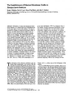
FIG. 1. Amino acid sequence comparisons between Xenopus and human caspases. Amino acid sequences of Xenopus caspase (xC)-2, -6, -7, -8, -9, and -10 are aligned with those of human caspase (hC)-2, -6, -7, -8, -9, and -10 (14, 54, 62– 69). The conserved residues are boxed and
Characterization of the Xenopus Caspases
10487
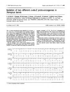
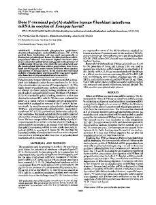
FIG. 2. Phylogenetic relationships of Xenopus and human caspases. Phylogenetic comparisons of Xenopus and human caspases were carried out based on the full-length proenzymes using the CLUSTAL W program (76). The dendogram was not affected (except for branch lengths) if the same analysis was carried out using the catalytic protease domains. Branch lengths are proportional to calculated values. xC, Xenopus caspase; hC, human caspase.
TABLE I Summary of amino acid sequence identities between Xenopus and human caspases The numbers represent the percentage of identity of the sequences compared in a prodomain and a catalytic protease domain.
Prodomain CPD a
XCa-1/ hC-1
XC-2/ hC-2
XC-3/ hC-3
XC-6/ hC-6
34.1 46.8
46.2 64.5
22.9 57.9
28.6 65.7
Comparison between: XC-7/ XC-8/ hC-7 hC-8
29.0 66.2
37.2 35.6
XC-8/ hC-10
XC-9/ hC-9
XC-10/ hC-10
XC-10/ hC-8
33.7 34.8
54.8 58.1
42.7 48.2
32.8 39.2
XC, Xenopus caspase; hC, human caspase; CPD, catalytic protease domain.
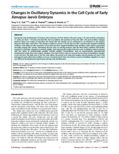
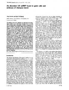
site. It is possible that Xenopus caspase-8 and -10 might be duplicated genes as a result of doubling chromosomes during evolution since X. laevis is allotetraploid. It seems unlikely because we have cloned two different caspase-1 and -2 cDNAs that share 79.5 and 85.6% identities, respectively,2 whereas Xenopus caspase-8 and -10 show only 31% identity. However, we cannot completely exclude the possibility that Xenopus caspase-8 and -10 do not correspond to the human counterparts. Expression of Xenopus Caspases—It has been shown that metamorphosis transforms most, if not all, organs and tissues in a tadpole especially after stage 58 (7) and that the stages at which maximal cell death is observed are different in different organs. The degeneration of tail muscle segments proceeds very rapidly after stage 63, and tail is resorbed completely until stage 66 (22), whereas the limbs differentiate and grow. In the intestine, the apoptotic degeneration of larval epithelial cells is most frequently observed at stages 59 – 61, and the proliferation of adult epithelial cells occurs in a predetermined order (23, 24). Although the central nervous system (brain and spinal cord) undergoes cell death of the mesencephalic fifth nucleus (the proprioceptors of the jaw musculature) at stage 62 (25), no conspicuous change takes place except topographical changes during the climax of metamorphosis (22). Caspase mRNA ex-
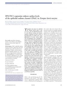
pression was examined in these organs during metamorphosis and in T3-treated myoblast cell lines derived from tadpole tail (Fig. 3). As has been observed, caspase probes hybridized to transcripts of 2.0 kb (caspase-1), 2.5 kb (caspase-2), 3.7 and 1.6 kb (caspase-3), 2.2 kb (caspase-6), 1.5 kb (caspase-7), 2.5 kb and 3.0 kb (caspase-8), 2.7 kb (caspase-9), and 2.3 kb (caspase-10). Expression of all cloned caspases increased in the intestine. Expression of all caspases except caspase-9 and -10 increased in the tail, whereas caspase expression did not change in the hind limb or the central nervous system. Both XLT-15 and the subline XLT-15-11 expressed constant levels of caspase mRNAs despite T3 treatment, except for caspase-3 mRNA, which increased in T3-treated XLT-15 cells (1). Although the expression of caspase-1 fluctuated during tail regression,2 in general, it increased somewhat. Overexpression of Xenopus Caspases—Many reports have demonstrated that the elevated expression of a caspase by transfection of the gene into cultured cells induces apoptotic cell death (26, 27). To compare the cytotoxic activity of caspases, Xenopus caspase genes in expression vector were transfected into XLT-15-11 cells with a reporter gene (pEGFPN2), and apoptotic cells were identified by the morphology of GFP-positive cells (Fig. 4). The overexpression of caspases with a long prodomain (caspase-1, -2, -8, -9, and -10) caused greater
shaded. The active sites (pentapeptide QACXG) are indicated by dots. Asp cleavage sites of human caspases are indicated by vertical arrows below the sequences (42, 70). Caspase-2 cleavage sites are predicted as equivalent sites being present in Nedd2 (71, 72). Based on the crystal structure of the interleukin-1-converting enzyme, open diamonds below the sequences represent residues adjacent to the substrate P2–P4 amino acids (73). In A and E, the caspase recruitment domain is underlined (74). In E, closed diamonds below the sequence indicate residues whose mutations inactivate or reduce the interface interaction with Apaf-1 (21). Primers used for the anchor PCR method are shown by horizontal arrows labeled RT and Amp. The EcoRI site where the amplified 5⬘-fragment is adjoined to the 3⬘-partial Xenopus caspase-9 cDNA is indicated by an arrowhead labeled Eco. In D and F, two death effector domains are underlined (42, 75).
10488
Characterization of the Xenopus Caspases cluded a small amount of apoptotic floating cells, in which the overexpressed caspase was activated, modified, and detected by affinity labeling. On the other hand, the extracts from cells introduced with the caspase-2, -8, -9, or -10 gene showed no additional signal as compared with the vector-transfected cell extract (Fig. 6A). In each case, the affinity labeling reagent Z-EK(bio)D-aomk might not bind to the active caspase efficiently, yet cell death might be induced before sufficient amounts of endogenous caspase-3, -6, and -7 for detection are processed. To estimate which caspases are activated in T3-treated XLT-15 and XLT-15-11 cells, their cell extracts were reacted with the affinity labeling reagent. The T3 treatment augmented the intensity of several signals, including two polypeptides that comigrated with processed caspase-6 and -7 (Fig. 6B). The intensity of a signal (17.1 kDa) that might correspond to processed caspase-3 increased in the T3-treated XLT-15 cell extract, but not in dying XLT-15-11 cells. DISCUSSION
FIG. 3. Expression of Xenopus caspase mRNAs. Each Xenopus caspase-specific probe (Xenopus caspase (xC)-1, -2, -3, -6, -7, -8, -9, or -10) was hybridized to 5 g of total RNAs isolated from X. laevis tadpole organs (hind limb (H.L.), brain, intestine (Int.), and tail) at the indicated stages and in two cultured cell lines, XLT-15 (T15) and XLT-1511(T15-11), which were maintained in the absence (control (C)) or presence of 10 nM T3 for 2 days. Control hybridization of the blot with the Xenopus elongation factor-1␣ probe (EF) is shown below. Stage numbers were assigned according to Nieuwkoop and Faber (22).
cell death than that of caspases with a short prodomain (caspase-3, -6, and -7) (Fig. 5), as reported previously (28). Affinity Labeling of Active Caspases—To identify an activated caspase in the caspase-overexpressing cells, we used an affinity labeling method that enabled us to detect the large subunits of processed caspases without preparing antibodies against Xenopus caspases. If an caspase overexpressed by transfection of its gene is activated during the cell death process (including spontaneous cell death) and the active form of this caspase has an affinity for Z-EK(bio)D-aomk, the caspase should be modified covalently by the reagent. The extracts from cells transfected with the caspase-1, -3, -6, or -7 gene showed signals by labeled active caspases (Fig. 6A). We interpreted these signals as follows based on the predicted cleavage sites by comparison of human and Xenopus caspase sequences (Fig. 1) and the molecular masses of the large subunits from the affinity labeling experiment. The single bands at 17.7 and 19.0 kDa (Fig. 6A, lanes 6 and 7) observed in the extracts of cells transfected with the caspase-6 or -7 gene correspond to the large subunits of transfected gene products processed by cutting at Asp35 and Asp192 of caspase-6 and at Asp31 and Asp209 of caspase-7, respectively. Cleavage at three sites (Asp16, Asp36, and Asp186) of caspase-3 produced 18.8- and 17.1-kDa polypeptides from the large subunit (Fig. 6A, lane 5). The processing at Asp104 and Asp296 of caspase-1 generated two polypeptides, 29.8 kDa (a prodomain plus a large subunit) and 21.4 kDa (a large subunit). The other signal (19.0 kDa) might represent cleavage of an endogenous caspase-7 (Fig. 6A, lane 3). The extracts from adherent cells overexpressing caspase-1, -3, -6, or -7 contained much less labeled active caspase than the extracts from adherent plus floating cells,2 which indicates that most of the signal was derived from floating dead cells (29). Cells transfected with the caspase-3, -6, or -7 gene in-
Numerous lines of evidence have established that caspases play a central role in apoptosis and that this cell death pathway has been conserved throughout the evolution of eukaryotes. Many caspase genes have been characterized, including ced-3 from C. elegans (8); dcp-1 (30), drICE (31), dredd (32), and dronc (33) from Drosophila; 10 mouse caspase genes (34 – 41); and 12 human caspase genes (42, 43). We have cloned cDNAs of Xenopus caspases corresponding to all human members that are involved in apoptosis. The sequences of Xenopus caspases enable us to compare the corresponding caspases in distant vertebrates and to delineate evolutionarily conserved regions that might be functionally important. The caspase family members are conserved from frog to human (Figs. 1 and 2), suggesting that the same two caspase cascades triggered by activation of caspase-8 and -9 as apical caspases previously described in mammalian apoptosis are similar in Xenopus. In fact, when Xenopus egg cytosol is incubated with isolated mitochondria, cytochrome c is released from mitochondria, leading to the activation of DEVD-specific caspases and nuclear apoptosis in vitro (44). This might correspond to the cell death pathway caused by the activation of caspase-9 that is mediated through the association of Apaf-1 and cytochrome c. On the other hand, there is so far no report on Xenopus that describes the characterization of the death receptors belonging to the tumor necrosis factor receptor superfamily or that suggests the death receptor signaling pathway. The Xenopus caspase cascade involving caspase-8 activation via its complex with FADD remains to be examined. Caspase-1, -8, and -10 are more divergent between frog and human than the other caspases (Table I). However, it does not mean that the latter caspases are more essential to normal development than the former because caspase-2 knockout mice reach adulthood without any gross abnormalities (45), and homozygous targeted disruption of the mouse caspase-8 gene results in death in utero (46). Caspase-2 is important to survival after birth since apoptosis mediated by granzyme B and perforin is defective in caspase-2-deficient B lymphocyte (45). A lower degree of identity of caspase-8 below 40% (Table I and Fig. 2) between human and frog might reflect a developmental change in the immune system during evolution in vertebrates (47). In regard to caspase-10, only a human gene other than Xenopus has been cloned. The human caspase-10 gene is located between the CASH (a proteolytically inactive homolog of caspase-8 and -10) and caspase-8 genes on the chromosome, whereas the mouse caspase-8 gene is located 30 kb downstream of the CASH gene in a genomic clone (46), implying a possibility
Characterization of the Xenopus Caspases
10489
FIG. 4. Morphological changes induced by overexpression of Xenopus caspase. XLT-15-11 cultured cells were cotransfected with the GFP expression construct (pEGFP-N2) and pcDNA1/Amp (Vec.) or pcDNA1/Amp-Xenopus caspase (xC)-1, -2, -3, -6, -7, -8, -9, or -10 and viewed 2 days later with a Zeiss Axiovert 135 TV inverted fluorescence microscope.
FIG. 5. Cell death-inducing activity by overexpression of Xenopus caspase in XLT-15-11 cultured cells. The GFP expression construct (pEGFPN2) with a vector or one of the Xenopus caspase (xC) gene expression constructs was transiently transfected into XLT15-11 cells as described in the legend to Fig. 4. The percentage of apoptotic round cells in GFP-expressing cells was determined 2 days after transfection. More than 200 cells were counted for each determination. Data are the means ⫾ S.E.
that there is no caspase-10 gene in mouse chromosomal DNA. The existence of Xenopus and human caspase-10, however, suggests that a vertebral animal at least from Amphibia basically possesses the caspase-10 gene. Human caspase-9 is inactivated by Akt phosphorylation (48) and contains sites that conform to the consensus Akt phosphorylation motif RXRXX(S/T) (49) at Ser183 (RTRTGS) and Ser196 (RRRFSS). The latter site is recognized and phosphorylated by Akt. The corresponding sequences of Xenopus caspase-9 (STRTGS and ANRMRS) do not, however, fulfill the requirements of the consensus sequence (Fig. 1). There are two possibilities to explain this. First, the regulation of caspase-9 by Akt phosphorylation may not be conserved in Xenopus. Second, Xenopus Akt may have a different recognition sequence compared with the human protein. This seems unlikely because strong amino acid sequence conservation is observed from Dictyostelium to human (50), and bovine Akt can rescue Drosophila Akt mutants (51). On the other hand, since RGD-containing peptides induce procaspase-3 autoprocessing and activation directly in a cell-free system without any requirement for in-
tegrin-mediated cell clustering or signals and since human procaspase-3 contains an RGD-binding motif (DDX), it has been proposed that RGD peptides exert their effects by the interaction with a potential RGD-binding site of procaspase-3, leading to the conformational changes that promote its autoprocessing and activation (52). However, the region of Xenopus caspase-3 corresponding to an RGD-binding motif is not conserved (1). Moreover, Tyr277 and Ser401 of Xenopus caspase-10 correspond to Leu242 and Val367 of human caspase-10, respectively, missense mutations of which are found in two kindreds with autoimmune lymphoproliferative syndrome type II, decrease caspase activity, and interfere with death receptor-induced apoptosis (53). Leu242 is substituted by Phe in the one family, and Val367 is changed to Ile in the other. Although Xenopus caspase-10 contains substitutions by quite different amino acids, its overexpression showed a strong cytotoxic activity as compared with other caspases (Fig. 5). The regulation of caspases might be different between human and frog in some details. Our results clearly show that the enforced expression of
10490
Characterization of the Xenopus Caspases
FIG. 6. Detection of active caspase proteins. A, comparison of active caspases labeled with Z-EK(bio)D-aomk in the extracts of cells transfected with Xenopus caspase (xC) gene expression constructs. XLT-15-11 cultured cells were transfected with a vector (lanes 1 and 2) or one of the Xenopus caspase gene expression constructs (lanes 3–10) and harvested 1 day after the cytotoxic assay (Fig. 5). Their extracts were prepared and incubated without (lane 1) or with (lanes 2–10) Z-EK(bio)D-aomk. 20 g of total proteins were resolved on a 12.5% SDS-polyacrylamide gel. The active caspases that reacted to Z-EK(bio)D-aomk were visualized by an avidin-biotin complex method. B, active caspases in extracts from T3-treated cultured cells. Samples containing 20 g of extract protein from XLT-15 or XLT-15-11 cells cultured in the absence (⫺) or presence (⫹) of 10 nM T3 for 3 days were labeled with Z-EK(bio)D-aomk, subjected to SDS-polyacrylamide gel electrophoresis, and detected for A. Arrowheads indicate the estimated positions of large subunits of caspase-3 (17.1 kDa), -6 (17.7 kDa), and -7 (19.0 kDa).
XLT-15-11 is derived from XLT-15 and maintains T3 sensitivity, although the expression profile of caspase genes was a little different from the pattern of its parental cell line (Fig. 3). After T3 treatment, caspase-3 mRNA was up-regulated in XLT-15 cells, but not in XLT-15-11 cells, whereas the expression of the other caspase genes did not increase in XLT-15 or XLT-15-11 cells. This result indicates that the increase in expression of any caspase gene is not necessary for T3-induced cell death of XLT-15-11 cells. On the other hand, the up-regulation of caspase expression was observed in a regressing and a remodeling organ during metamorphosis, but not in an organ that grows or undergoes minimal changes (Fig. 3). In addition, the ced-3 mRNA is most abundant during nematode embryogenesis, when most programmed cell deaths occur (8). In late third instar larvae of Drosophila, dronc mRNA is dramatically up-regulated in salivary glands and midgut before histolysis of these tissues (33). The expression of the caspase-1 gene is induced in post-lactational involution of mouse mammary gland (57). Since T3 treatment causes apoptosis of XLT-15-11 cells without an increase in any caspase mRNAs, it is reasonable that the increase in caspase expression promotes the apoptotic process efficiently, but is not essential to cell death. However, we cannot rule out the possibility that caspase expression is up-regulated in the cultured cell lines treated with T3 by translational regulation. Signals of several polypeptides in the affinity labeling assay were increased in the extracts of T3-treated XLT-15 and XLT15-11 cells (Fig. 6B). Two main molecules among them comigrated with large subunits of caspase-6 and -7, although it has been reported that caspase-3 and -6 are the major active caspases in apoptotic cells in many cases (58, 59). It is possible that the apical caspase (caspase-8 or -9) that is supposed to process procaspase-3 (60) can afford to activate procaspase-7 in T3-treated XLT-15 and XLT-15-11 cells (61) because these cells express only a small amount of caspase-3. In T3-treated XLT-15 cells, a faint signal that might correspond to a large subunit of caspase-3 appeared in the affinity labeling assay (Fig. 6B, lane 2) as caspase-3 expression was induced. The polypeptides other than caspase-3, -6, and -7 observed in the extract of T3-treated cells remain to be identified. Our data show that the caspase family is conserved at least from Amphibia to mammals in terms of amino acid sequence and function and that programmed cell death in regressing organs does not depend on the transcription of caspase genes. The sequence information on Xenopus caspases might enable us to clone caspase genes of other animals, including fishes and ascidians, by utilizing the sequences of the conserved regions between frog and human caspases and to elucidate the duplication and diversification process of caspase genes during evolution. Acknowledgments—We thank Dr. D. D. Brown for critical comments and Dr. P. A. Krieg for Xenopus elongation factor-1␣ cDNA.
caspases with a long prodomain induced stronger cytotoxic activity than that of caspases with a shorter prodomain. This is in good agreement with the idea that the function of the long prodomain is to promote homodimerization, which leads to autoprocessing of caspase (28). In fact, it has been proposed that caspase-8 is proteolytically autoactivated in the deathinducing signaling complex after oligomerization through its death effector domain during Fas-mediated apoptosis (54 –56). The observation that mRNAs of caspases with a long prodomain were up-regulated in a regressing organ might raise the possibility that these caspases are autoactivated and kill cells. However, this seems unlikely because mRNAs of caspases with a long prodomain were not increased in the cultured cells that were induced to die by T3 treatment (Fig. 3).
REFERENCES 1. Yaoita, Y., and Nakajima, K. (1997) J. Biol. Chem. 272, 5122–5127 2. Saunders, J. W., Jr. (1966) Science 154, 604 – 612 3. Lockshin, R. A., and Zakeri, Z. F. (1991) Apoptosis: The Molecular Basis of Cell Death, pp. 47– 60, Cold Spring Harbor Laboratory, Cold Spring Harbor, NY 4. Yaoita, Y., Shi, Y. B., and Brown, D. D. (1990) Proc. Natl. Acad. Sci. U. S. A. 87, 7090 –7094 5. Yaoita, Y., and Brown, D. D. (1990) Genes Dev. 4, 1917–1924 6. Kerr, J. F., Harmon, B., and Searle, J. (1974) J. Cell Sci. 14, 571–585 7. Dodd, M. H. I., and Dodd, J. M. (1976) in Physiology of the Amphibia (Lofts, B., ed) Vol. III, pp. 467–599, Academic Press, New York 8. Yuan, J., Shaham, S., Ledoux, S., Ellis, H. M., and Horvitz, H. R. (1993) Cell 75, 641– 652 9. Li, P., Nijhawan, D., Budihardjo, I., Srinivasula, S. M., Ahmad, M., Alnemri, E. S., and Wang, X. (1997) Cell 91, 479 – 489 10. Nagata, S. (1997) Cell 88, 355–365 11. Nagata, S. (1996) Curr. Biol. 6, 1241–1243 12. Srinivasula, S. M., Ahmad, M., Fernandes Alnemri, T., and Alnemri, E. S.
Characterization of the Xenopus Caspases (1998) Mol. Cell 1, 949 –957 13. Johnson, A. L., Bridgham, J. T., Bergeron, L., and Yuan, J. (1997) Gene (Amst.) 192, 227–233 14. Fernandes Alnemri, T., Litwack, G., and Alnemri, E. S. (1995) Cancer. Res. 55, 2737–2742 15. Frohman, M. A., Dush, M. K., and Martin, G. R. (1988) Proc. Natl. Acad. Sci. U. S. A. 85, 8998 –9002 16. Chirgwin, J. M., Przybyla, A. E., MacDonald, R. J., and Rutter, W. J. (1979) Biochemistry 18, 5294 –5299 17. Krieg, P. A., Varnum, S. M., Wormington, W. M., and Melton, D. A. (1989) Dev. Biol. 133, 93–100 18. Samuels, H. H., Stanley, F., and Casanova, J. (1979) Endocrinology 105, 80 – 85 19. Sa´nchez, I., Xu, C. J., Juo, P., Kakizaka, A., Blenis, J., and Yuan, J. (1999) Neuron 22, 623– 633 20. Yamashita, K., Takahashi, A., Kobayashi, S., Hirata, H., Mesner, P. W., Jr., Kaufmann, S. H., Yonehara, S., Yamamoto, K., Uchiyama, T., and Sasada, M. (1999) Blood 93, 674 – 685 21. Qin, H., Srinivasula, S. M., Wu, G., Fernandes Alnemri, T., Alnemri, E. S., and Shi, Y. (1999) Nature 399, 549 –557 22. Nieuwkoop, P. D., and Faber, J. (1956) Normal Tadpole of Xenopus laevis, North-Holland Publishing Co., Amsterdam 23. Ishizuya Oka, A., and Shimozawa, A. (1987) Anat. Anz. 164, 81–93 24. Ishizuya Oka, A., and Shimozawa, A. (1992) J. Morphol. 213, 185–195 25. Kollros, J. J., and Thiesse, M. L. (1985) J. Comp. Neurol. 233, 481– 489 26. Miura, M., Zhu, H., Rotello, R., Hartwieg, E. A., and Yuan, J. (1993) Cell 75, 653– 660 27. Alnemri, E. S. (1997) J. Cell. Biochem. 64, 33– 42 28. Colussi, P. A., Harvey, N. L., Shearwin-Whyatt, L. M., and Kumar, S. (1998) J. Biol. Chem. 273, 26566 –26570 29. Samejima, K., Tone, S., Kottke, T. J., Enari, M., Sakahira, H., Cooke, C. A., Durrieu, F., Martins, L. M., Nagata, S., Kaufmann, S. H., and Earnshaw, W. C. (1998) J. Cell Biol. 143, 225–239 30. Song, Z., McCall, K., and Steller, H. (1997) Science 275, 536 –540 31. Fraser, A. G., McCarthy, N. J., and Evan, G. I. (1997) EMBO J. 16, 6192– 6199 32. Chen, P., Rodriguez, A., Erskine, R., Thach, T., and Abrams, J. M. (1998) Dev. Biol. 201, 202–216 33. Dorstyn, L., Colussi, P. A., Quinn, L. M., Richardson, H., and Kumar, S. (1999) Proc. Natl. Acad. Sci. U. S. A. 96, 4307– 4312 34. Nett, M. A., Cerretti, D. P., Berson, D. R., Seavitt, J., Gilbert, D. J., Jenkins, N. A., Copeland, N. G., Black, R. A., and Chaplin, D. D. (1992) J. Immunol. 149, 3254 –3259 35. Molineaux, S. M., Casano, F. J., Rolando, A. M., Peterson, E. P., Limjuco, G., Chin, J., Griffin, P. R., Calaycay, J. R., Ding, G. J., Yamin, T. T., Palyha, O. C., Luell, S., Fletcher, D., Miller, D. K., Howard, A. D., Thornberry, N. A., and Kostura, M. J. (1993) Proc. Natl. Acad. Sci. U. S. A. 90, 1809 –1813 36. Kumar, S., Kinoshita, M., Noda, M., Copeland, N. G., and Jenkins, N. A. (1994) Genes Dev. 8, 1613–1626 37. Van de Craen, M., Vandenabeele, P., Declercq, W., Van den Brande, I., Van Loo, G., Molemans, F., Schotte, P., Van Criekinge, W., Beyaert, R., and Fiers, W. (1997) FEBS Lett. 403, 61– 69 38. Van de Craen, M., Van Loo, G., Declercq, W., Schotte, P., Van den Brande, I., Mandruzzato, S., van der Bruggen, P., Fiers, W., and Vandenabeele, P. (1998) J. Mol. Biol. 284, 1017–1026 39. Wang, S., Miura, M., Jung, Y., Zhu, H., Gagliardini, V., Shi, L., Greenberg, A. H., and Yuan, J. (1996) J. Biol. Chem. 271, 20580 –20587 40. Ahmad, M., Srinivasula, S. M., Hegde, R., Mukattash, R., Fernandes Alnemri, T., and Alnemri, E. S. (1998) Cancer Res. 58, 5201–5205 41. Van de Craen, M., Van Loo, G., Pype, S., Van Criekinge, W., Van den Brande, I., Molemans, F., Fiers, W., Declercq, W., and Vandenabeele, P. (1998) Cell Death Differ. 5, 838 – 846 42. Cohen, G. M. (1997) Biochem. J. 326, 1–16 43. Humke, E. W., Ni, J., and Dixit, V. M. (1998) J. Biol. Chem. 273, 15702–15707 44. Kluck, R. M., Martin, S. J., Hoffman, B. M., Zhou, J. S., Green, D. R., and Newmeyer, D. D. (1997) EMBO J. 16, 4639 – 4649 45. Bergeron, L., Perez, G. I., Macdonald, G., Shi, L., Sun, Y., Jurisicova, A., Varmuza, S., Latham, K. E., Flaws, J. A., Salter, J. C., Hara, H., Moskowitz, M. A., Li, E., Greenberg, A., Tilly, J. L., and Yuan, J. (1998) Genes Dev. 12, 1304 –1314 46. Varfolomeev, E. E., Schuchmann, M., Luria, V., Chiannilkulchai, N., Beckmann, J. S., Mett, I. L., Rebrikov, D., Brodianski, V. M., Kemper, O. C., Kollet, O., Lapidot, T., Soffer, D., Sobe, T., Avraham, K. B., Goncharov, T.,
10491
Holtmann, H., Lonai, P., and Wallach, D. (1998) Immunity 9, 267–276 47. Kaufman, J., Skjoedt, K., and Salomonsen, J. (1990) Immunol. Rev. 113, 83–117 48. Cardone, M. H., Roy, N., Stennicke, H. R., Salvesen, G. S., Franke, T. F., Stanbridge, E., Frisch, S., and Reed, J. C. (1998) Science 282, 1318 –1321 49. Alessi, D. R., Caudwell, F. B., Andjelkovic, M., Hemmings, B. A., and Cohen, P. (1996) FEBS Lett. 399, 333–338 50. Meili, R., Ellsworth, C., Lee, S., Reddy, T. B. K., Ma, H., and Firtel, R. A. (1999) EMBO J. 18, 2092–2105 51. Staveley, B. E., Ruel, L., Jin, J., Stambolic, V., Mastronardi, F. G., Heitzler, P., Woodgett, J. R., and Manoukian, A. S. (1998) Curr. Biol. 8, 599 – 602 52. Buckley, C. D., Pilling, D., Henriquez, N. V., Parsonage, G., Threlfall, K., Scheel Toellner, D., Simmons, D. L., Akbar, A. N., Lord, J. M., and Salmon, M. (1999) Nature 397, 534 –539 53. Wang, J., Zheng, L., Lobito, A., Chan, F. K., Dale, J., Sneller, M., Yao, X., Puck, J. M., Staus, S. E., and Lenardo, M. J. (1999) Cell 98, 47–58 54. Muzio, M., Chinnaiyan, A. M., Kischkel, F. C., O’Rourke, K., Shevchenko, A., Ni, J., Scaffidi, C., Bretz, J. D., Zhang, M., Gentz, R., Mann, M., Krammer, P. H., Peter, M. E., and Dixit, V. M. (1996) Cell 85, 817– 827 55. Muzio, M., Stockwell, B. R., Stennicke, H. R., Salvesen, G. S., and Dixit, V. M. (1998) J. Biol. Chem. 273, 2926 –2930 56. Yang, X., Chang, H. Y., and Baltimore, D. (1998) Mol. Cell 1, 319 –325 57. Lund, L. R., Romer, J., Thomasset, N., Solberg, H., Pyke, C., Bissell, M. J., Dano, K., and Werb, Z. (1996) Development (Camb.) 122, 181–193 58. Faleiro, L., Kobayashi, R., Fearnhead, H., and Lazebnik, Y. (1997) EMBO J. 16, 2271–2281 59. Martins, L. M., Kottke, T., Mesner, P. W., Basi, G. S., Sinha, S., Frigon, N., Jr., Tatar, E., Tung, J. S., Bryant, K., Takahashi, A., Svingen, P. A., Madden, B. J., McCormick, D. J., Earnshaw, W. C., and Kaufmann, S. H. (1997) J. Biol. Chem. 272, 7421–7430 60. Stennicke, H. R., Jurgensmeier, J. M., Shin, H., Deveraux, Q., Wolf, B. B., Yang, X., Zhou, Q., Ellerby, H. M., Ellerby, L. M., Bredesen, D., Green, D. R., Reed, J. C., Froelich, C. J., and Salvesen, G. S. (1998) J. Biol. Chem. 273, 27084 –27090 61. Slee, E. A., Harte, M. T., Kluck, R. M., Wolf, B. B., Casiano, C. A., Newmeyer, D. D., Wang, H. G., Reed, J. C., Nicholson, D. W., Alnemri, E. S., Green, D. R., and Martin, S. J. (1999) J. Cell Biol. 144, 281–292 62. Wang, L., Miura, M., Bergeron, L., Zhu, H., and Yuan, J. (1994) Cell 78, 739 –750 63. Duan, H., Chinnaiyan, A. M., Hudson, P. L., Wing, J. P., He, W. W., and Dixit, V. M. (1996) J. Biol. Chem. 271, 1621–1625 64. Fernandes Alnemri, T., Takahashi, A., Armstrong, R., Krebs, J., Fritz, L., Tomaselli, K. J., Wang, L., Yu, Z., Croce, C. M., Salveson, G., Earnshaw, W. C., Litwack, G., and Alnemri, E. S. (1995) Cancer Res. 55, 6045– 6052 65. Lippke, J. A., Gu, Y., Sarnecki, C., Caron, P. R., and Su, M. S.-S. (1996) J. Biol. Chem. 271, 1825–1828 66. Boldin, M. P., Goncharov, T. M., Goltsev, Y. V., and Wallach, D. (1996) Cell 85, 803– 815 67. Srinivasula, S. M., Fernandes-Alnemri, T., Zangrilli, J., Robertson, N., Armstrong, R. C., Wang, L., Trapani, J. A., Tomaselli, K. J., Litwack, G., and Alnemri, E. S. (1996) J. Biol. Chem. 271, 27099 –27106 68. Duan, H., Orth, K., Chinnaiyan, A. M., Poirier, G. G., Froelich, C. J., He, W. W., and Dixit, V. M. (1996) J. Biol. Chem. 271, 16720 –16724 69. Fernandes Alnemri, T., Armstrong, R. C., Krebs, J., Srinivasula, S. M., Wang, L., Bullrich, F., Fritz, L. C., Trapani, J. A., Tomaselli, K. J., Litwack, G., and Alnemri, E. S. (1996) Proc. Natl. Acad. Sci. U. S. A. 93, 7464 –7469 70. Srinivasula, S. M., Ahmad, M., Fernandes Alnemri, T., Litwack, G., and Alnemri, E. S. (1996) Proc. Natl. Acad. Sci. U. S. A. 93, 14486 –14491 71. Allet, B., Hochmann, A., Martinou, I., Berger, A., Missotten, M., Antonsson, B., Sadoul, R., Martinou, J. C., and Bernasconi, L. (1996) J. Cell Biol. 135, 479 – 486 72. Harvey, N. L., Trapani, J. A., Fernandes Alnemri, T., Litwack, G., Alnemri, E. S., and Kumar, S. (1996) Genes Cells 1, 673– 685 73. Wilson, K. P., Black, J. A., Thomson, J. A., Kim, E. E., Griffith, J. P., Navia, M. A., Murcko, M. A., Chambers, S. P., Aldape, R. A., Raybuck, S. A., and Livingston, D. J. (1994) Nature 370, 270 –275 74. Hofmann, K., Bucher, P., and Tschopp, J. (1997) Trends Biochem. Sci. 22, 155–156 75. Sakamaki, K., Tsukumo, S., and Yonehara, S. (1998) Eur. J. Biochem. 253, 399 – 405 76. Thompson, J. D., Higgins, D. G., and Gibson, T. J. (1994) Nucleic Acids Res. 22, 4673– 4680