Apr 26, 1993 - From the Department of Clinical Chemistry, Bispebjerg Hospital, Bispebjerg Bakke 23, DK-2400 Copenhagen, ... function of the insulin receptor tyrosine kinase in signal ... internalization and stimulation of thymidine incorpora-.
Vol. 268, No. 31,Issue of November 5,pp. 23435-23440, 1993 Printed in U.S.A.
THE JOURNALOF BIOLOGICAL CHEMISTRY 0 1993 by The American Society for Biochemistry and Molecular Biology, Inc.
Structure-Function Relationshipof the Insulin-likeGrowth Factor-I Receptor %osine Kinase* (Received forpublication, April 26, 1993)
Mette Gr~nborg,Birgitte S. Wulff,Jesper S . RasmussenS, Thomas KjeldsenS, and Steen Gammeltoftl From the Department of Clinical Chemistry, Bispebjerg Hospital, Bispebjerg Bakke 23, DK-2400 Copenhagen, Denmark and $Novo-Nordisk Industries, DK-2880 Bagsurerd, Denmark Insulin-like growth factorI (IGF-I) and insulin recep- deficient receptors are biologically inactive. Insulin receptor phosphorylation of tors are structurally similar with ligand-stimulated ty- and IGF-I receptor activationresultsin rosine kinase activity in their cytoplasmic domains. The pp185 or insulin receptor substrate-1 (IRS-l), which appears to function of the insulin receptor tyrosine kinase in signal be a common element in insulin andIGF-I signaling (Sunet al., transduction has been studied extensively in contrast1991; to Myers et al., 1993). The physiological effects of insulin the IGF-I receptor tyrosine kinase. In the present study and IGF-I are different, but it is not clear whether this is due we have analyzed the regulatory function of the IGF-I to differential expression and distribution of the two receptors receptortyrosinekinaseandcarboxyl-terminaldoin vivo or to differences in the molecular mechanism of signal mains in mitogenic signalingby overexpression of mu- transduction (Czech, 1989). tant IGF-I receptors in mouse NIH-3T3fibroblasts.A muThe structure-function relationship of the insulin receptor 3 tyrosines ("yr1lS1,"yrllss, tant IGF-I receptor, in which tyrosine kinase has been studied extensively, in contrast tothe and "yrllsa) analogoustothethreemqjorautophoshighly homologous IGF-I receptor kinase.Five autophosphoryphorylation sites in the insulin receptor kinase were replaced by phenylalanines, was devoid of kinase activ-lation sites have been identified in two regions of the insulin receptor P-subunit (Tornqvist et al.,1987; White et al., 1988a; ity in uiuo and in vitro and inactive with respect to IGF-I Myers et al., 1991).Three of these sites,2 Tyr1146, Tyr1150, and internalization and stimulation of thymidine incorporation. Another mutant IGF-I receptor, which lacks 49 the Tyr1151, are clustered in the tyrosine kinase domain, and two carboxyl-terminal amino acids (residues 1289-1337) of sites are in the carboxyl terminus: Tyr1316 and Tyr13". Phosthe P-subunit, was fully active. Our data suggest that thephorylation of the 3 tyrosine residues in the kinase domainof structure-function relationship of the IGF-I receptor ty- the insulin receptor is requiredfor full activation of the tyrorosine kinase activation and signal transduction is simi- sine kinase and thebiological responses since insulin receptor mutants with replacement of tyrosine by phenylalanines are lar to that of the insulin receptor. inactive (Ellis et al., 1986; Murakami and Rosen, 1991; Zhang et al., 1991; Wilden et al., 1992a, 1992b). The functionalrole of In mammalian organisms, insulin regulates cell metabolism, the carboxyl terminus of the insulin receptor P-subunit is not whereas insulin-like growth factor I (IGF-IIl is an important clear. Autophosphorylation of the two carboxyl-terminal tyroregulator of cell growth (Gammeltoft,1989; Humbel, 1990). The sine sites does not appearto regulate kinase activity (Maegawa biological effects of insulin and IGF-I are mediated by distinct et al. 1988; Myers et al.,1991).Insulin receptors with deletion but structurally similar receptorscomposed of two a-subunits of 43 carboxyl-terminal amino acids or mutations of the 2 tyand two P-subunits linked together by disulfide bridges (U11- rosine residues of the carboxyl terminus exhibit enhancedmirich et al., 1985, 1986). Both receptors belong to the superfam- togenic signaling and impairedmetabolic responses (Maegawa ily of receptortyrosinekinases (Ullrich and Schlessinger, et al., 1988; Thies et al., 1989; Takata et al.,1991; Ando et al., 1990). Ligand binding to the extracellular domain leads toac- 1992). Others have found, however, that the insulin receptor functions normally after truncation of the carboxyl terminus or tivation of the intracellular tyrosine kinase, receptor autophosRosen, 1991; Myers phorylation, and phosphorylation of cellular proteins. The ki- mutation of the 2 tyrosines (Murakami and nase activity is essential for signal transduction as kinase- et al., 1991). A comparison between insulin receptor and IGF-I receptor amino acid sequences reveals 84% identity in their tyrosine *This work was supported by DanishHealth ResearchCouncil Grants 12-9295 and 12-0180, Danish Cancer Society Grant 90-076, the kinase domains and 44% identity in their carboxyl-terminal Novo-Nordisk Foundation, and the Danish Biotechnology Center for domains (Ullrich et al., 1985, 1986). The tyrosine autophosSignal Peptide Research. The costs of publication of this article were phorylation sitesof the IGF-I receptor have not yet been idendefrayed in part by the payment of page charges. This article must tified. The 3 tyrosines in the IGF-I receptor kinase (Tyr1131, "advertisement" in accordancewith18 therefore beherebymarked U.S.C. Section 1734 solely to indicate this fact. Tyr1135 and Tyr1136) are homologous to the autophosphoryla5 To whom all correspondence should be addressed Dept. of Clinical tion sites in the insulin receptor kinase, itand is possible that Chemistry, Bispebjerg Hospital, DK 2400 Copenhagen W,Denmark. their phosphorylation could be involved in the activationof the Tel.: 45-35312646; Fax: 45-35313955. IGF-I receptor kinase. The carboxyl-terminal domains of the The abbreviations used are: IGF-I,insulin-likegrowthfactor-I; IRS-1, insulin receptor substrate-1;WT, wild-type IGF-I receptor; FFF, P-subunits of the two receptors show a low degree of amino acid IGF-I receptor with replacement of Tyr1I3l, 'Q7r1136, and Tyr1lS6 with sequence identity, and only one of the two carboxyl-terminal phenylalanines;DCT, IGF-I receptor truncatedat residue 1289 lacking autophosphorylation sites in the insulin receptor, Tyr1316, is the 49 carboxyl-terminal amino acids of the P-subunit; NEO, endogenous IGF-I receptor in mouse NIH-3T3 fibroblasts transfected with phospSV2neo;DMEM, Dulbecco's modifiedEagle'smedium;PBS, ' h o variants of the insulin receptor exist. Numbering of amino acids phate-buffered saline; NCS, neonatal calf serum; WGA, wheat germ in this paper correspondsto the sequence of Ullrich et al. (1985), which agglutinin; bp, base paids). differs from that of Ebina et al. (1985) by being 12 lower.
23435
IGF-IReceptor Tyrosine Kinase
23436
present in the IGF-I receptor. It is possible that the carboxylterminal domain of the IGF-I receptorcouldbeinvolved in specifying the signal leading to IGF-I-stimulatedcell proliferation. Thesignificanceof the three putative tyrosine autophosphorylation sites of the IGF-I receptor kinase domain and the roleof the IGF-I receptor carboxyl-terminal domain has not been studied previously.In the present study we have addressed two questions concerning the structural basis for activation and signaling by the IGF-I receptor tyrosine kinase: 1) whether the identity between the 3 tyrosine autophosphorylation sites in the kinase domains of the insulin receptor and IGF-I receptor is reflected by similarity in their functional role, and 2) whether the difference between the carboxyl-terminal domains of the insulin receptor and IGF-I receptor is significant in determining their mitogenic signaling potential. Two IGF-I receptormutants were constructed and overexpressedin mouse NIH-3T3 fibroblasts. One mutant IGF-I receptor carrying phenylalanine substitutions of all three putative autophosphorylation sites in the kinase domain (FFF mutant) was biologically inactive, whereas another mutant receptor lacking the carboxyl-terminal 49 amino acids of the P-subunit (DCT mutant) was fully active.
genesis system (Promega) and themutagenic primer 5"CCTGAGTGTCAGTCGGGCAGT-3'. The introduced mutation was verified by DNA sequencing. After insertion of a 2211-bp SphI fragmentof IGF-I receptor sequences in correct orientation 5' to the mutant fragment, the mutant IGF-I receptor sequences were recombined into pBW 87 containing the full-length IGF-I receptor cDNA. Cell Culture a n d Dansfection-Mouse NIH-3T3 fibroblasts from American Type Culture Collection were grown in DMEM supplemented with 10% NCS, penicillin, streptomycin, and 2 mM glutamine. Transfection was carried out by calcium phosphate coprecipitation (Graham and Van der Eb, 1973), where 20 pg of mutant IGF-I receptor or wildtype IGF-I receptor cDNA were cotransfected with 2 pg of pSV2neo. Neomycin-resistant cells were selected in medium supplemented with 600 pg/ml G418. Stably transfected mass culturesconsisting of approximately 50 clones and several subcultures consisting of one t o four clones were analyzed for lZ5I-IGF-I binding and used for further studies. All experiments wererepeated with several different selectionsof mass cell cultures or subcultures in order to prove the reproducibility of the results. lZ5I-IGF-Ibinding of the different cell cultures was routinely determined before the experiments, and thereceptor number was constant during at least 10cell passages. IGF-I Binding-Subconfluent cell cultures growing on culture dishes (55 cm2) were washed once with PBS and incubated with EDTA (0.2 glliter) in PBS at 0 "C for 5 min. T h e cells in suspension were centrifuged for 5 min a t 1,000 rpm andresuspended in Krebs-Ringer-HEPES buffer (124mM NaC1,3.56 n m KCl, 1.19 mM MgSO,, 2.5 mM CaCI,, 1.19 mM KH2Po4, 25 mM HEPES, pH 7.4) supplemented with 10 mg/ml bovine serum albumin. Cell suspensions (4 x IO4 celldml) were incuEXPERIMENTAL PROCEDURES bated for 20 h at 4 "C with 40,000 c p d m l lZ5I-IGF-I(15 PM) together Materials-HumanrecombinantIGF-I was obtained from Amer- with different concentrationsof unlabeled IGF-Iin a total volume of 250 sham, United Kingdom and [ [ 1 2 5 1 ] m ~ n ~ i ~ d ~ - ~ 1 1(2 1 GpCi/pmol) F-I pl. Bound 'T-IGF-1 was measured and corrected for nonspecific bindprepared by the IODO-GEN method and purified by high performance ing determined in the presence of 100 nM unlabeled IGF-I (Verland and liquid chromatography (Drejer et al., 1991) was a gift from Dr. U. D. Gammeltoft, 1989). Larsen, Novo-Nordisk, Bagsverd, Denmark. Apolyclonal anti-phosphoI n Vivo IGF-I Receptor a n d pp185 prosine Phosphorylation tyrosine antibody (Giorgino et al., 1992) was a gift from Dr. Robert J . -Subconfluent monolayers of cells in six-well dishes starvedfor 2 days Smith, Joslin Diabetes Center, Boston, MA. Cell culture reagents and in DMEM with 2% NCS were incubated in theabsence or the presence newborn calf serum (NCS) were from Biologicals Industries, culture of 50 nM IGF-I for 3 min at 37 "C. The medium was rapidly removed and dishes from Greiner, and G418 from Sigma. [y-32PlATP(3000 Cifmmol), the cells lysed in 150pl of 2 x Laemmli sample buffer (100 mM Tris-HC1, [methyl-1',2'-3H]thymidine (120 Cifmmol), and lZ5I-protein A (30mCi/ pH 6.8, 20% glycerol, 2% SDS, 0.1% bromphenol blue) with 100 mM mg) were from Amersham, United Kingdom. dithiothreitol and 1mM sodium orthovanadate andboiled for 2 min. The Expression Plasmid Construction-The expression vector, pBW 87, is lysates were triturated with a syringe needleto shear the DNA, and the derived from pUC 19 and contains theconstitutive promotor from the proteins were separated on a 7.5% SDS-PAGE mini-gel (Bio-Rad). The human ubiquitinC geneand polyadenylation signal sequencesfrom the proteins were then transferred to nitrocellulose using a semi-dry elechuman growth hormone gene.pBW 87 wasconstructed in thefollowing troblot apparatus from JKA Biotech. The nitrocellulose membrane was manner. The promotor from the humanubiquitin gene UbC (Wiborg et probed with anti-phosphotyrosine antibody and 1251-labeledprotein A al., 1985) was subcloned as a Hind111 fragment intoa HindIII-digested followed by autoradiography as previously described (Wilden et al., pUC19 plasmid, resulting in pHD 183. A blunt-ended SmaIIEcoRI frag- 1992a). ment containing the polyadenylation signal from the human growth In Vitro prosine Kinase Activity-Cells from confluent culture dishes hormone gene was subcloned into a n EcoRI-digested pHD 183, which (150 cm2) starved by culture for 2 days in DMEM with 2% NCS were was rendered blunt, resulting in pBW 87 with the following unique solubilized in 150 mM NaCI, 50 mM HEPES, pH 7.4, 1% Triton X-100,4 restriction enzyme sites in the polylinker: XbaI, SalI, BamHI, KpnI, mM EDTA, 400 kallikrein-inhibitory unitdml aprotinin,1mM pepstatin, and SacI. cDNA encoding the human IGF-I receptor (Andersen et al., 1mM leupeptin, and 1 mM phenylmethylsulfonyl fluoride and partially 1990) and 15 base pairs constituting a putative ribosome binding site purified on wheat germ agglutinin-agarose. Kinase activity of the partially purified IGF-I receptors was determined by phosphorylation of a derived from the human insulin receptor cDNA were cloned ona 4125-bp BamHI fragment into the expression vector using the unique synthetic substrate as previously described for insulin receptors by Shoelson et al.(1992) with some modifications. Equal amountsof WGABamHI site in the polylinker. Construction of the Mutants-The polymerase chain reaction was purified IGF-I receptors were incubated for 60 min a t 23 "C in the used t o construct the FFF mutantIGF-I receptor with Tyr1131, Tyr1135, presence or absence of different concentrations of IGF-I and addition of 5 mM MnCI2C. Substrate phosphorylation was measured ina total voland ' 9 4 1 3 6 replaced by phenylalanine. A mutant IGF-I receptor fragment of 633 bpcorresponding to the3' end of the IGF-I receptor cDNA ume of 50 pl by incubation for 5 min a t 23 "C following the simultaneous addition of 0.28 mg/mlpoly(Glus,-Tyrzo) and 50 p~ (6 pCifnmol) between the EcoRV and the BamHI sites was generated using the IGF-I [ys2P1ATP.The reaction was terminated by spotting 40 pl of the reacreceptor cDNA as template and a mutagenic oligonucleotide, 5'-ATGAATTCAGATATC'M'I'GAGACAGAC'M'CT"CCG-3', with mis- tion mixture on phosphocellulose paper (Whatman) andprecipitation of the proteinson the filters in10% trichloroacetic acidwith 10m~ sodium matches underlined, anda n amplifying oligonucleotide complementary to 3' sequences in the IGF-I receptor cDNA, 5'-TGGGCACTGGAGTG pyrophosphate. The filters were washed extensively in trichloroacetic acid with 10 mM sodium pyrophosphate, dried, and counted for 32P. GCAACTTCCAGGG-3' as primers in the polymerase chain reaction In Vitro Autophosphorylation of the IGF-I Receptor-Equal amounts reaction. Themutantfragmentwasinsertedinto phagemidPBS+ (Stratagene) andDNA sequenced to confirm the presence of the muta- of WGA-purified IGF-I receptors were incubated for 15 min at 23 "C in the absence or presence of 100 n~ IGF-I and addition of 5 m~ MnCI,. tions. The 5' end of the IGF-I receptor cDNA was then inserted to generate the full-length cDNA, encoding the three amino acid substi- The in vitro autophosphorylation of IGF-I receptors was performed as tutions, which was cloned into pBW 87. The DCT mutant IGF-I receptor previously described for insulin receptors by Wilden et al.(1992a) with with deleted carboxyl terminus was generated by changing the arginine some modifications. The phosphorylation reaction was initiated by the 2 minby codon in position 1289 to a stop codon. A 1012-bp SphI fragment of the addition of 50 p~ (6 pCi/nmol) [y-32PlATPand terminated after the addition of one volume of 2 x Laemmli sample buffer with 100 mM IGF-I receptor cDNA (from base 3168 to the the SphI site in the polylinker of the vector PBS+) wassubcloned into thepSelectm vector dithiothreitol and boiling for 10 min. The phosphoproteins were analyzed by SDS-polyacrylamide gel electrophoresis and autoradiography. (Promega). Single-stranded DNA was grown in Escherichia coli strain IGF-I Internalization and Degradation-Cell monolayers in 24-well JM109 superinfected with helperphage R408. The single-stranded temdishes (5 x lo4 celldwell) were washed once in PBS and incubated at plate DNA was mutagenized using the Altered SitesTMin vitro muta"
IGF-I Receptor nrosine Kinase 37 "C in 250 pl of DMEM with addition of 50 mM HEPES, pH 7.4, 1 mglml bovine serum albumin, and 80,000 c p d m l 1251-IGF-I(30 PM). M e r 60 min, internalized and surface-bound 1251-IGF-I wasdetermined by acid washing. Degraded 12sI-IGF-Ireleased to the medium was determined as soluble Iz5I activity after precipitation of proteins from the medium with 20% trichloroacetic acid. The amountof trichloroacetic acid-soluble Iz5I activity in medium incubated a t 37 "C in the absence of cells was less than 1% (Nielsen et al., 1991). IGF-I-stimulated DNA Synthesis-Subconfluent monolayers of cells in 96-well dishes were starved in DMEM with 2% NCS for 2 days to achieve quiescence. IGF-I or NCS was added in different concentrations; after 17 h, the medium was aspirated and thecells pulse-labeled a t 37 "C with 0.2 pCi/well [methyl-l',2'-3Hlthymidine in fresh medium for 3 h. Finally, the cells were solubilized in 0.2 M NaOH, harvested on Whatman glassmicrofiber filters usinga BetaplateTM96-well harvester (Pharmacia), andcounted for radioactivity (Nielsen et al., 1991). RESULTS
ZGF-Z Binding-The affinity of the IGF-I receptor and number of IGF-I binding sites in transfected cell cultures were determined by Scatchard analysis of [ [12511monoiodoW ' I I G F - I binding at steady state. Scatchardplots of transfected human wild-type receptors (WT1 and WT2) and mutant receptors (FFF and DCT) were parallel, indicating that their binding affinities are identical (Fig. 1).The calculated K d values varied from 0.15 to 0.34 nM, but the difference was not statistically significant (Table I). In comparison, the Kd value of the endogenous mouse IGF-I receptor, NEO, was 0.40 nM as measured in thecells transfected with theneomycin resistance plasmid alone. The total number of IGF-I receptors in transfected cell cultures: WT1, FFF, WT2, and DCT, varied from 0.87 to 3.90 x lo6 binding sitedcell, while the numberof endogenous IGF-I receptors was determined in N E 0 cells as 0.17 x lo6 binding sitedcell. The number of IGF-I receptors in NIH-3T3 fibroblasts isabout 2-fold higher thanpreviously determined in ourlaboratory due to theuse of a homogeneous tracer, [[1251]monoiodo-Tyr31JIGF-I, which binds with 2-fold higher binding affhity compared with a heterogeneous tracer IGF-I tracer (data notshown). We conclude that thetransfected cells express high numbers of wild-type and mutant receptors with
23437
TABLEI IGF-1 receptor binding characteristics derived Fom Scatchardplots 12sI-IGF-Ireceptor binding to intact NIH-3T3 fibroblasts stably transfected with WT, FFF, or DCT receptors or with pSV2neo was determined and analyzed as described in the legend to Fig. 1.Kd and IGF-I-binding sitedcell are mean values f S.D. of three exDeriments. IGF-I receptor
IGF-I-binding sitedcell"
Kd
nu
WTlb wT2h FFF DCT NE0
x
106
0.87 f 0.27 2.70 f 0.75 1.00 f 0.45 3.90 f 1.80 0.17 f 0.23
0.21 f 0.15
0.28 f 0.05 0.22 0.05 0.34 f 0.07 0.40 0.07
0 The number of IGF-I-binding sitedcell is 2-fold higher than the number of IGF-I receptordcell under the assumption of a bivalent receptor. WT1 and WT2 remesent two transfected cell cultures expressing different levels of wild-type IGF-I receptors.
M, (~10-3)
WTI 1
200
-
97
-
68 43
2
FFF 3
4
NEO 5
6
Wn 7
8
D m 9 1 0
-
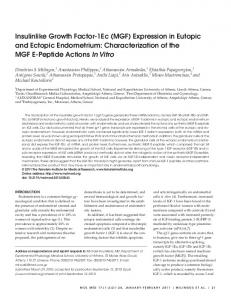
IGF-I - + - + - + - + - + FIG.2. In uiuo IGF-I stimulationof tyrosine phosphorylationof IGF-I receptor and ppl85. Subconfluent cells expressing IGF-I receptors WT1, FFF, NEO, WT2, or DCT were cultured in 6-well dishes, starved for 2 days in DMEM with 2% NCS, and incubated 3 min in the absence (-; lanes I , 3,5, 7, and 9) or in the presence of 50 nM IGF-I (+; lanes 2, 4, 6, 8, and 10). Cellular proteins were extracted in 2% SDS with 100 nM dithiothreitol, separated by SDS-polyacrylamide gel electrophoresis, and transferred to nitrocellulose. Qrosine-phosphorylated proteins were detected after incubation with anti-phosphotyrosine antibody and 1251-labeledprotein A followed by autoradiography.
normal binding affinity. Zn vivo ZGF-Z Receptor and pp185 prosine Phosphorylation -Cells expressing wild-type or mutant IGF-I receptors were incubated in theabsence or in thepresence of IGFrI (50 nM) for 3 min at 37 "C and cellular proteins were extractedand immuOB0 noblotted with anti-phosphotyrosine antibody. A protein of M , 120,000, with the same phosphotyrosine content, was present in all cell lines and showed no IGF-I stimulation (Fig. 2). The tyrosine phosphorylation of the p-subunit of the WT1, WT2, and DCT receptors was stimulated 10-20-fold by 50 nM IGF-I, whereas the FFF receptor showed no tyrosine phosphorylation. IGF-I stimulation of NE0 cells resulted in %fold increased phosphorylation of the p-subunit of the IGF-I receptor. A phosphotyrosine-containing protein of M, 185,000 was seen after IGF-I stimulation of cells with WT1, WT2, and DCT receptors and probably represents IRS-1, which is also phosphorylated by IGF-I receptors (Myers et al., 1993). The increase in 1%-1 phosphorylation afterstimulation with IGF-I is less pro0 25 50 75 nounced in WT1 cells than inWT2 and DCT cells, as expected lGF4 bound (fmo1/10,000 cells) from their 3-4-fold lower level of receptor expression (cf Table FIG.1. Scatchard plotsof lmI-IGF-Ibinding to NIH-3T3 fibro- I). IRS-1 phosphorylation could not be detected in IGF-I-stimublasts transfected with wild-type and mutant humanIGF-I re- lated FFF cells. Finally, a very faint phosphoprotein with M , ceptor cDNA. Suspension of cells expressing IGF-I receptors WT1 (V), 185,000 was detectable in IGF-I-stimulated N E 0 cells on the WT2 (O),DCT (A),FFF (O),or NE0 (0) were incubated for 20h at 4 "C with 15 PM 1251-IGF-I(40,000 c p d m l ) in the absence or presence of autoradiogram (data not shown). In IGF-I-stimulated WT1, unlabeled IGF-I. Receptor-bound 12"I-IGF-Iwas determined afterwash- WT2, and DCT cells, several other tyrosine-phosphorylated ing the cell pellets twice, counting of radioactivity, and correction for proteins with M , values of40,000-90,000 were seen. These nonspecific binding determined in the presence of 100 nM unlabeled phosphoproteins may either be degradation products of the IGF-I. Data were analyzed in a Scatchard plot of boundfree IGF-I versus bound IGF-I (Gammeltoft, 1990). The data arerepresentative of tyrosine-phosphorylated IGF-I receptors or still unidentified three experiments, which were used for calculation of Kd and IGF-I- endogenous substrates for the IGF-I receptor tyrosine kinase. binding sitedcell in Table I. In conclusion, the IGF-I receptor mutant lacking the three
hP \. \
IGF-IReceptor nrosine Kinase
23438
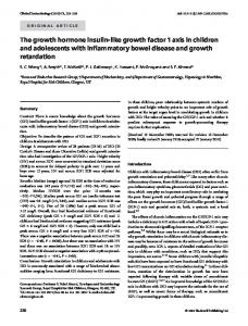
putative autophosphorylation sites shows abolished receptor and substratephosphorylation in vivo, suggesting thatat least one of the three tyrosines is necessary for tyrosine kinase activation, whereas the carboxyl terminus of the IGF-I receptor P-subunit is not involved in autophosphorylation of the IGF-I receptor and activation of the kinase towards cellular substrates. In Vitro Kinase Activity-IGF-I receptors were solubilized from W T 1 , DCT, FFF, and NE0 cells and partially purified by WGA chromatography. The kinase activity was assayed as the ability toincorporate 32Pinto a synthetic substrate,poly(GlusoTyrzo), after stimulation withIGF-I. As seen inFig. 3, partially purified WT1 and DCT receptors showed significantIGF-Istimulatedkinase activitytoward poly(Gluao-Tyrzo)with a maximal stimulation of 3.5- to 4.5-fold relative to the basal level and EDs0 values about 0.3 m. The kinase activity of purified NE0 receptors was stimulated 2.5-fold by IGF-I with ED50 of about 0.3 nM. In contrast, theFFF receptor kinase was not stimulated by IGF-I. From these resultswe conclude, that at least one of the three putative autophosphorylation sites (Tyr1131,Tyr1135, and Tyr1136) in the kinase domain of the IGF-I receptor is crucial for IGF-I-dependent activation of the kinase against exogenous substrates, whereas the carboxyl-terminal domain is not necessary for activation. Zn Vitro Autophosphorylation of the ZGF-Z Receptor -Autophosphorylation of equal amounts of partially purified WT1, DCT, and FFF receptors was measured during a 2-min incubation with [y-32P]ATP.32Pincorporation into the P-subunit of the WT1 receptor (M,95,000)was stimulated approximately 10-fold by addition of IGF-I, whereas the phosphorylation of the DCT receptor P-subunit (M,90,000) was stimulated approximately &fold by IGF-I (Fig. 4). FFF receptors showed low basal phosphorylation and no IGF-I-stimulated autophosphorylation. These datashow that thelevel of IGF-I-stimulated autophosphorylation of the IGF-I receptor is dependent on the presence of at least one of the tyrosines Tyr1131, Tyr1135, and Tyr"36, but not of the carboxyl-terminal domain. ZGF-Z Internalization and Degradation-The ability of the transfected wild-tme and mutant IGF-I recentors to mediate
8
determined as the amountof trichloroacetic acid-soluble radioactivity in the incubation medium. As seen in Fig. 5, cells expressing WT1, WT2, or DCT receptors internalized 1 5 2 0 % of added 1251-IGF-I.In contrast, incells with FFF receptors, only about 3% of added lZ5I-IGF-Iwas internalized after 60 min of incubation, whereas most of the cell-associated radioactivity was presenton the cell surface. Receptor-mediated internalization of IGF-I was followed bydegradation of IGF-I. After 60 min of incubation of cells expressing W T 1 , WT2, or DCT receptors about 20% of added lZ5I-IGF-Iwas trichloroacetic acid-soluble in theincubation medium,whereas only about 2% trichloroacetic acid-soluble lZ5Iactivity could be detected in the medium after incubation with FFF cells. N E 0 cells internalized and degraded only about 5%of added lZ5I-IGF-Iin accordance with their lower receptor number compared with W T 1 , WT2, and DCT cells (cf. Table I). We conclude that receptor-mediated internalization and degradation of IGF-I is dependent on a functional tyrosine kinase, whereas the carboxyl-terminal domain is dispensable. ZGF-I-stimulated DNA Synthesis-The stimulation of [3H]thymidine incorporationinto DNA by IGF-I was measured after exposing stanred cells to IGF-I for 17 h (Fig. 6). The M, (x10")
1
200
-
116
-
DCT 3 4
WTI 2
FFF 5 6
n
92
U
66 IGF-I
-
+
-
+
-
+
A
20
v
El
.O
I5
m 0
P
."9
6
10
5
0
0
WTl 0
0
PPF 0.3
DCT [=3 3
NE0
=
WT2 WTl 100
FIG.3. In v i h l n a s e activity of partially purified IGFd receptors toward synthetic substrate. Equal amounts of WGA-puntied IGF-I receptors W T 1 , FFF, DCT, and N E 0 were incubated for 60 min a t 23 "C in the absence ( 0 ) or presence of IGF-I in nanomolar concentrations (0.3,3,or 100). The phosphorylation reaction was initiated by simultaneous addition of 50 PM [y-32PlATP and 0.28 mg/ml p~ly(Glu~~-'Q continued r ~ ~ ) , for 5 min at 23 "C, and stopped by precipitation of proteins on filterpaper in10% trichloroacetic acid with sodium pyrophosphate. After washing in 5% trichloroacetic acid with sodium pyrophosphate, the filterswere counted in a p-counter. The results are expressed as incorporated 3zPactivity in cpm. Data are mean values* S.D. of three experiments.
m W a fe 8
FFT Ewlhtoh
DCT
NE0
o d e -
FIG. 5. IGF-I internalization and degradation byNlH-STS fibroblasts expressing wild-typeandmutant IGF-Ireceptors. Cells expressing IGF-I receptors WT1, WT2, FFF, DCT, or NE0 were incubated for 60 min at 37 "C with 30 PM 12sI-IGF-I(80,000 cpdml). The medium was collected and degraded 1251-IGF-Iwas determined as 1251activity soluble in 20% trichloroacetic acid (degraded). Surfacebound lzaI activity was released by incubating thecells for 5 min a t 0 "C with 0.2 M acetic acid and 0.5 M NaCI, pH 2.5 (surface). Internalized 12sI activity was extracted in0.2 M NaOH (intern.).Results were expressed as percentage of total added llSI activity. Data are mean values * S.D. of three experiments.
IGF-I Receptor nrosine Kinase
23439
tyrosines (Tyr1131, Tyr1135, and T y r 1 1 3 9 in thekinase domain of the IGF-I receptor is needed for activation of the tyrosine kinase and phosphorylation of cellular substrates in the signal pathway. Our observations with the IGF-I receptor are in agreement with previous studies of the insulin receptor where mutations of the threeautophosphorylation sites in the kinase domain resulted in an inactive receptor (Ellis et al., 1986; Zhang et ul., 1991; Murakami and Rosen, 1991; Wildenet al., 1992a, 1992b).Thus, the role of the threehomologous tyrosines in the kinase domains of the insulin receptors and IGF-I receptors is similar with respect to kinase activation and signaling. In arecent study, Kat0 et al. (1993)demonstrated that a kinasedeficient IGF-I receptor mutant in which Lys1003in the ATP" 0 binding site of the kinase domain had been replaced by alanine 0 0.1 1 10 or arginine was inactive with respect to induction of biological [IOF-11 (nM) responses. These data are in agreement with our conclusion FIG.6. IGF-I-stimulatedDNA synthesis in N"3T3 fibroblasts that thetyrosine kinase activity of the IGF-I receptor is necesexpressing wild-type and mutant human IGF-I receptors. Subconfluent cells expressing IGF-I receptors W T 1 (V),WT2,).( DCT (A), sary for activation of the IGF-I-stimulated signal transduction FFF (O),and NE0 (0) were cultured in 96-well dishes and starved for cascade. 2 days in DMEM with 2% NCS. Cells were cultured for 17 h at 37 "C in The mutant IGF-I receptor truncated at amino acid 1289and the absence or presence of 0.1-10 nM IGF-I or 5% NCSfollowedby lacking the carboxyl-terminal 49 amino acids of the P-subunit pulse-labeling for 3 h at 37 "C with [3Hlthymidine.Cells were solubiwas fully active, suggesting that the IGF-I receptor carboxyl lized in 0.2 M NaOH and harvested, and the DNA was collected on terminus is not involved in regulation of kinase activity or filters. After washing, the incorporated radioactivity was counted in a p-counter. The results are expressed as percentage of t3H1thymidine stimulation of mitogenesis. Our data are inagreement with a incorporation relative to stimulation with 5% NCS. Data are mean recent study of a mutant insulin receptor truncated at residue values * S.D. of four experiments. 1301 and lacking the carboxyl-terminal 43 amino acids of the P-subunit, which was fully active with respect to kinase actipassage of NIH-3T3 fibroblasts used for transfection showed vation and internalization as well as metabolic and mitogenic almost no IGF-I-stimulated DNA synthesis under the assay responses in Chinese hamster ovary cells (Myers et al., 1991). conditions used when measured in NE0 cells transfected with In contrast, other studies have reported that this truncated the neomycin resistance plasmid alone. Overexpression of WT insulin receptor shows impaired metabolic signaling and augreceptors increased the ability of the cells to respond to IGF-I mented mitogenic response in Rat-1 fibroblasts, suggesting with an EDE0of approximately 1 m. The maximal response that the carboxyl terminus may play a role in discriminating depended on the number of receptors, inasmuch as WT2 cells between metabolic and mitogenic actions of insulin (Maegawa expressing 2.7 x lo6 IGF-I binding sitedcell showed 2.5-fold et al., 1988; Thies et al., 1989). Furthermore, substitution of higher maximal response than WT1 cells expressing 0.87 x lo6 two carboxyl-terminal autophosphorylation sites, T y r 1 3 1 6 and IGF-I binding sitedcell. Overexpression of FFF receptors did Tyr1322, in the insulin receptor by phenylalanines enhanced not increase the responsiveness to IGF-I of [3Hlthymidine in- insulin-induced mitogenesis in both Rat-1fibroblasts and Chicorporation, inasmuch as FFF cells expressing 1.1x lo6 IGF-I nese hamster ovary cells suggesting that the phosphorylation binding sitedcell were unresponsive. Finally, cells expressing of these tyrosines may play an inhibitory role in the mitogenic the mutantDCT receptor with 3.9 x lo6 IGF-I binding sitedcell signaling of the insulin receptor (Takata et al., 1991; Ando et showed the same sensitivity and responsiveness to IGF-I as al., 1992). On the other hand, Murakami and Rosen (1991) insulin receptor with Phe1316 and WT2 cells expressing an equivalent amount of receptors. From found thatthemutant these results we conclude that at least one of the tyrosines Phe1322 showednormal biological activity in Chinese hamster (Tyr1131,Tyr1135, and Tyr1136) in the tyrosine kinase domain of ovary cells. At present these conflicting data with the insulin the IGF-I receptor, but not the carboxyl-terminal domain, is receptor are difficult to reconcile. In the IGF-I receptor a tyrosine residue, Tyr1316, is conserved in thehomologous positionto important for the mitogenic response of IGF-I. Tyr1322 in the insulin receptor, whereas a phenylalanine, DISCUSSION Phe1310, is found in the homologous position to Tyr1316in the The molecular basis of signal transduction by insulin recep- insulin receptor. It is not known whether Tyr1316 in the IGF-I tor and IGF-I receptor tyrosine kinases remains an enigma. receptor is autophosphorylated after IGF-I stimulation. The two receptors show remarkable structural similarity in We interpret the dataof the present study to mean that the their cytoplasmic domains and shareat least one common sub- structural basis for activation of tyrosine kinase and signaling strate pp185 or IRS-1 (Izumi et al., 1987; Kadowakiet al., 1987; of the IGF-I receptor is strikingly similar to that of the insulin Shemer et ul. 1987;Myers et al., 1993). Insulin and IGF-I receptor. Like other receptor tyrosine kinases, phosphorylation nevertheless elicit different effects on cellular metabolism and of tyrosine residues in thekinase domain has at least one of two growth in vivo. In the present study we have investigated the functions: modulation of kinase activity and recognition of celregulatory functions of the tyrosine kinase domain and the lular substrates in the signal pathway. Three putative autocarboxyl-terminal domain in the IGF-I receptor with respect to phosphorylation sites in insulin receptors and IGF-I receptors kinase activation and mitogenic signaling. Replacement of are located at homologous positions in an almost identical kiTyr1131, Tyr1135, and Tyr1136in the kinase domain by phenyl- nase domain and seem to serve the same function in theIGF-I alanines abolished IGF-I-stimulated kinase activity, IN-1 receptor with respect to kinase activation and biological actions phosphorylation, receptor internalization, and DNA synthesis, as in theinsulin receptor (Wilden et al., 1992a, 1992b).Mutawhereas truncation of the 49 carboxyl-terminal amino acids tion of the ATP-binding site in insulin receptors and IGF-I was without effect on these receptor functions. receptors blocks kinase activity and most, if not all, of the The lack of kinase activity and biological activity of the FFF actions of insulin and IGF-I, respectively (Ebina et al., 1987; mutant suggests that autophosphorylation of at least one ofthe Kat0 et al., 1993). The carboxyl-terminal domains in the
23440
IGF-I Receptor KinaseDrosine
@-subunitsof insulin receptors and IGF-I receptors are divergent but seem,overall, to play a minimal role in regulation of receptor kinase activity and cellular effects. It is possible, however, that autophosphorylation of 'I'yr1316 in the insulinreceptor carboxyl terminus may exert an inhibitory role on the mitogenic activity, depending on the cell type used for the analysis (Takata et al., 1991; Ando et al., 1992). Analysis of a mutant IGF-I receptor with replacementof Phe1310 bytyrosine, making it similar to the insulin receptor, may elucidate thequestion of whether autophosphorylation of this site serves as negative regulation of mitogenic activity. Thejuxtamembrane domain of theinsulin receptor and IGF-I receptor show 50% amino acid sequence identity, including theautophosphorylation site Tyrg60 in the insulinreceptor, receptor (Ullrichet al., which corresponds to TyrS5* in the IGF-I 1985, 1986). Mutation of 'I'yr960 to phenylalaninein the insulin receptor abolishes signal transduction and IRS-1phosphorylation, whereas receptor autophosphorylation is normal (White et al., 1988b; Murakami and Rosen, 1991). The same phenotype has been described recently for the IGF-I receptor mutated a t the homologous position, Tyrg50 (Yamasaki et al., 1992). Although the structure-function relationship of insulin and IGF-I receptor kinases seems to be similar, a previousstudy by Lammers et al. (1989) using chimerasof the extracellularportion of the insulin receptor and the intracellular portion of the IGF-I receptor indicated that the IGF-Ireceptor @-subunit issignificantly more potent than the insulinreceptor @-subunitin mitogenic signaling. This observation suggests that domains in the IGF-I receptor P-subunit other than the juxtamembrane domain, the ATP-binding site, the regulatory kinase domain, and the carboxyl terminus are associated with the higher mitogenic potency. The similarity in the molecular mechanism of signal transduction of insulin receptors and IGF-I receptors extends beyond the receptor tyrosine kinase to the major endogenous substrate, IRS-1 or pp185, which is phosphorylated by both receptors and is a common element in the activation of phosphoinositol3'-kinase by insulin and IGF-I (Myers et al., 1993). Thus, the initial steps in the signal transduction of insulin and IGF-I show striking homology. Taken together, these datasuggest that thedifference in thebiological actions of insulin and IGF-I in vivo may be due to the differential expression and different ligand binding of their receptors rather than divergent signal pathways.
Ando, A., Momomura, K., Tobe, K., Yamamoto-Honda, R., Sakura, H., Tamori, Y., Kaburagi, Y., Koshio, 0..Akanuma, Y., Yazaki, Y.,Kasuga, M., and Kadowaki, T.(1992)J. Biol. Chem. 267, 1278S12796 Czech, M. P. (1989)Cell 69, 235-238 Drejer, K., Kruse, V., Larsen, U. D., Hougaard, P., Bj~irn,S., and Gammeltoft, S. (1991)Diabetes 40, 148&1495 Ebina, Y., Ellis, L., Jarnagin, K., Edery, M., Graf, L., Clauser, E., Ou, J.-h., Masiarz, E , Kan, Y W., Goldfine, I. D., Roth, R. A., and Rutter, W. J. (1985)Cefl 40, 747-758 Ebina, Y., Araki, E., Taira, M., Shimada, F., Mori, M., Craik, C. S., Siddle, K., Pierce, S. B., Roth, R. A,, and Rutter, W. J. (1987)Proc. Natl. Acad.Sci. U. S. A . 84, 704-708 Ellis, L., Clauser, E., Morgan,D. O., Edery, M., Roth, R. A,, and Rutter,W. J. (1986) Cell 46, 721-732 Gammeltoft, S. (1989)in Peptkde Hormones as Prohormones: Processing,Biological (Martinez, J., ed) pp. 17G210, Ellis Honvood, ActivityandPharmacology Chichester, United Kingdom Gammeltoft, S. (1990)in Peptide Hormone Action (Siddle, K., and Hutton, J. C., eds) pp. 1 4 1 . IRL Press, Oxford Giorgino, F., Chen,J.-H., and Smith, R. J. (1992)Endocrinology 130, 1433-1444 Graham, F. L., and Van der Eb, A. J. (1973)virology 62,456467 Humbel, R. F. (1990)Eur. J . Biochem. 190,445462 Izumi, T., White, M. F., Kadowaki, T., Takaku, F., Akanuma, Y., and Kasuga, M. (1987)J. Biol. Chem. 262, 1282-1287 Kadowaki, T., Koyasu, S., Nishida, E., Tobe, K., Izumi, T., Takaku, F., Sakai, H., Yahara, I., and Kasuga, M. (1987)J. Biol. Chem. 262, 7342-7350 Kato, H., Faria, T. N., Stannard, B., Roberts, C. T., Jr., and LeRoith, D. (1993)J. Biol. Chem. 268, 2655-2661 Lammers, R.,Gray, A,, Schlessinger, J., and Ullrich, A. (1989)EMBO J. 8, 13691375 Maegawa, H., McClain, D. A,, Freidenberg, G . , Olefsky, J. M., Napier, M., Lipari, T.,Dull, T. J., Lee, J., and Ullrich, A. (1988)J. B i d . Chem. 263, 891243917 Murakami, M. S., and Rosen, 0.M. (1991)J . Biol. Chem. 266,22653-22660 Myers, M. G., Jr., Backer, J. M., Siddle, K., and White, M. F. (1991)J.Biol. Chem. 266, 10616-10623 Myers, M. G., Jr., Sun,X.J., Cheatham,B., Jachna, B. R., Glasheen, E. M., Backer, J. M., and White, M. F. (1993)Endocrinology 132, 1421-1430 Nielsen, F. C., Wang, E., and Gammeltoft,S. (1991)J . Neurochem. 66, 12-21 Shemer, J.,Adamo, M., Wilson, G . L., Heffez, D., Zick, Y.,and LeRoith, D. J. (1987) J . Biol. Chem. 262, 1547615462 Shoelson, S. E., Chatterjee,S., Chaudhuri, M., and White, M. F. (1992)Proc. Natl. Acad. Sci. U. S. A. 89,2027-2031 Sun, X. J., Rothenberg, P., Kahn, C. R., Backer, J . M., Araki, E., Wilden, P. A., Cahill, D. A,, Goldstein, B. J., and White, M. F. (1991)Nature 362, 73-77 Takata, Y.,Webster, N. J. G . , and Olefsky, J. M. (1991)J. B i d . Chem. 266, 91359139 Thies., R. S., Ullrich, A,, and McClain, D.A. (1989)J. Biol. Chem. 264, 1282012825 Tornqvist, H. E., Pierce, M. W., Frackelton, A. R., Nemenoff, R. A., and Avruch,J. (1987)J. Biol. Chem. 262, 10212-10219 Ullrich, A., and Schlessinger, J. (1990)Cell 61,203-212 Ullrich, A,,Bell, J . R., Chen, E.Y., Herrera, R., Petruzzelli, L. M., Dull, R. J.,Gray, A., Coussens, L., Liao, Y.-C., Tsubokawa, M., Mason, A., Seeburg, P. H., Grunfeld, D., Rosen, 0. M., and Ramachandran, J. (1985)Nature 313,756761 Ullrich, A., Gray, A. Tam, A. W., Yang-Feng, T., Tsuhokawa, M., Collins, C., Henzel, W., Bon, T.L., Kathuria, S., Chen, E., Jacobs, S., Francke, U., Ramachandran, J., and Fujita-Yamaguchi, Y. (1986)EMBO J. 5,2503-2512 Verland, S., and Gammeltoft, S. (1989)Mol. Cell. Endocrinol. 67,207-216 White, M.F., Shoelson, S. E., Keutmann, H., and Kahn, C.R. (1988a)J . B i d . Chem. 263,2969-2980 White, M. F., Livingston, J. N . , Backer, J. M., Lauris, V., Dull, T., Ullrich, A., and Acknowledgments-We thank Stina Olsen and Birte Kofoed f o r inKahn, C. R. (1988b)Cell 64,641449 valuable technical assistance. We gratefully acknowledge Dr. Robert J. Wiborg, O., Pedersen, M. S., Wind, A,, Berglund,L. E., Marcker, K. A., and Vuust, Smith for the gift of anti-phosphotyrosine antibody and Dr. Ulla DahlJ. (1985)EMBO J. 4,755-759 Larsen for the gift of [['2511monoiodo-Tyl-31]IGF-I.We thank Drs. Wilden, P. A,, Siddle, K., Haring, E., Backer, J. M., White, M. F., and Kahn, C. R. (1992a)J. Biol. Chem. 267, 13719-13727 Jonathan M. Backer and Bentley Cheatham for critical review of the Wilden, P. A., Kahn, C. R., Siddle, K., and White, M. F. (1992b)J. Biol. Chen. 267, manuscript. 16660-16668 Yamasaki, H., Prager, D., Gebremedhin, S., and Melmed, S. (1992)J. B i d . Chem. REFERENCES 267,20953-20958 Zhane, B., Tavare, J. M., Ellis, L., and Roth, R.A. (1991)J. Biol. Chem. 266, Andersen, A. S., Kjeldsen, T., Wiberg, F. C., Christensen, P. M., Rasmussen, J. S., Noms, K., Mdler, K. B.. and Mdler, N.P. H. (199O)Biochemistry 29.7363-7366 99&996